Exam #3 NURS230
 Call Kai
 Learn
Learn Practice Test
 Spaced Repetition
 Match
Match Flashcards
 Knowt Play
1/20

Earn XP
Description and Tags
LAST ONE YAY
Name | Mastery | Learn | Test | Matching | Spaced | Call with Kai | Chat |
|---|
No analytics yet
Send a link to your students to track their progress
21 Terms
Postpartum infection
-Defined as presence of a fever of 38 C or 100.4 F in the first 10 postpartum days (not including the first 24 hours after birth)
Common Types
Endometritis is the most common postpartum infection, occurring in ~2% after vaginal birth and 10-15% after cesarean birth. The highest incidence is associated with cesarean birth after prolonged labor and rupture of membranes.
Infection of the lining of the uterus
Fundala tenderness, febrile
Prolonged rupture of membranes, manual introduction of bacteria
Wound infections are also common, often developing after discharge home. Rates after cesarean birth are 3-5%. Women can also develop infection in perineal lacerations or episiotomy sites.
Key Signs and Symptoms
General indicators:
Fever and temperature elevation
Chills
Localized tenderness or pain
Discoloration and swelling
Endometritis-specific:
Fever (usually >38°C)
Increased pulse
Anorexia, nausea, fatigue, lethargy
Pelvic pain and uterine tenderness
Foul-smelling lochia
Wound infection-specific:
Erythema, edema, warmth
Tenderness and pain
Seropurulent drainage
Wound separation
Treatment
Endometritis: IV broad-spectrum antibiotics (clindamycin and gentamicin, cephalosporins, or penicillins) with supportive care. Antibiotic therapy is usually discontinued 24 hours after the client is afebrile and asymptomatic.
Wound infections: IV antibiotic therapy. When pus or significant drainage is present, the wound is opened, drained, and irrigated with normal saline; healing occurs by secondary intention.
Prevention
Most effective treatment is prevention:
Good prenatal nutrition to reduce anemia risk
Proper maternal perineal hygiene with thorough hand hygiene
Strict aseptic technique by all healthcare professionals
Prophylactic antibiotics during labor and cesarean surgery
Critical Nursing Considerations
Early discharge challenge: Women are usually discharged by 48 hours after vaginal birth, often before signs of infection are evident. You must:
Identify at-risk clients
Provide anticipatory teaching before discharge
Teach signs of infection and when to contact the provider
Teaching priorities:
Hygienic care techniques (changing pads front to back, hand hygiene before/after perineal care)
Signs of worsening condition
Adherence to treatment plan
Need for follow-up care
Endometritis
Endometritis is an infection of the uterine lining that usually begins as a localized infection at the placental site but can spread to the entire endometrium 1.
Incidence
Most common puerperal infection1
~2% after vaginal birth 1
10-15% after cesarean birth 1
Highest incidence: Cesarean birth after prolonged labor and rupture of membranes 1
Signs and Symptoms
Fever (usually >38°C) 1
Increased pulse 1
Chills 1
Anorexia, nausea 1
Fatigue and lethargy 1
Pelvic pain 1
Uterine tenderness 1
Foul-smelling lochia1
Laboratory Findings
Leukocytosis 1
Markedly increased RBC sedimentation rate 1
Anemia may be present 1
Blood cultures or intracervical/intrauterine bacterial cultures are not clinically useful and increase cost of care 1
Treatment
Medical management 12:
IV broad-spectrum antibiotic therapy: clindamycin and gentamicin, cephalosporins, or penicillins
Supportive care: hydration, rest, pain relief
Antibiotics discontinued 24 hours after client is afebrile and asymptomatic12
Comfort measures 2:
Cool compresses
Warm blankets
Perineal care
Sitz baths
Prevention
Prophylactic antibiotics administered during labor and during cesarean surgery can help reduce incidence and severity 1.
Ongoing Nursing Care
Throughout treatment 12:
Assess lochia
Monitor vital signs
Evaluate changes in client's condition
Patient teaching 2:
Side effects of therapy
Prevention of infection spread
Signs/symptoms of worsening condition
Adherence to treatment plan
Need for follow-up care
Additional support 2:
Maintain mother-infant interactions
Support breastfeeding continuation
Key Nursing Consideration
Assessments and monitoring continue throughout the entire treatment period, not just until fever resolves
Wound infection
Wound infections are common postpartum infections that often develop after discharge home. Rates after cesarean birth are 3-5%. Women can also develop infection in the perineum at a repaired laceration or episiotomy site.
Risk Factors
Predisposing factors are similar to those for endometritis, including:
Perineal lacerations and episiotomies (disruption of skin integrity) and cellulitis (abscess of wound)
Signs and Symptoms
Fever
Erythema (redness)
Edema (swelling)
Warmth
Tenderness and pain
Seropurulent drainage
Wound separation
Diagnosis
Wound exudate may be cultured to identify the causative organism.
Treatment
Medical management:
IV antibiotic therapy
When pus or significant serosanguineous effusion is present, the wound is opened and drained
Wounds are irrigated with normal saline and redressed several times daily
Healing occurs by secondary intention
In some cases, a wound vacuum device is used
Antibiotic treatment continues until the base of the wound appears clear and there are no signs of cellulitis
Nursing Care
In-facility care:
Frequent assessments of temperature and vital signs
Wound assessment and care
Comfort measures: analgesics, sitz baths, warm compresses, perineal care
Patient teaching:
Hygienic care techniques:
Changing perineal pads front to back
Cleansing from front to back (urethra to anus)
Hand hygiene before and after perineal care
Self-care measures
Signs of worsening conditions to report to the provider
Post-discharge care:
Wound care and assessment continue after discharge
Woman and family instructed in wound care and dressing changes
Home visits by nurses may be provided to assess wound, reinforce teaching, and offer support
Prevention
Proper perineal care helps prevent infection and aids healing :
Wipe front to back after voiding or defecating
Use squeeze bottle with warm water or antiseptic solution after each voiding
Change perineal pad front to back with each void or bowel movement
Wash hands thoroughly before and after
Endometritis
Endometritis is an infection of the uterine lining that usually begins as a localized infection at the placental site but can spread to the entire endometrium.
Incidence
Most common puerperal infection
~2% after vaginal birth
10-15% after cesarean birth
Highest incidence: Cesarean birth after prolonged labor and prolonged rupture of membranes and manual introduction of bacteria
Signs and Symptoms
Fever (usually >38°C)
Increased pulse
Fundal tenderness, febrile
Chills
Anorexia, nausea
Fatigue and lethargy
Pelvic pain
Uterine tenderness
Foul-smelling lochia
Laboratory Findings
Leukocytosis
Markedly increased RBC sedimentation rate
Anemia may be present
Blood cultures or intracervical/intrauterine bacterial cultures are not clinically useful and increase cost of care
Don’t usually culture, just treat
Treatment
Medical management:
IV broad-spectrum antibiotic therapy: clindamycin and gentamicin, cephalosporins, or penicillins
Supportive care: hydration, rest, pain relief
Antibiotics discontinued 24 hours after client is afebrile and asymptomatic
Comfort measures:
Cool compresses
Warm blankets
Perineal care
Sitz baths
Prevention
Prophylactic antibiotics administered during labor and during cesarean surgery can help reduce incidence and severity.
Ongoing Nursing Care
Throughout treatment:
Assess lochia
Monitor vital signs
Evaluate changes in client's condition
Patient teaching:
Side effects of therapy
Prevention of infection spread
Signs/symptoms of worsening condition
Adherence to treatment plan
Need for follow-up care
Additional support:
Maintain mother-infant interactions
Support breastfeeding continuation
Key Nursing Consideration
Assessments and monitoring continue throughout the entire treatment period, not just until fever resolves
Urinary Tract Infections (UTIs)
-Important to note:
Get UA
Foley increases risk of UTI
UTIs are a common medical complication of pregnancy, occurring in up to 20% of all pregnant women and responsible for 10% of all hospitalizations during pregnancy .
Most common causative organisms :
Escherichia coli: Responsible for at least 80% of initial cases and ~70% of recurrent cases
Klebsiella pneumoniae and Proteus species (especially in women with recurrent infections)
Gram-positive organisms (group B streptococci, enterococci, staphylococci): Up to 10% of infections
Signs and Symptoms
Although UTIs can be asymptomatic, typical symptoms include:
Frequency
Urgency
Dysuria
Burning
Suprapubic pain
Dribbling
Hesitancy
Gross hematuria (can occur)
Women should be instructed to inform their healthcare provider promptly if they experience these symptoms.
Complications
Pyelonephritis (renal infection) is the most frequent serious nonobstetric medical complication of pregnancy and a leading cause of septic shock during pregnancy. Maternal complications include sepsis, acute respiratory distress syndrome, and preterm labor.
Prevention Strategies
Hygiene practices:
Proper hand hygiene before and after urinating
Wipe perineum front to back
Use soft, absorbent, white, unscented toilet tissue (harsh/scented paper can cause irritation)
Avoid bubble bath or bath oils (can irritate urethra)
Clothing choices:
Wear all-cotton undergarments and cotton-lined pantyhose
Avoid tight-fitting slacks or jeans for long periods
Prevent heat and moisture buildup in genital area
Fluid intake and urination habits:
Drink at least 2.5 L daily (8-10 glasses of 8 oz each), preferably water
Do not limit fluids to reduce urination frequency
Increase fluids if urine appears dark/concentrated
Do not ignore the urge to urinate (holding urine allows bacteria to multiply)
Urinate before bed at night
Urinate before and after sexual activity, then drink a large glass of water
Treatment
Oral antibiotics are commonly prescribed. Complete the entire course rather than stopping when feeling better to prevent drug-resistant organisms
What are other reasons for fever/infection symptoms?
-Pneumonia, flu or other virus, DVT/PE, mastitis
Mastitis will be febrile, flu-like symptoms, true infection
-Misoprostol can cause transient fever, not too high and no other symptoms
Postpartum bleeding and hemorrhage
All women who have given birth are at risk for excessive bleeding that can progress to postpartum hemorrhage (PPH). Excessive blood loss can occur during different time periods and from various causes.
Primary Causes
Uterine atony (failure of the uterine muscle to contract firmly) is the most frequent cause of excessive bleeding after birth. When atony occurs:
The relaxed uterus distends with blood and clots
Blood vessels in the placental site are not clamped off
Excessive bleeding results
Though the cause isn't always clear, uterine atony often results from retained placental fragments.
High Risks
High parity
Polyhydramnios
Macrosomia
Uterine fatigue/long labor
Tocolytic drugs
Other causes:
Vaginal or vulvar hematomas
Trauma (lacerations or genital tract or hematomas)
Unrepaired lacerations of the vagina or cervix
Incomplete placental separation
Excessive fundal manipulation or cord traction
Uterine prolapse
Late PPH: subinvolution, infection, retained placental fragments, or coagulopathy
Inversion of the uterus (potentially life threatening but rare)
Key Assessment Finding
Suspect lacerations or hematomas if excessive vaginal bleeding occurs with a firmly contracted uterine fundus.
Measuring Blood Loss
Quantification by weighing (1 mL = 1 g) is the most accurate method for objectively determining blood loss. Visual estimation alone is inaccurate.
Time factor is critical: A woman saturating a pad in 1 hour is bleeding much more heavily than one saturating a pad in 8 hours. Always ask when the pad was last changed.
Prevention - Two Most Important Interventions
Maintaining good uterine tone
Preventing bladder distention
Initial Management of Uterine Atony
Firm massage of the uterine fundus (FIRST THING)
Expression of clots
Get help, call code hemorrhage
Elimination of bladder distention
Continuous IV infusion of 10-40 units oxytocin in 1000 mL lactated Ringer's or normal saline
Additional Medications if Oxytocin Fails
Misoprostol (Cytotec): Can be given rectally, sublingually, or orally
Methylergonovine: IM for sustained contractions. Contraindicated with hypertension
Hemabate: 0.25 mg IM, every 15 minutes maximum 2 mg or 8 doses. Contraindicated with asthma
Tranexamic acid (TXA): Antifibrinolytic agent if initial therapy ineffective
Mastitis
-Important note from slides:
Usually stap aureus can be treated with antibiotics (ex. dicloxacillin, cephalexin)
Mastitis will be febrile, flu-like symptoms, true infection
Mastitis is an infectious process of the breast characterized by sudden onset of influenza-like symptoms.
Signs and Symptoms
Fever
Chills
Malaise
Body aches
Headache
Nausea and vomiting
Localized breast pain and tenderness
Hot, reddened area on the breast
Location and Timing
Most commonly occurs in the upper outer quadrant of the breast
One or both breasts can be affected
Most cases occur during the first 2 to 4 weeks postpartum, although mastitis can occur at any time
Causative Organisms
The most common pathogens are:
Staphylococcus
Streptococcus
Escherichia coli
Risk Factors
Inadequate emptying of the breasts is common and can be related to:
Engorgement
Plugged ducts
Sudden decrease in number of feedings
Abrupt weaning
Wearing underwire bras
Other predisposing factors:
Sore, cracked nipples (provide portal of entry for organisms)
Stress and fatigue
Maternal illness
Ill family members
Breast trauma
Poor maternal nutrition
Treatment
Bedrest
Antibiotics: dicloxacillin, cephalexin, or clindamycin
Pain and swelling reduction: anti-inflammatory medications and cold compresses
Continued lactation
Complete emptying of the breasts through breastfeeding, hand expression, or pumping
Adequate fluid intake and balanced diet
Important Points
The infection cannot be transmitted to the infant
Breastfeeding should continue during treatment
Mothers should be taught signs of mastitis before discharge from the birth facility
Women need to know to call their healthcare provider promptly if symptoms occur
Complications
If not treated early, complications can include:
Breast abscess
Chronic mastitis
Fungal infections of the breast
Most complications can be prevented by early recognition and treatment
Breastfeeding positions
Traditional Breastfeeding Positions
Four traditional positions:
Football or clutch hold (under the arm)
Across the lap (cross-cradle or modified cradle)
Cradle
Side-lying
Semireclining Position
For initial feedings, a semireclining position can be advantageous:
Mother is in a semireclining position
Newborn lies prone, skin-to-skin on mother's bare chest
Mother's body supports the baby
Benefits: Mother is more relaxed, nipple pain is reduced or eliminated, more freedom of movement, baby can use inborn reflexes to latch and feed effectively
Some mothers prefer this position even after early days
Football or Clutch Hold
Often recommended for early feedings because the mother can see the baby's mouth easily as she guides the infant onto the nipple
Especially helpful for:
Mothers who gave birth by cesarean (often prefer this position)
Late preterm and early term infants (helps avoid flexing the head, which can impede breathing and cause positional apnea)
Modified Cradle or Across-the-Lap Hold
Works well for early feedings, especially with smaller babies
Side-Lying Position
Allows the mother to rest while breastfeeding
Preferred by women with:
Perineal pain and swelling
Cradle Position
The most common breastfeeding position for infants who have learned to latch easily and feed effectively
Key Positioning Principles
Regardless of position:
Mother holds infant securely at the level of the breast
Supported by firm pillows or folded blankets
Baby facing toward mother
Baby's mouth is directly in front of the nipple
Mother supports baby's neck and shoulders with her hand (not pushing on the occiput)
Baby's body held in alignment (ears, shoulders, and hips in a straight line)
Recommendation
The mother should be encouraged to use the position that most easily facilitates latch while allowing maximal comfort. Before discharge, nurses can help mothers try all positions so they feel confident at home
Engorgement
Engorgement is a common response of the breasts to the sudden change in hormones and the onset of significantly increased milk volume during lactogenesis stage II. It usually occurs 3 to 5 days after birth as milk transitions from colostrum to mature milk.
What Happens Physiologically
Increased blood flow to the breasts
Increased uptake of glucose and oxygen
Milk production rapidly increases, potentially exceeding storage capacity of the alveoli
If milk is not removed, alveoli become distended, impairing capillary blood flow
Blood vessels become congested, fluid leaks into surrounding tissue causing edema
Milk ducts become compressed by tissue edema, preventing easy milk flow
Signs and Symptoms
Breasts become firm, tender, and hot
Appear shiny and taut
Areolae become edematous
Nipples can flatten, making it difficult for infant to latch
Risk Factors
Primiparas (first-time mothers)
Women who received large amounts of IV fluids during labor and birth
Women who had previous breast surgery
Infrequent or ineffective feedings during first 2-3 days after birth
Prevention
Early and frequent feedings (8-12 times in 24 hours)
Keeping mother and infant together to recognize and respond to feeding cues
Management for Breastfeeding Mothers
Feed frequently without limiting time on first breast
Use hand expression or breast pump to reduce engorgement
Apply ice packs to breasts
Apply cabbage leaves (replace when wilted)
Wear well-fitted support bra
Take antiinflammatory medications (e.g., ibuprofen) for discomfort
Management for Formula-Feeding Mothers
Wear well-fitted support bra continuously for at least 72 hours
Avoid breast stimulation (warm water, expressing milk)
Apply ice packs periodically
Use cabbage leaves
Take mild analgesic or antiinflammatory medication
Do NOT express breast milk (stimulates production and worsens engorgement)
Important Points
Engorgement is temporary, usually resolving within 24 hours
Back pressure on full milk glands inhibits milk production - if milk isn't removed, supply can diminish
Jaundice
-Important to note from slides:
RBCs have a shorter lifespan
Bili metabolism is initiated in the reticuloendothelial system, primarily liver and spleen
Bili binds tightly to albumin, its carrier protein enters the liver for conjugation and excretion
Can be treated with Phototherapy: UVB light, an overhead lights and blanket
Absorbed through skin, causes photochemical reaction with Bilirubin, facilitating dissolution of it for excretion
Types of Neonatal Jaundice
Physiologic jaundice occurs in approximately 60% of term newborns, appears after 24 hours of age, and usually resolves without treatment 3. Total serum bilirubin (TSB) levels peak at 5-6 mg/dL by 72-96 hours of life, then gradually decrease.
Two types related to breastfeeding:
Breastfeeding-associated jaundice (early-onset)
Occurs during the first week of life
Related to insufficient feeding and infrequent stooling
NOT caused by breastfeeding itself, but by lack of effective breastfeeding
Breast milk jaundice (late-onset)
Develops between 5-10 days of age
Infants typically thrive, gain weight, and stool normally
Bilirubin can remain elevated for 3-12 weeks
Etiology uncertain, possibly related to factors in breast milk that inhibit bilirubin conjugation
Why Jaundice Occurs
Bilirubin is excreted primarily through the intestines. When stooling is infrequent, bilirubin in stool is reabsorbed back into the infant's system, increasing bilirubin levels.
Prevention of Early-Onset Jaundice
Frequent breastfeeding: at least 8-12 times or more in 24 hours during first several days
Colostrum has a natural laxative effect and promotes early passage of meconium
Increased feeding frequency is associated with decreased bilirubin levels
Assessment
Any breastfeeding infant with jaundice should be evaluated for:
Weight loss greater than 7%
Decreased milk intake
Infrequent stooling or delayed stool transition
Decreased urine output (fewer than 4-6 wet diapers per day)
Bilirubin levels should be assessed by serum testing or transcutaneous monitoring.
Treatment of Early-Onset Jaundice
Evaluate breastfeeding: frequency, length, positioning, latch, milk transfer
Address factors interfering with effective feeding (sleepy infant, breast engorgement)
If infant not feeding effectively: mother can pump and feed expressed milk
Some cases may need formula supplementation
In presence of other risk factors, phototherapy may be required
Key Point
For breast milk jaundice, no intervention is usually necessary. These infants have no signs of hemolysis or liver dysfunction despite elevated bilirubin
Fetal Loss Communication
Caring Theory Framework
Swanson's caring theory provides a model for nursing practice with five key concepts:
Knowing: Assess how the woman, partner, and family perceive the loss and what it means to them
Being with: Provide caring presence that conveys acceptance of various feelings and perceptions
Doing for: Perform activities that provide physical care, comfort, and safety
Enabling: Offer options for care, provide information, articulate choices, and give support to help family feel more in control 5
Time and Presence
Create adequate time to engage with bereaved families without being rushed 1. Nurses may need adjusted assignments to provide sufficient attention to newly bereaved individuals.
Active Listening
Women who experienced stillbirth or infant death have a particularly strong need to share details about the birth 3. Actively listen and allow women to work through their birth and loss stories as they try to make sense of the experience.
Helping Parents See and Hold Their Baby
Prepare the baby with special care:
Bathe, apply lotion, comb hair
Place identification bracelets
Dress in diaper and special outfit
Wrap in soft blanket
Model appropriate behavior: Hold the baby close, touch a hand or cheek, use the baby's name, and talk about the infant's special features—this conveys it's okay for parents to do likewise.
If the baby has a congenital anomaly, help explain what they are seeing.
Respect Family Decisions
Nursery and baby items: Bereaved parents should make decisions about when and by whom these items are stored or given away 3. Well-meaning relatives who rush to disassemble the nursery may compound grief if parents weren't involved in the decision.
Follow-Up Care
A grief conference typically occurs as follow-up, where:
The loss is discussed in detail
Autopsy and genetic study results are shared
Parents can ask questions
Events of hospitalization are reviewed
Assessment of family coping occurs
Information about grief support groups is provided
This helps parents understand the cause of loss or accept that the cause may never be known
Neonatal Heat Loss
Four Mechanisms of Heat Loss
1. Convection Heat loss from warm body surface to cooler surrounding air 2. Air flow and velocity affect the rate of loss.
Occurs when drafts come from open doors, air conditioning, or even air currents created by people moving about
2. Conduction Heat loss from direct contact between the infant's skin and cooler surfaces.
Conduction occurs when the infant comes in contact with cold objects or surfaces such as a scale, a circumcision restraint board, cold hands, or a stethoscope
3. Radiation Heat loss to cooler surfaces not in direct contact with the infant.
Heat is loss by radiation when the infant is near cold surfaces. Thus, heat is lost from the infant’s body to the sides of the crib or incubator and to the outside walls and windows
4. Evaporation Heat loss when water on the skin surface converts to vapor.
Can occur during birth or bathing from moisture on skin, as a result of wet linens or clothes, and from insensible water loss
Why Newborns Are at Risk
Large body surface area in relation to weight
Limited fat stores and calorie reserves
Immature skin (especially preterm infants <28 weeks) causes greater transepidermal water loss
Goal: Neutral Thermal Environment (NTE)
The ideal environmental temperature that allows the newborn to maintain normal body temperature (36.5°C-37°C or 97.9°F-99°F) while minimizing oxygen and glucose consumption.
Prevention Strategies
Dry the infant quickly after birth
Skin-to-skin contact with mother reduces conductive and radiant heat loss
Place naked newborn on mother's bare chest, cover with warm blanket
Cap on infant's head helps conserve heat
For preterm infants: use prewarmed incubator or radiant warmer
For extremely low birth weight infants: place in polyethylene bag to decrease heat and water loss
Consequences of Hypothermia and Cold Stress
Early signs:
Pale, mottled skin
Cool extremities
Acrocyanosis
Physiologic effects:
Increased oxygen consumption and respiratory rate
Increased glucose consumption → risk of hypoglycemia
Vasoconstriction to conserve heat
Metabolic acidosis (if prolonged)
Apnea, bradycardia, central cyanosis (severe cases)
Diverts energy from normal brain/cardiac function and growth to heat production
Rewarming
If hypothermia occurs, slow rewarming is recommended:
External heat sources slightly warmer than skin temperature
Increase gradually to avoid apnea and acidosis
Use radiant heaters or heated mattresses
Cold Stress - Condensed
Definition: Metabolic and physiologic demands from uncorrected hypothermia.
Pathophysiology:
Norepinephrine → vasoconstriction → pale, mottled, cool skin
Increased O₂ consumption and metabolic rate → energy diverted from growth/normal function to heat production
Respiratory Effects:
↓ O₂ tension → pulmonary vasoconstriction → ↓ perfusion
↓ PO₂, ↓ pH, altered surfactant
Can cause/worsen respiratory distress
Metabolic Complications:
Metabolic acidosis: Anaerobic glycolysis → ↑ acid production
Hypoglycemia: Depleted glucose stores
Hyperbilirubinemia: Fatty acids displace bilirubin from albumin
Clinical Signs:
Pale, mottled, cool extremities
Acrocyanosis, respiratory distress
Apnea, bradycardia, central cyanosis (severe)
Key Point: Prevention is critical nursing role.
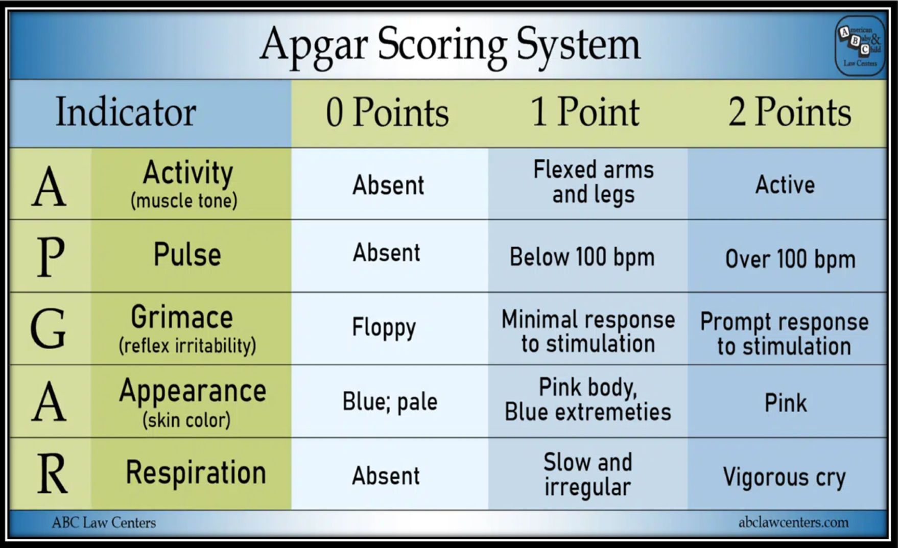
APGAR Score
What the Apgar Score Measures
The Apgar score is a routine rapid assessment of the newborn's overall status and response to resuscitation. It evaluates five signs indicating the physiologic state of the neonate:
Heart rate - auscultated with stethoscope or by palpating umbilical cord
Respiratory effort - observed chest wall movement
Muscle tone - degree of flexion and movement of extremities
Reflex irritability - presence of grimace, crying, or active withdrawal
Generalized skin color - described as pallid, cyanotic, or pink
Scoring and Interpretation
Score ranges:
0-3: Severe distress
4-6: Moderate difficulty
7-10: Minimal or no difficulty adjusting to extrauterine life
Timing of Assessment
Assigned at 1 and 5 minutes after birth
For scores less than 7 at 5 minutes, repeat assessment every 5 minutes for up to 20 minutes
Important Considerations
Who performs it: Nurse or birth attendant, depending on facility policy
What it does NOT do: Apgar scores do not predict future neurologic outcome for the newborn
What it DOES do: Useful in describing the newborn's transition to extrauterine environment and response to resuscitative efforts
Critical timing note: If resuscitation is required, it should be initiated before the 1-minute Apgar score is determined
Adjunct Assessment
When there has been an abnormal or confusing fetal heart rate tracing during labor or neonatal depression at birth, umbilical cord blood acid-base determination is a useful adjunct to the Apgar score 3. Cord blood values should be obtained when a newborn has an Apgar score of 5 or less at 5 minutes of age
Head to Toe Assessment Findings
Head and Skull
Proportions: Head is approximately one-fourth of total body length at term; appears large and heavy with face appearing small in relation to skull.
Molding: Shaping of fetal head by overlapping cranial bones during labor to facilitate birth canal passage.
Caput succedaneum: Generalized edematous area of scalp (usually occiput), present at birth, crosses suture lines, resolves spontaneously in 3-4 days. Results from sustained pressure against cervix causing venous congestion.
Cephalhematoma: Blood collection between skull bone and periosteum, does not cross suture lines, firmer and better defined than caput, resolves in 2-8 weeks. Can cause hyperbilirubinemia as it resolves. May be present in up to 25% of newborns with linear skull fractures.
Subgaleal hemorrhage: Bleeding beneath galea aponeurosis, associated with difficult operative vaginal birth (especially vacuum extraction). Results from traction/shearing forces pulling scalp away from bony calvaria.
Extremities
Symmetry: Should be symmetric and equal length.
Digits: Five fingers on each hand, five toes on each foot with nails present.
Oligodactyly: Missing digits
Polydactyly: Extra digits
Syndactyly: Fused digits
Hip assessment for Developmental Dysplasia of Hip (DDH):
More common in: breech presentations, firstborn infants, female infants, family history of DDH
Signs: Asymmetric gluteal/thigh skinfolds, uneven knee levels, positive Ortolani test, positive Barlow test
Gluteal and thigh skinfolds should be equal and symmetric
Legs should be equal length
Knee levels in flexion should be equal
Skeletal Injuries
Skull fractures:
Linear fractures: Most common, from compression during birth, usually no treatment needed
Depressed fractures: "Ping-pong ball" indentations from pressure on bony pelvis or forceps
Occipital osteodiastasis: Separation of occipital bone cartilaginous joint from traumatic breech births
Clavicle: Most commonly fractured bone during birth, typically in middle third
Cardiovascular System
Heart rate: Normal is 110-160 beats/min when awake; can range from 85-100 during deep sleep to up to 180 during crying 1.
Heart sounds: Should be clear and regular; murmurs are common in first few hours and usually benign 1.
Blood pressure: Average is 60-80 mmHg systolic and 40-50 mmHg diastolic at birth.
Peripheral pulses: Should be palpable and equal bilaterally (brachial, radial, femoral).
Respiratory System
Respiratory rate: Normal is 30-60 breaths/min; counted for full minute.
Breathing pattern: Primarily diaphragmatic and abdominal; may be shallow and irregular with brief pauses (less than 20 seconds).
Chest: Should be symmetric with equal bilateral breath sounds.
Signs requiring attention: Nasal flaring, grunting, retractions, tachypnea (>60 breaths/min), or apnea (pauses >20 seconds).
Integumentary System
Color: Pink or ruddy; acrocyanosis (bluish hands/feet) is normal in first 24 hours.
Vernix caseosa: Protective white cheesy substance, more abundant in term infants.
Lanugo: Fine downy hair, especially on shoulders, back, and forehead.
Milia: Small white papules on nose, chin, forehead from sebaceous gland retention.
Erythema toxicum: Pink papular rash with vesicles on trunk/extremities, appears 24-48 hours after birth, resolves spontaneously.
Neurologic Assessment
Muscle tone: Should have flexed posture with resistance to extension.
Key reflexes:
Moro (startle): Arms extend then flex with crying when startled
Rooting: Turns toward touch on cheek
Sucking: Strong coordinated suck
Grasp: Fingers curl around object placed in palm
Babinski: Toes fan outward when sole stroked
Abdomen
Shape: Rounded, soft, moves with respirations.
Umbilical cord: Should have three vessels (two arteries, one vein); clamp should be secure.
Bowel sounds: Present within first hour after birth
Genitourinary System
Female genitalia:
Labia majora may be swollen from maternal hormones
Vernix caseosa between labia folds is normal
Pseudomenstruation: Small amount of blood-tinged mucus discharge from maternal hormone withdrawal (normal finding)
Hymenal tag may be visible
Male genitalia:
Testes should be descended into scrotum (palpate if not visible)
Scrotum may appear large and edematous
Urethral opening should be at tip of glans penis
Hypospadias: Urethral opening on ventral (underside) surface
Epispadias: Urethral opening on dorsal (top) surface
Foreskin adheres to glans (if uncircumcised)
Voiding: First void should occur within 24 hours of birth; document timing.
Eyes, Ears, Nose, Mouth
Eyes:
Eyelids may be edematous
Subconjunctival hemorrhages common from birth pressure
Eye color typically slate gray, blue, or brown
Red reflex should be present bilaterally
Blink reflex present
Ears:
Top of ear should align with outer canthus of eye
Cartilage should be present and firm
Startle response to loud sounds
Nose:
Obligate nose breathers
Nares patent bilaterally (check for choanal atresia)
Mouth:
Palate intact (hard and soft)
Epstein pearls (small white cysts on palate) are normal
Tongue moves freely
Coordinated suck and swallow
Spine
Curvatures: Two primary forward concave curvatures in thoracic and sacral regions.
Appearance: Should appear straight and flat; can flex easily.
Mobility: Newborn can lift head and turn side to side when prone.
Pilonidal dimple: If noted, inspect for sinus; especially with hairy nevus can indicate spina bifida.
Skin Assessment for Problems
Concerning findings:
Pallor, plethora (deep purplish from increased RBCs)
Petechiae: Can indicate low platelets or infection if scattered over body
Central cyanosis
Jaundice
Birth injuries: forceps marks, fetal monitoring lesions
Bruising on head/neck/face with nuchal cord or face presentation
Neonatal Hypoglycemia
Definition and Thresholds
Hypoglycemia is defined as blood glucose concentration inadequate to support neurologic, organ, and tissue function. The lower limit for normal plasma glucose during the first 72 hours after birth is often cited as 40-45 mg/dL.
Timing
Most commonly occurs within the first 1-6 hours after birth.
At-Risk Populations
Infants of diabetic mothers (IDM) - especially macrosomic or large-for-gestational-age (LGA) infants
Intrauterine growth restriction (IUGR) infants
Preterm infants
Late preterm infants (LPIs)
All high-risk infants
Pathophysiology in Infants of Diabetic Mothers
When the umbilical cord is clamped, the maternal glucose supply stops abruptly, but the infant's fetal hyperinsulinism persists. High insulin levels cause:
Inhibition of gluconeogenesis and glycogenolysis → hypoglycemia
Can take several days for the newborn to regulate insulin secretion
Pathophysiology in IUGR/Preterm Infants
Decreased glycogen stores
Decreased rate of gluconeogenesis
Inadequate intake and increased metabolic demands from illness
Clinical Signs and Symptoms
Common signs:
Jitteriness
Tremors
Apnea
Tachypnea
Hypotonia
Decreased activity/lethargy
Cyanosis
Poor feeding
Hypothermia
Diaphoresis
Weak cry
Floppy posture
Severe hypoglycemia: Seizures or coma
Critical concern: Many infants remain asymptomatic. Asymptomatic hypoglycemia is just as concerning as symptomatic and has been associated with increased risk of poor executive function and visual motor function.
Physiologic Relationships
Direct correlation exists between temperature regulation, glucose homeostasis, and respiratory distress.
Screening and Monitoring
Point-of-care testing (Accu-Chek, One-Touch) or laboratory confirmation
Screen all high-risk infants soon after birth, preferably after feeding
Monitor frequently during first few hours until glucose levels stabilize
Most nurseries have protocols for frequency of monitoring in at-risk infants
Treatment Threshold
Blood glucose values less than 40 or 45 mg/dL or presence of symptoms should be treated
Skin to skin care in Neonate
Skin-to-Skin Care (Kangaroo Care)
Definition: Parent is bare-chested or wears loose-fitting, open-front top. Undressed infant (diaper only) is placed vertically on parent's bare chest, allowing direct eye contact, skin-to-skin contact, and close proximity.
Who Can Provide: Mothers, fathers, or co-parents.
When to Initiate:
Immediately after birth during first hour (recommended standard of care)
Stable preterm infants
Even with mechanically ventilated infants
Benefits for Infant:
Maintains thermal stability and oxygen saturation
Reduces conductive and radiant heat loss
Decreases stress in preterm infants
Increased feeding vigor and enhanced breastfeeding
Maintains organized behavioral state
Decreased pain perception during painful procedures (heelsticks)
Improved transitional circulation in preterm infants
Benefits for Parents:
Facilitates bonding and affectionate behaviors
Positive healing effect for mothers with high-risk pregnancies
Promotes maternal behavior through oxytocin release with breastfeeding
Standard of Care: Recommended as standard practice due to many benefits and no adverse effects.
Clinical Application:
Routine assessments and procedures can be completed with newborn on mother's abdomen or chest
Initial newborn bath often delayed to allow skin-to-skin time
Viable option for high-risk/preterm infants due to maintenance of appropriate body temperature
Infant may wear cap to conserve additional heat
Reassurance for Delayed Contact: Parents who cannot have early contact (e.g., infant in NICU) can be reassured that immediate contact is not essential for optimal parent-infant interactions. The parent-infant relationship is a process that develops over time
Neonatal infection
-Important to note from slides:
Birth of baby exposed to TORCH infections (ex. Hepatitis B, HIV, and Herpes)
S/S: Respiratory distress, apnea, decreased tone, difficulty with thermoregulation
Neonatal Sepsis
Definition: Systemic inflammatory response syndrome (SIRS) secondary to infection. Diagnosed by ≥2 signs: hypothermia/fever, tachypnea/hyperventilation, tachycardia, bradycardia, proven/suspected sepsis, or high/low WBC count.
Classification by Timing:
Early-Onset Sepsis (within first 72 hours):
Common organisms: Group B streptococci (GBS), E. coli, coagulase-negative staphylococci
Risk factors: Prematurity, low birth weight (<2500 g), preterm labor, ROM >18 hours, maternal fever >38°C (100.4°F), chorioamnionitis
Note: Intrapartum antibiotics for GBS+ mothers have significantly reduced early-onset GBS infections
Late-Onset Sepsis (after 72 hours up to 30 days):
Common organisms: Coagulase-negative staphylococci, Klebsiella, enterobacter, E. coli, Candida
Risk factors: Prematurity (most significant), invasive procedures (intubation, catheterization, surgery)
Entry sites: Umbilical stump, skin, mucous membranes, respiratory/nervous/urinary/GI systems
Clinical Presentation
Signs are nonspecific and nonlocalizing:
Temperature instability (usually hypothermia)
Respiratory distress
Lethargy
Feeding difficulties
Abdominal distention
Purpura
Seizures
Challenge: Signs mimic noninfectious problems (anemia, hypoglycemia).
Diagnosis
Laboratory studies:
Cultures: Blood, CSF, urine
CBC with differential
Total neutrophil count, immature-to-total neutrophil ratio, absolute neutrophil count, platelet count, procalcitonin, C-reactive protein
Note: Tests are adjuncts; combination of labs + clinical signs + history guide treatment
Treatment
Early-onset: Ampicillin + aminoglycoside
Late-onset: Vancomycin + aminoglycoside
Duration: Depends on organism, infection site, clinical response
Antivirals: Acyclovir, ganciclovir for viral infections
Breastfeeding Benefits
Encouraged for protective mechanisms:
IgA in colostrum protects GI tract
Iron-binding protein has bacteriostatic effect on E. coli
Contains macrophages and lymphocytes
Passive immunity transfer reduces vulnerability to respiratory pathogens
Differences of Early-onset and Late-onset Sepsis
Quick Differentiation: Early vs. Late-Onset Sepsis
Feature | Early-Onset Sepsis | Late-Onset Sepsis |
|---|---|---|
Timing | Within first 72 hours of life | After 72 hours up to 30 days of life |
Source | Maternal/perinatal transmission | Maternally derived OR health care-acquired |
Common Organisms | • Group B streptococci (GBS) | • Coagulase-negative staphylococci |
Risk Factors | • Prematurity | • Prematurity (most significant) |
Entry Sites | Vertical transmission during labor/delivery | Umbilical stump, skin, mucous membranes, respiratory/nervous/urinary/GI systems |
Antibiotic Treatment | Ampicillin + aminoglycoside | Vancomycin + aminoglycoside |
Key Clinical Point
Signs and symptoms are nonspecific and similar for both types:
Temperature instability (usually hypothermia)
Respiratory distress
Lethargy
Feeding difficulties
Abdominal distention
Purpura
Seizures
Memory Tip
Early = "E" for External/Environmental exposure during birth
Think: maternal/labor factors (ROM, fever, chorioamnionitis)
Late = "L" for Life-sustaining procedures
Think: hospital-acquired from invasive interventions
Neonatal Skin Findings
Normal Skin Findings
At Birth:
Erythematous (red) for first few hours, then fades to normal color
Blotchy or mottled appearance, especially over extremities
Acrocyanosis: Slight cyanosis of hands and feet from vasomotor instability; common first 48 hours, intermittent for 7-10 days, especially with cold exposure
Vernix caseosa: Cheeselike, whitish protective covering with emollient, antimicrobial, antioxidant properties; prevents fluid loss; present after 35 weeks gestation
Skin Structure:
All structures present at birth but epidermis and dermis are loosely bound and extremely thin
Desquamation (peeling) follows vernix removal in most infants
Benign Variations
Periauricular papillomas (skin tags): Unilateral or bilateral; usually familial trait with no consequence
Signs Requiring Assessment
Birth-Related Trauma:
Erythema, ecchymoses, petechiae, abrasions, lacerations, edema on face, head, buttocks, extremities
Localized discoloration over presenting/dependent parts
Forceps marks: Linear configuration across both sides of face
Nuchal cord: Bruises/petechiae on head, neck, face; skin over entire head can be ecchymotic
Face presentation: Bruising over face
Breech presentation: Bruising and swelling over buttocks and genitalia
Petechiae:
Benign if: Disappear within 2 days, no new lesions appear
Report to provider if: Scattered over body (may indicate low platelet count or infection)
Concerning Findings to Document:
Pallor
Plethora (deep purplish color from increased RBCs)
Central cyanosis
Jaundice
Note: Bruising increases risk for hyperbilirubinemia
Newborn Vital Signs
-Important note from slides:
Pulse oximetry: 95%+
Blood sugar: 42-50 minimum
Weight: 2700-4100 G (6-9 lbs)
Length: 46-56 cm (18-21 in)
Head circumference: 32-36 cm (12.6-14.2 in)
Newborn Vital Signs
Parameter | Normal Range | Key Points |
|---|---|---|
Heart Rate | 100-160 beats/min | • 80-100 beats/min during deep sleep |
Respiratory Rate | 30-60 breaths/minute | • Count for full minute due to irregular pattern |
Temperature | 97.7 F - 99.5 F 36.5 C - 37.5 C | • Hypothermia is common concern |
Blood Pressure | 60-80/40-50 | • MAP should equal weeks of gestation (e.g., 40 weeks = MAP 40 mmHg) |
Heart Rate Assessment Details
Location: Apical impulse at 4th intercostal space, left of midclavicular line
Characteristics:
Often visible and easily palpable (thin chest wall)
Higher pitch, shorter duration, greater intensity than adult
S₁ louder and duller than S₂ (which is sharp)
Irregular heart rate/sinus dysrhythmia common in first few hours
Emergency Assessment: During resuscitation, count for 6 seconds and multiply by 10; heart rate should be >100 beats/min
Blood Pressure Factors
Affected by:
Gestational age
Postconceptional age
Birth weight
Cuff size
State of alertness
Movement
Assessment Tips
When to Reassess Heart Rate:
If outside normal range (>160 or <100 beats/min)
When infant's activity state changes
After 30 minutes to 1 hour
Apgar Score Components 4: Heart rate is one of five signs assessed at 1 and 5 minutes after birth based on auscultation or umbilical cord palpation