YUCK EKGS
 Call Kai
 Learn
Learn Practice Test
 Spaced Repetition
 Match
Match Flashcards
 Knowt Play
1/25

There's no tags or description
Looks like no tags are added yet.
Name | Mastery | Learn | Test | Matching | Spaced | Call with Kai | Chat |
|---|
No analytics yet
Send a link to your students to track their progress
26 Terms
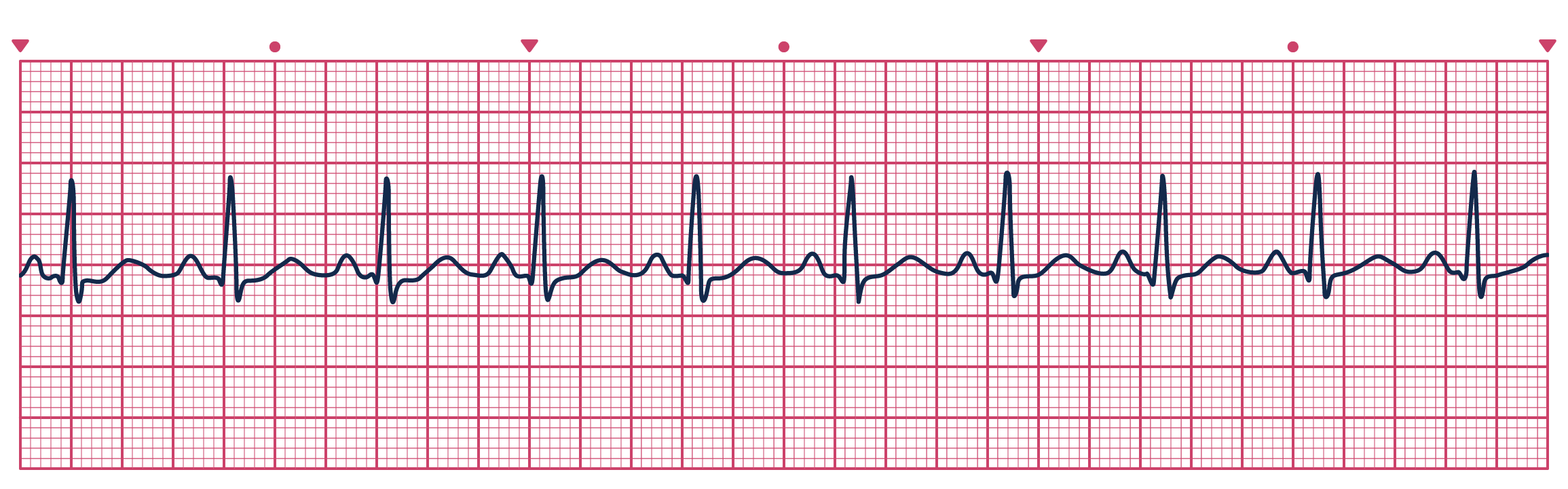
Guess the Rhythm>
A normal sinus rhythm is the natural rhythm of the heart, typically characterized by a heart rate of 60 to 100 beats per minute, where electrical impulses are generated from the sinoatrial node and spread uniformly through the atria and ventricles.
Intervention for NSR
None
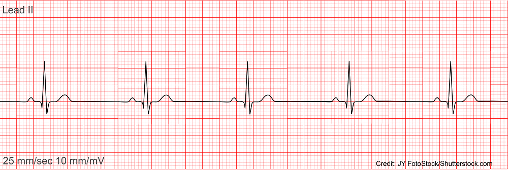
Guess the Rhythm>
Sinus Bradycardia A condition where the heart beats slower than normal, typically defined as a heart rate of less than 60 beats per minute, often resulting from increased vagal tone or medication effects.
Intervention for Sinus Bradycardia
The treatment for sinus bradycardia may include the use of atropine to increase heart rate, pacemaker insertion for persistent cases, or addressing underlying causes such as medication adjustment.
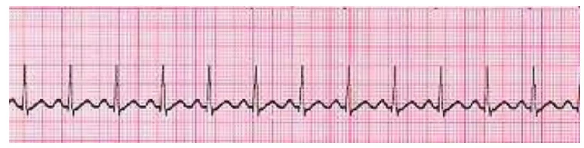
Guess the Rhythm>
Sinus Tachycardia A condition characterized by an elevated heart rate, typically defined as exceeding 100 beats per minute, often due to factors such as exercise, stress, pain, or underlying medical conditions.
Interventions of Sinus Tacyhycardia
Interventions for sinus tachycardia typically focus on addressing the underlying cause, such as managing stress or anxiety, adjusting medications, and, in some cases, using beta-blockers to reduce heart rate.
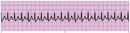
Guess the Rhythm>
Supraventricular tachycardia (SVT) is a rapid heart rhythm originating above the ventricles, often characterized by a heart rate greater than 150 beats per minute. It may result from various triggers, including stress or cardiac abnormalities.
Interventions of SVT
Interventions for supraventricular tachycardia (SVT) aim to terminate the rapid heart rhythm and may include vagal maneuvers, medication such as adenosine.

Guess the Rhythm>
Sinus Arrhythmia- Given Oxygen,

Guess the Rhythm>
Atrial Fibrillation A cardiac rhythm characterized by irregular, chaotic atrial electrical activity resulting in an irregularly irregular ventricular rhythm, with a ventricular rate that remains within an acceptable or controlled range.
Interventions for Atrial Fibrillation
amiodorine, cardioconversion

Guess the Rhythm>
Aflutter - A cardiac rhythm characterized by rapid, organized atrial activity, typically producing an atrial rate around 250–350 beats per minute, often with a characteristic “sawtooth” pattern. Ventricular response may be controlled or uncontrolled depending on the degree of AV conduction.
Guess the Rhythm>
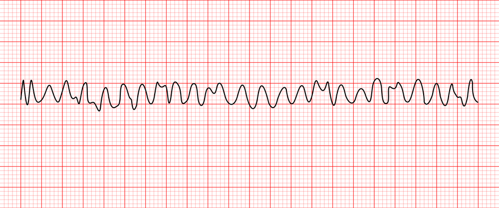
VFIB
A life-threatening cardiac rhythm characterized by chaotic, disorganized ventricular electrical activity resulting in ineffective ventricular contractions and no palpable pulse. It is a cardiac arrest rhythm requiring immediate intervention.
Interventions for VFIB
Interventions: Initiate CPR, activate emergency response/code, apply defibrillator and deliver unsynchronized shock, establish IV/IO access, administer epinephrine per ACLS protocol, and consider amiodarone or lidocaine for refractory VFib while treating reversible causes.
Interventions for Aflutter
Interventions: Assess hemodynamic stability, monitor ECG and vital signs, establish IV access, provide oxygen if hypoxemic, use synchronized cardioversion if unstable, and for stable patients consider rate control with medications such as beta-blockers or calcium-channel blockers and evaluate the need for anticoagulation.

Guess the Rhythm>
First Degree HB A cardiac conduction abnormality characterized by prolongation of the PR interval (>0.20 seconds), with every atrial impulse still conducted to the ventricles. It is often asymptomatic and may be associated with medications, increased vagal tone, or underlying cardiac disease.
Interventions for 1 degree HB
Interventions: Monitor

Guess the Rhythm>
Second-Degree Heart Block — Type I (Mobitz I/Wenckebach)
A cardiac conduction abnormality characterized by progressive prolongation of the PR interval followed by a dropped QRS complex, resulting from intermittent failure of atrial impulses to conduct to the ventricles
Interventions Second-Degree Heart Block
prepare for transcutaneous pacing, and arrange for transvenous pacing when indicated. Atropine may be attempted for symptomatic bradycardia
Guess the Rhythm>
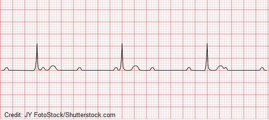
Third-Degree Heart Block (Complete Heart Block) A life-threatening conduction abnormality in which no atrial impulses are conducted to the ventricles, resulting in independent atrial and ventricular rhythms (AV dissociation) and often severe bradycardia.

Guess the Rhythm>
Second-Degree Heart Block — Type II (Mobitz II) A potentially serious cardiac conduction abnormality characterized by intermittent nonconducted P waves without progressive PR prolongation, resulting in dropped QRS complexes. It can progress to complete heart block.

Guess the Rhythm>
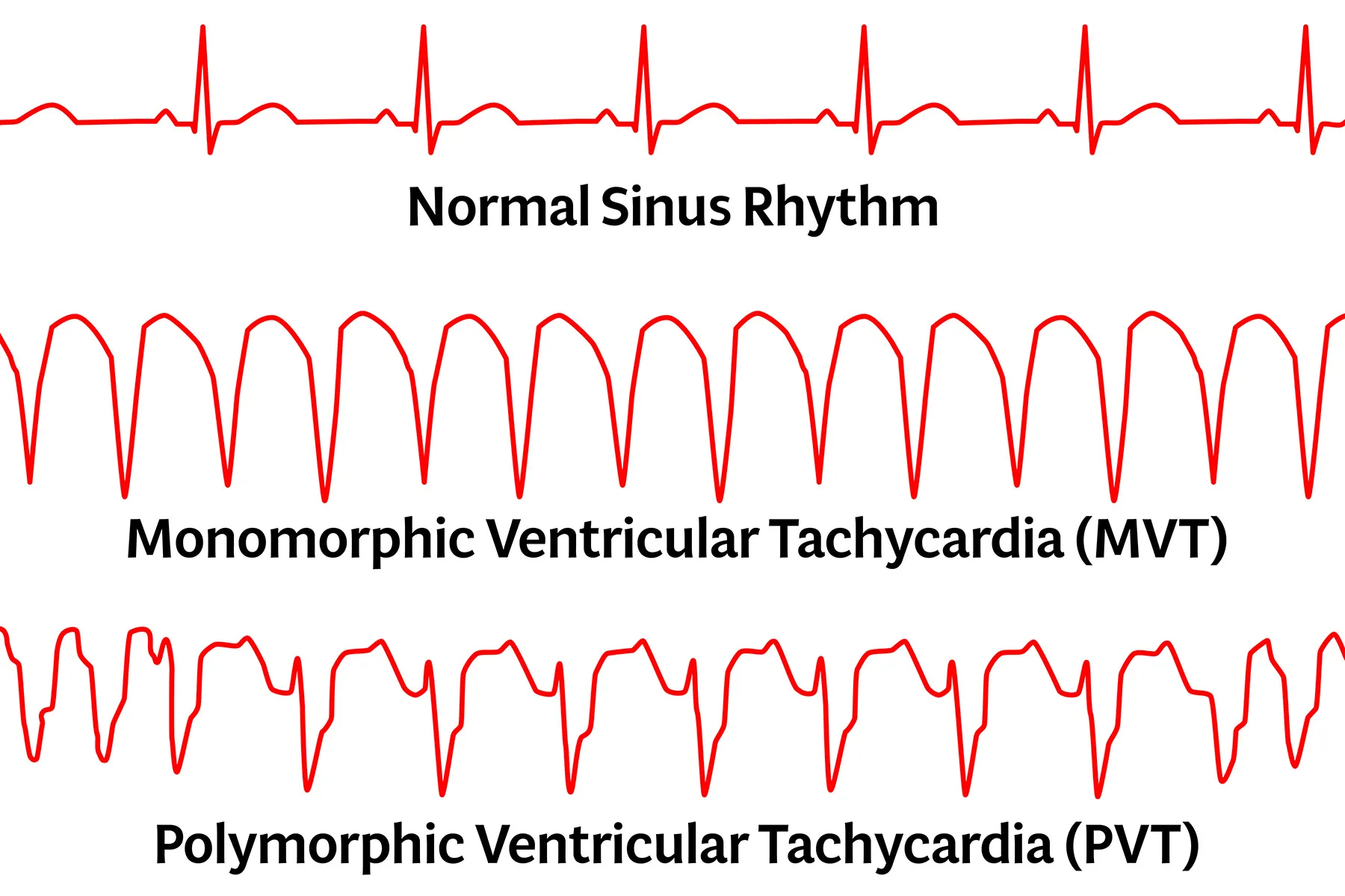
Ventricular Tachycardia (VTach) A potentially life-threatening cardiac rhythm characterized by three or more consecutive ventricular beats at a rapid rate, typically originating from the ventricles. VTach may occur with a pulse or without a pulse and can reduce cardiac output or progress to ventricular fibrillation.
Interventions Ventricular Tachycardia (VTach)
Interventions: Assess for a pulse and hemodynamic stability. Pulseless VTach: initiate CPR and perform immediate defibrillation according to ACLS. VTach with a pulse and instability: perform synchronized cardioversion. Stable VTach: consider antiarrhythmic
Guess the Rhythm>
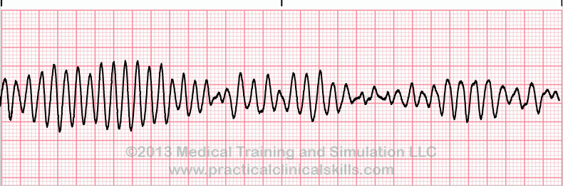
Torsade de Pointes A form of polymorphic ventricular tachycardia associated with a prolonged QT interval, characterized by QRS complexes that appear to twist around the ECG baseline. It can be transient or deteriorate into ventricular fibrillation and cardiac arrest.
Interventions for Torsades de Pointes
discontinue QT-prolonging medications when appropriate, correct electrolyte abnormalities—especially hypokalemia and hypomagnesemia—and administer IV magnesium sulfate.

Junctional Rhythm
A cardiac rhythm originating from the AV junction rather than the sinoatrial (SA) node, typically occurring when the SA node fails to initiate an impulse or when conduction from the atria is impaired. The rate is generally slower than normal sinus rhythm and may produce absent or inverted P waves.