Biology K103 Lab Exam 3 Diagram | Quizlet
 Call Kai
 Learn
Learn Practice Test
 Spaced Repetition
 Match
Match Flashcards
 Knowt Play
1/99

There's no tags or description
Looks like no tags are added yet.
Name | Mastery | Learn | Test | Matching | Spaced | Call with Kai | Chat |
|---|
No analytics yet
Send a link to your students to track their progress
100 Terms
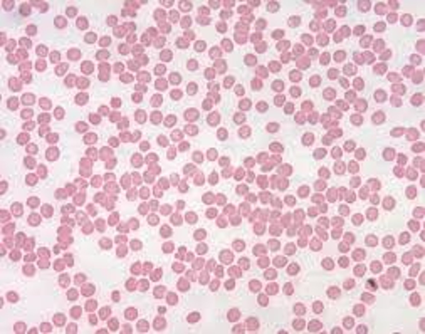
human blood smear
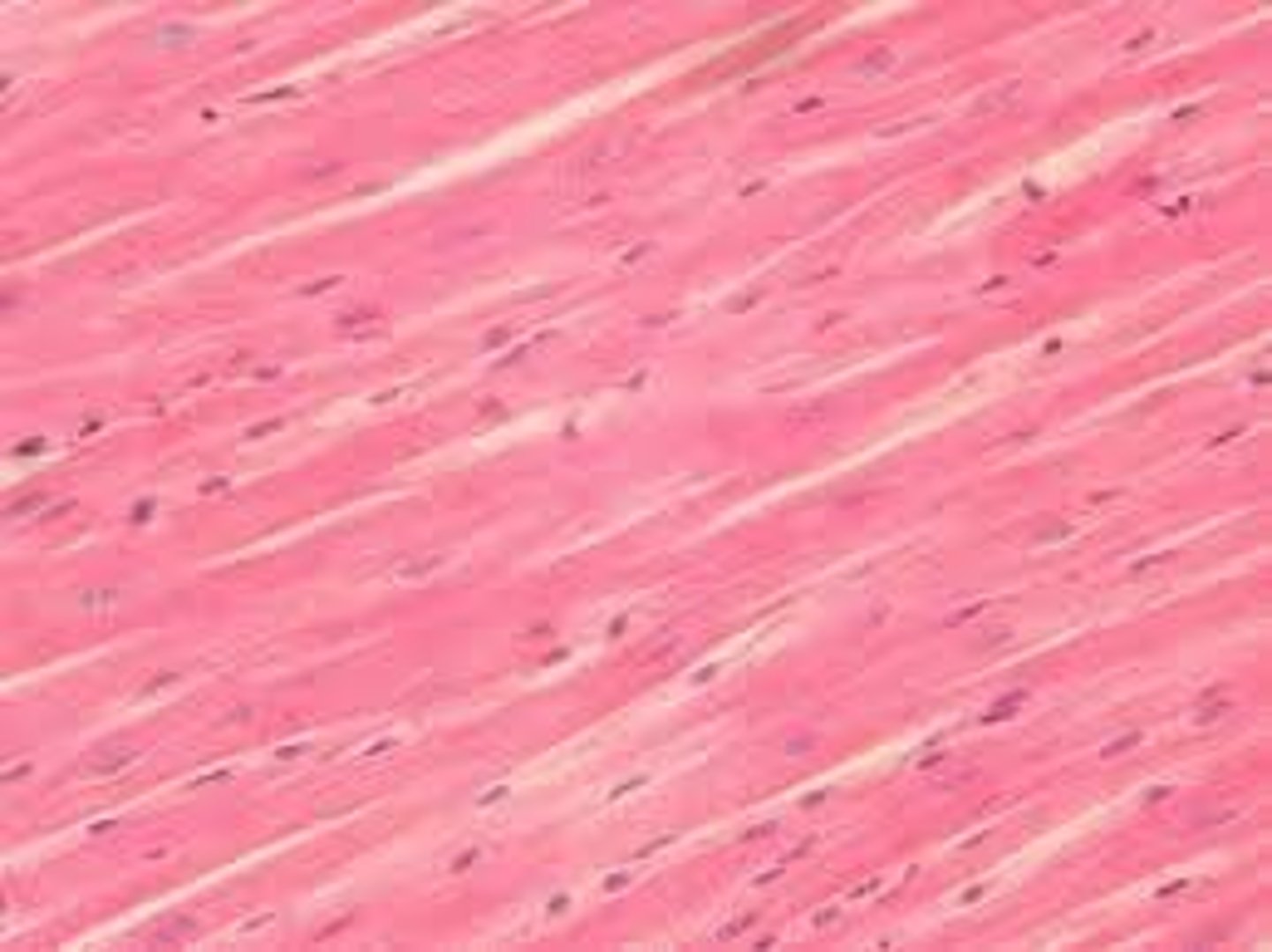
cardiac muscle
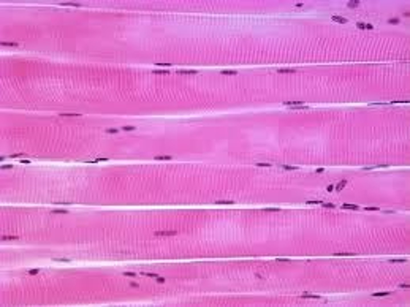
skeletal muscle
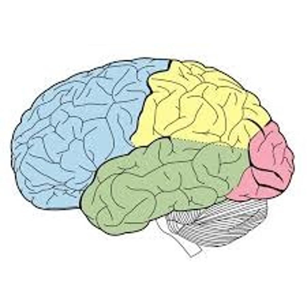
Blue
What color is the frontal lobe?
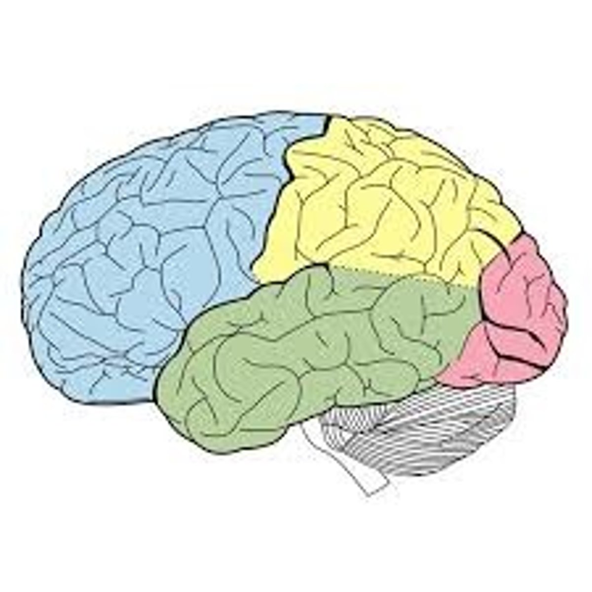
Yellow
What color is the parietal lobe?
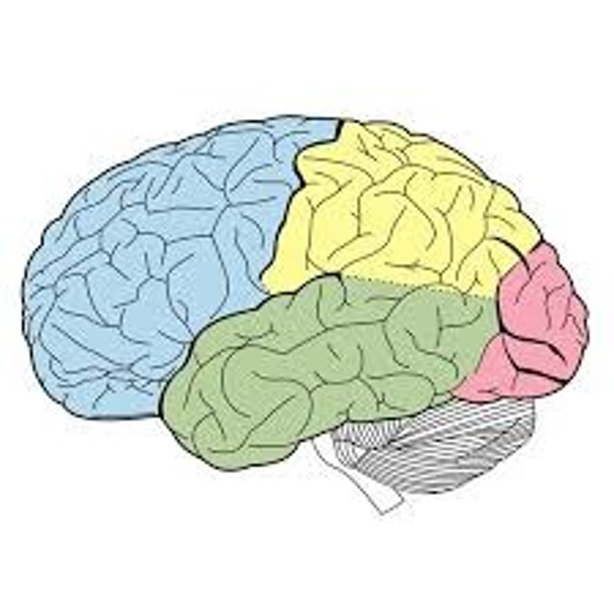
Green
What color is the temporal lobe
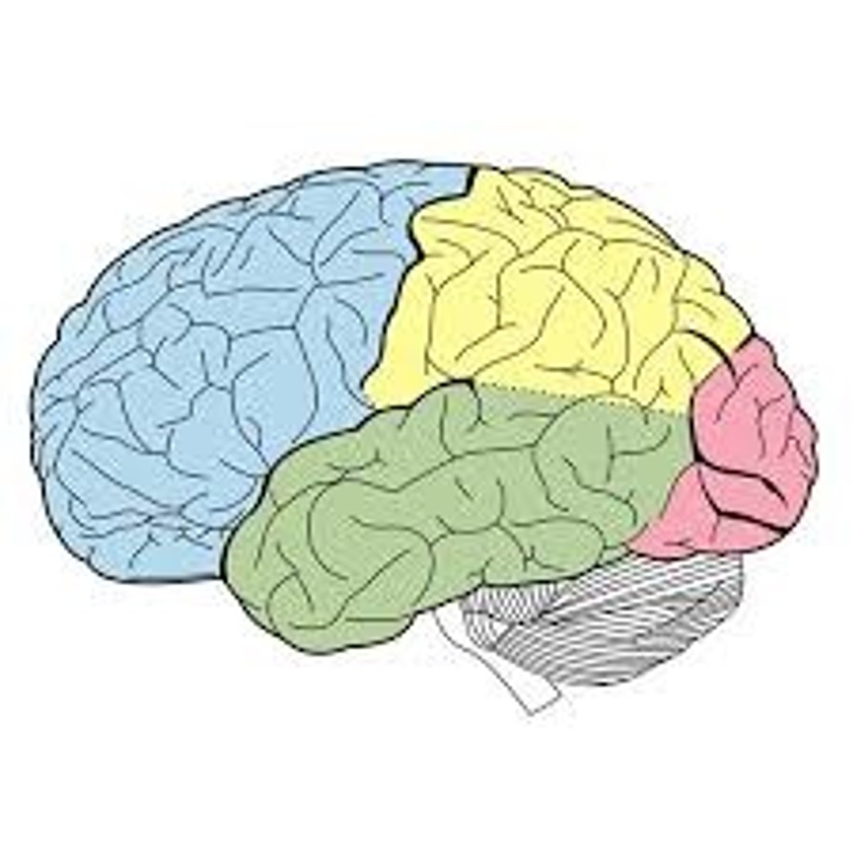
Pink
What color is the occipital lobe?
TERM
Longitudinal fissure
DEFINITION
Splits the brain into left and right hemispheres
TERM
Central Sulcus
DEFINITION
Separates the frontal and parietal lobes
TERM
Sulcus
DEFINITION
a groove or furrow on the surface of the brain
TERM
Postcentral Gyrus
DEFINITION
primary sensory area that perceives touch, pain, pressure and temperature
TERM
Pre-central Gyrus
DEFINITION
primary motor area that controls voluntary movements of the skeletal muscles
TERM
Gyrus
DEFINITION
a ridge or fold on the surface of the brain
optic chiasma
What part is circled in red?
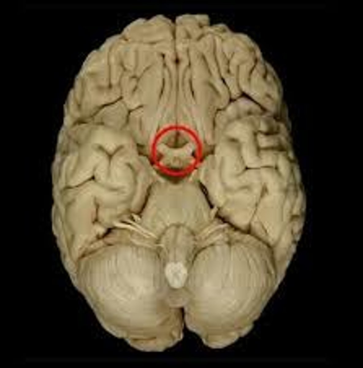
optic chiasma
point at which optic nerve fibers cross in the brain
Cerebellum
What is 1?
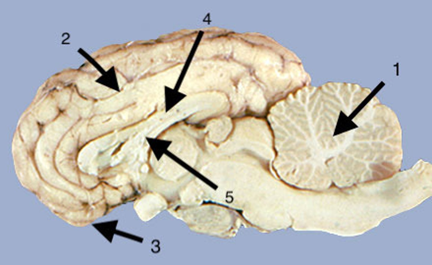
Cerebellum
A large structure of the hindbrain that controls fine motor skills.
Cerebrum
What is 2?
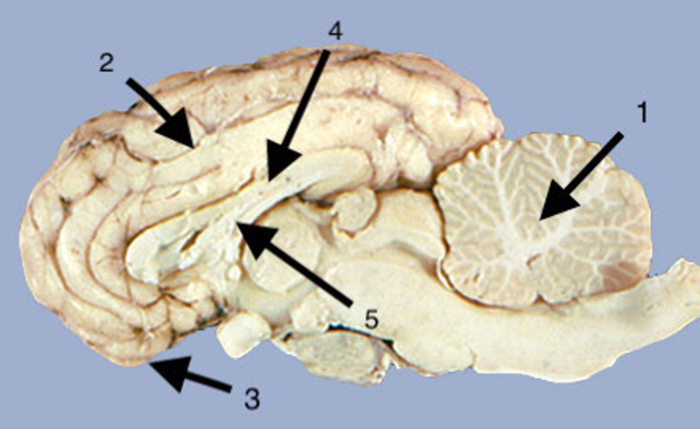
Cerebrum
Area of the brain responsible for all voluntary activities of the body
Cerebrum
largest part of the brain
Olfactory bulb
What is 3?
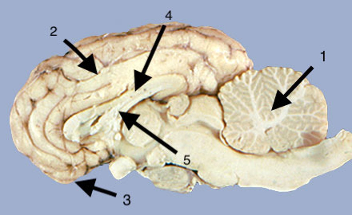
Olfactory bulb
a brain structure located above the nasal cavity beneath the frontal lobes
Corpus callosum
What is 4?
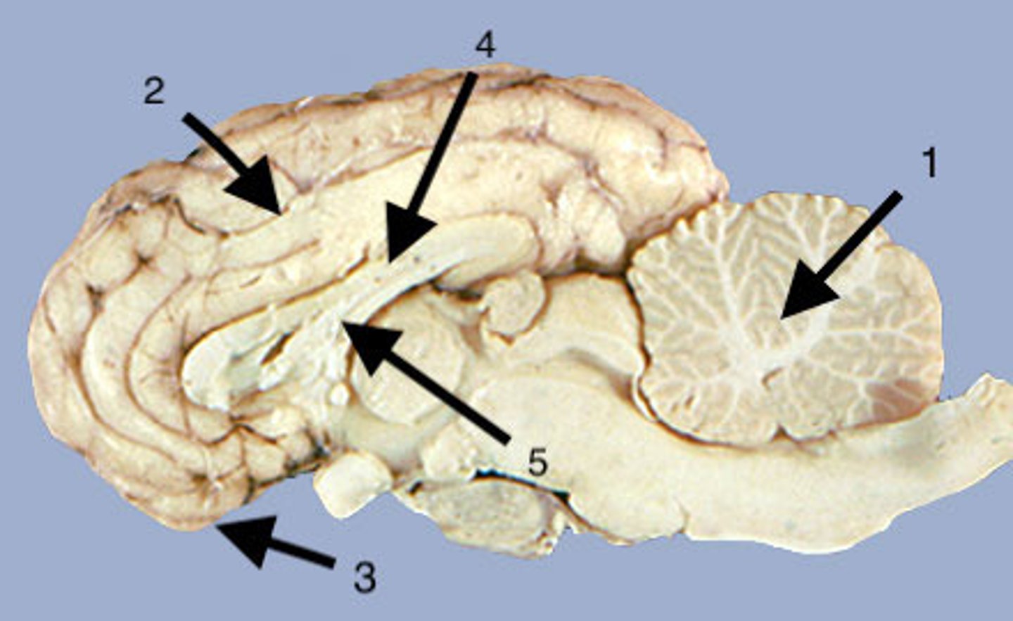
Corpus callosum
a broad band of nerve fibers joining the two hemispheres of the brain.
Corpus callosum
What carries messages between the two hemispheres of the brain?
Fornix
What is 5?
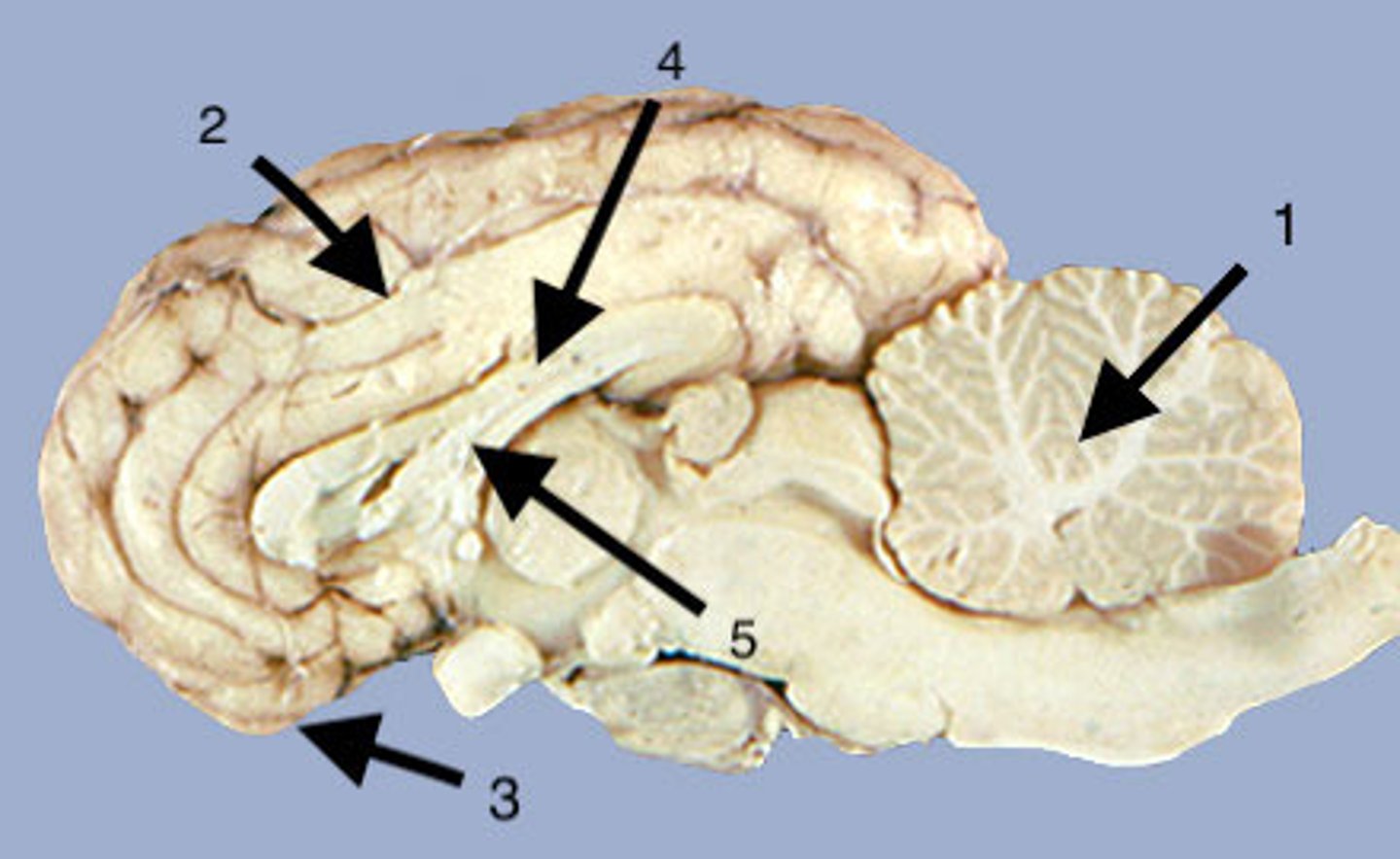
Fornix
C-shaped bundle of nerve fibers in the brain that acts as the major output tract of the hippocampus
Fornix
What lies below the Corpus callosum?
arbor vitae
What is the white matter of the cerebellum called?
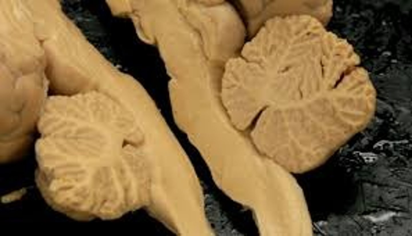
Vermis of cerebellum
What is 1?
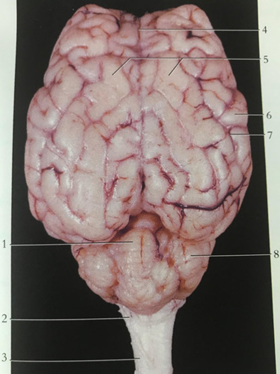
Vermis of cerebellum
Ridge that separates the cerebellum into 2 sections
Olfactory tract
What is 1?
Olfactory tract
the path along which the olfactory receptors send their electrical messages to the brain.
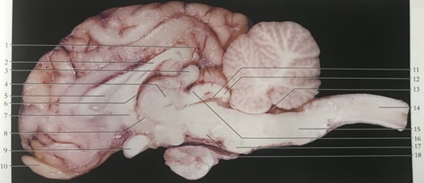
Pons
what is 17?
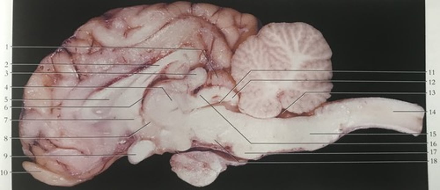
Pons
a brain structure that relays information from the cerebellum to the rest of the brain
Medulla
what is 15?
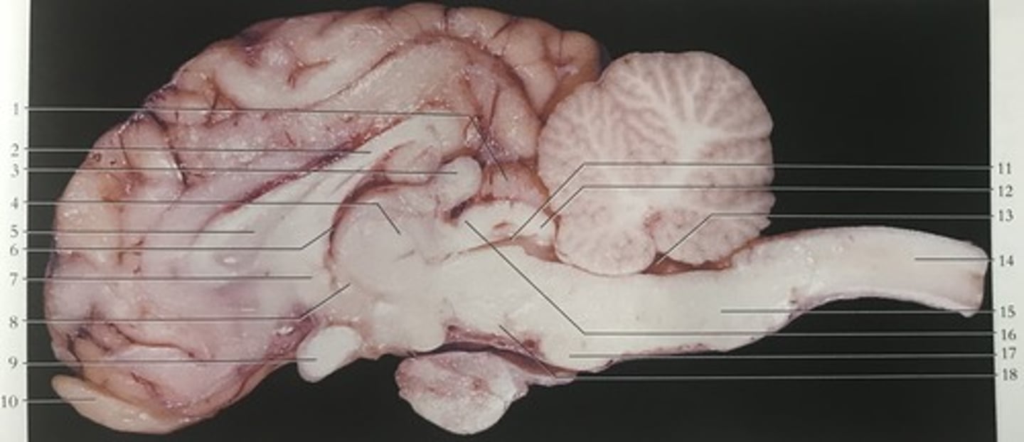
Medulla
the base of the brainstem; controls heartbeat and breathing
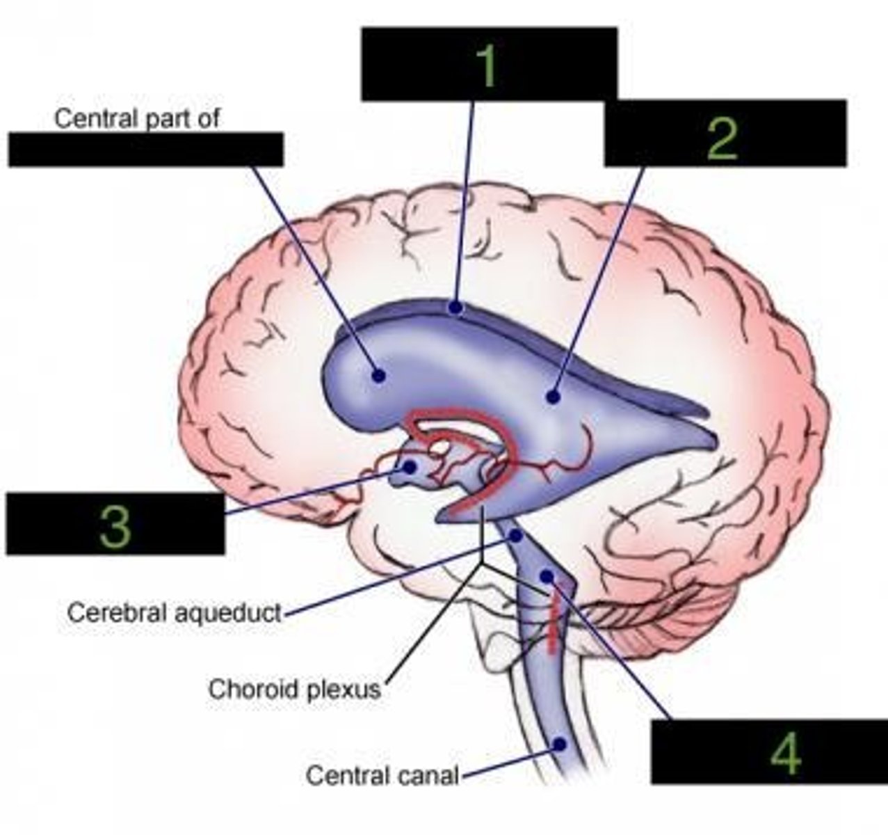
Four
how many ventricles does the brain have?
Right lateral ventricle
what ventricle is number 1?
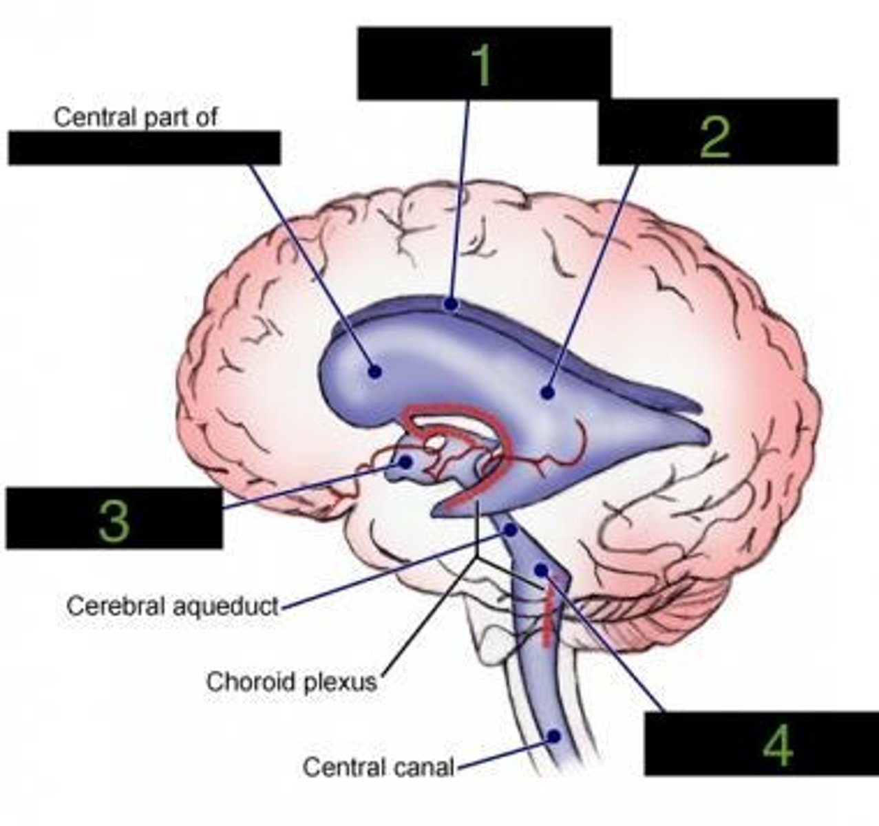
Left lateral ventricle
what ventricle is number 2?
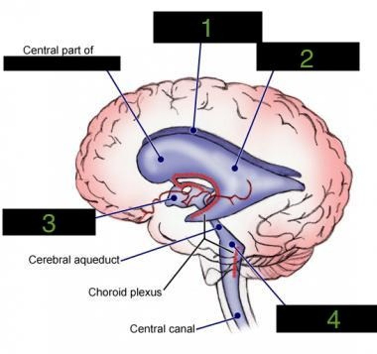
Third ventricle
what ventricle is number 3?
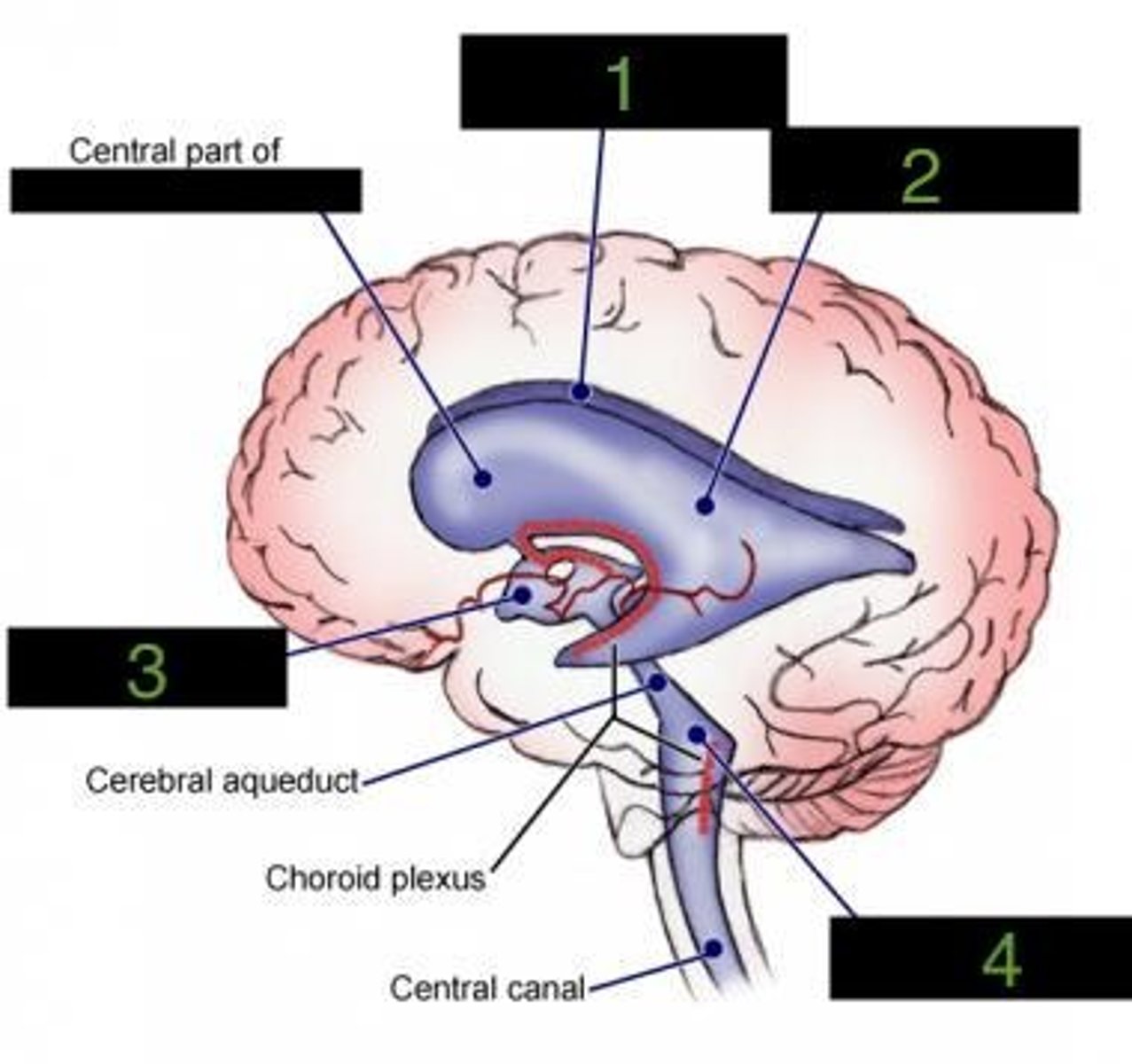
fourth ventricle
what ventricle is number 4?
cornea
what is F?
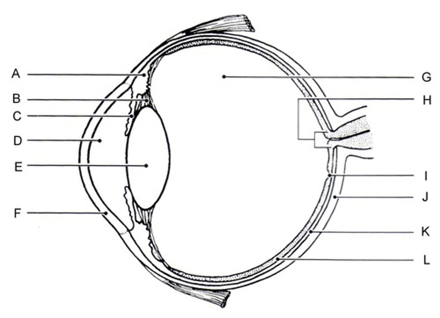
cornea
gathers and focuses incoming light
vitreous humor
what is the fluid between the retina and the lens?
vitreous humor
helps maintain the shape of the eye
sclera
what is J?
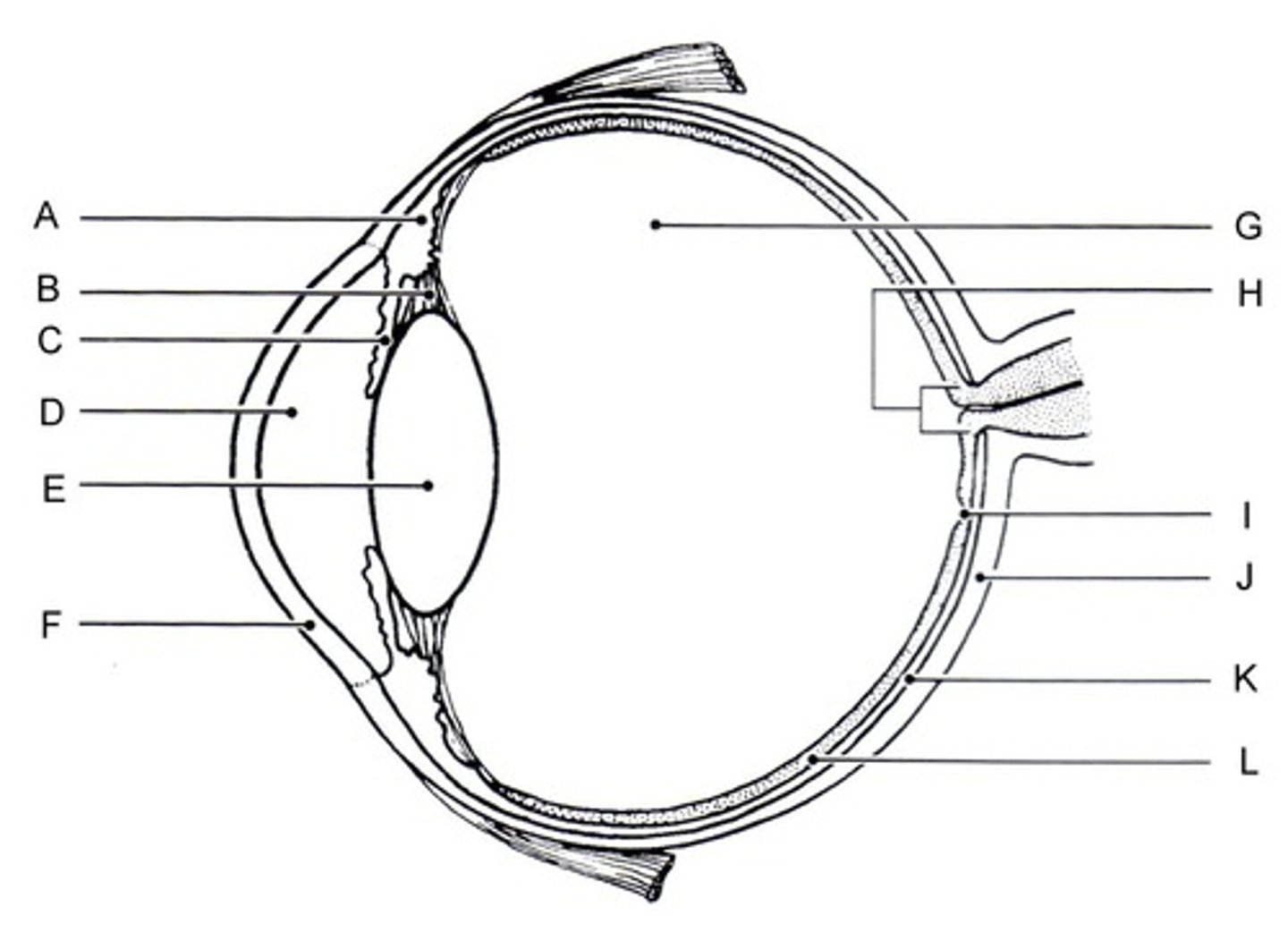
sclera
the white part of the eye that forms the cornea
lens
What part is circled in red?
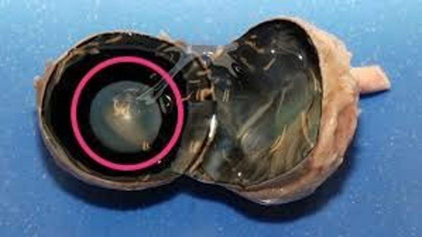
lens
the transparent structure behind the pupil that changes shape to help focus images on the retina
optic nerve
What part is circled?
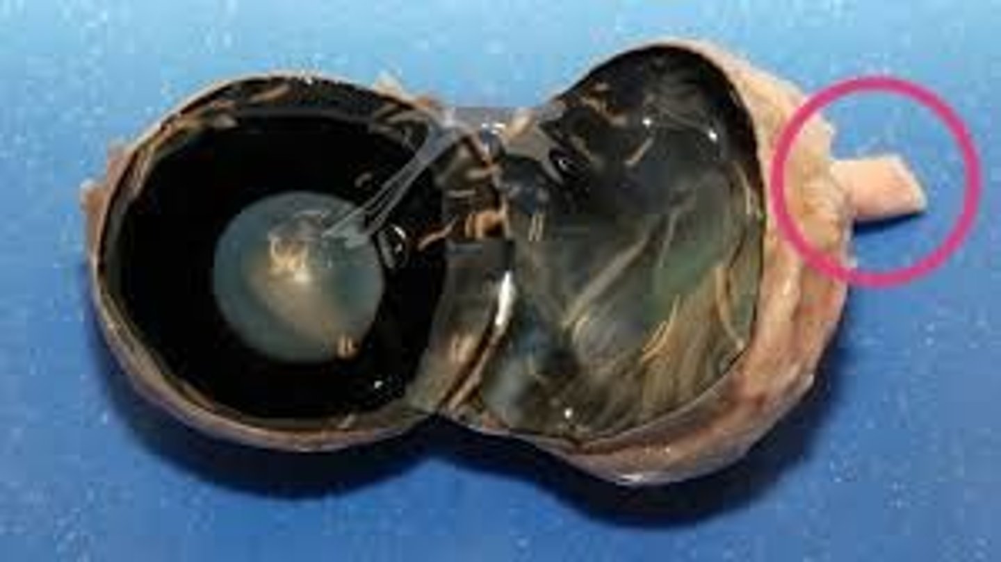
optic nerve
the nerve that carries impulses from the eye to the brain
ciliary body
What is the probe pointing to?
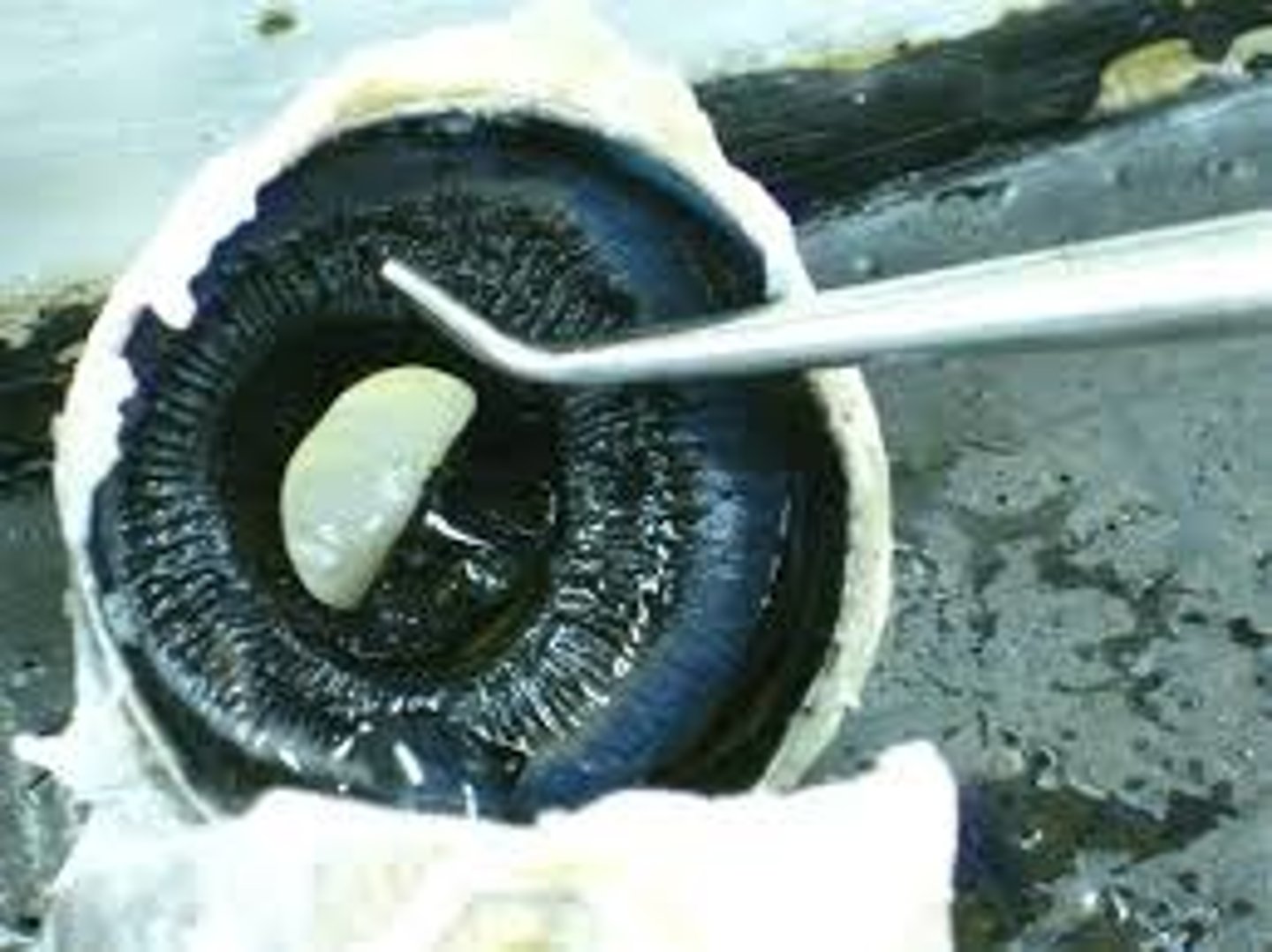
ciliary body
Structure surrounding the lens. It contains ciliary muscles, which control the shape of the lens.
retina
Contains sensory receptors that process visual information and sends it to the brain
pupil
the adjustable opening in the center of the eye through which light enters
pupil
What is C?
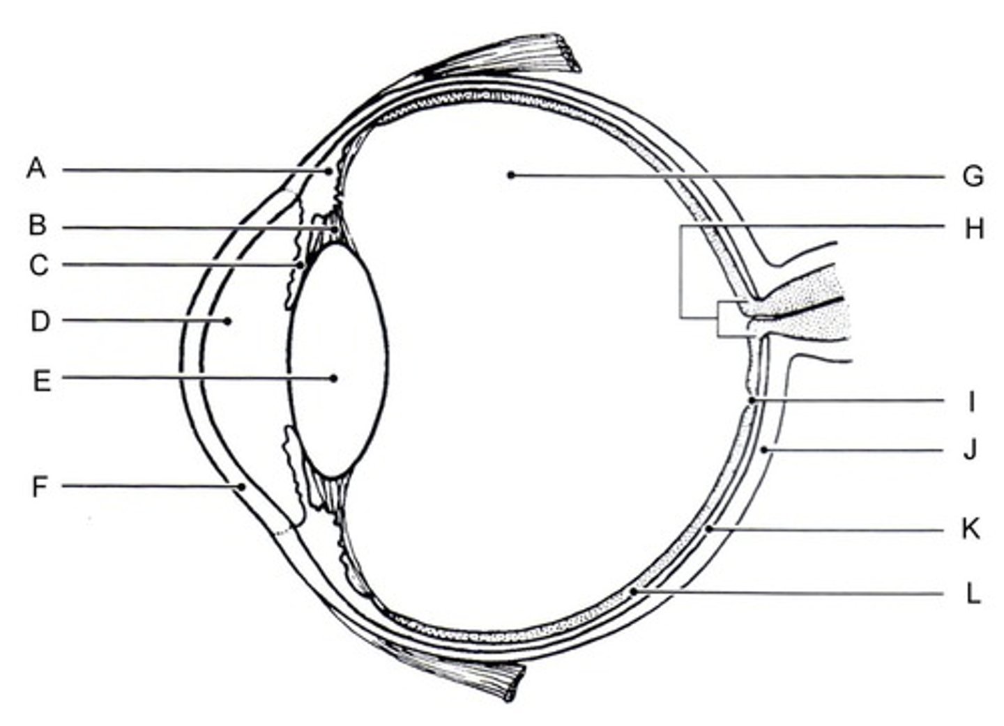
aqueous humor
What is D?
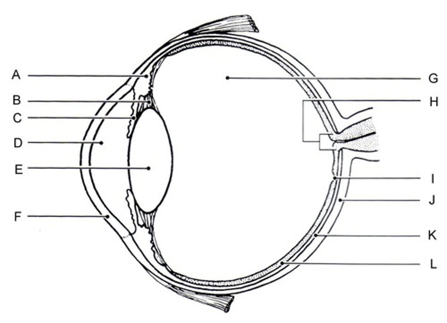
aqueous humor
fluid in the eye, found between the cornea and the lens
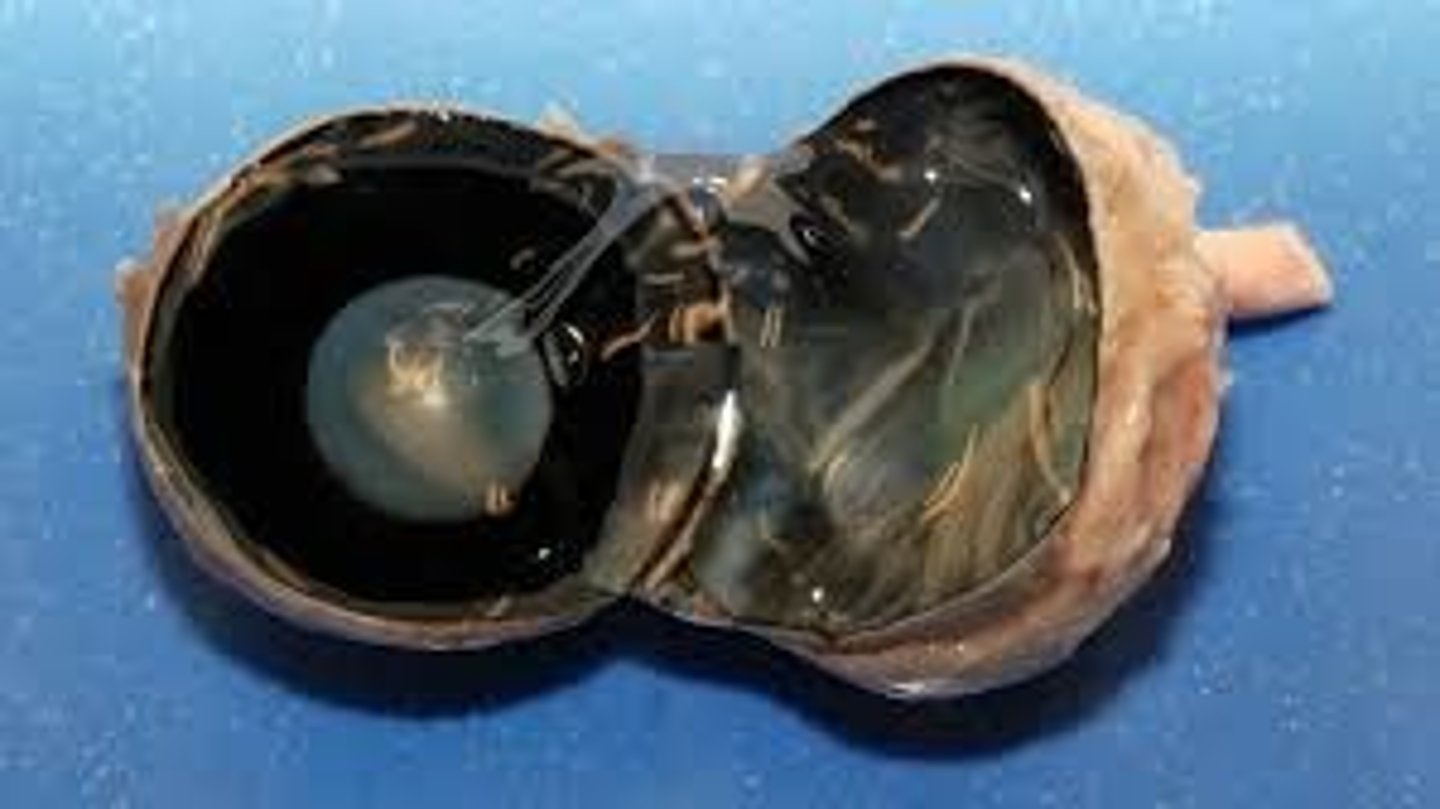
retina
What layer is being pulled away with the tweezers?
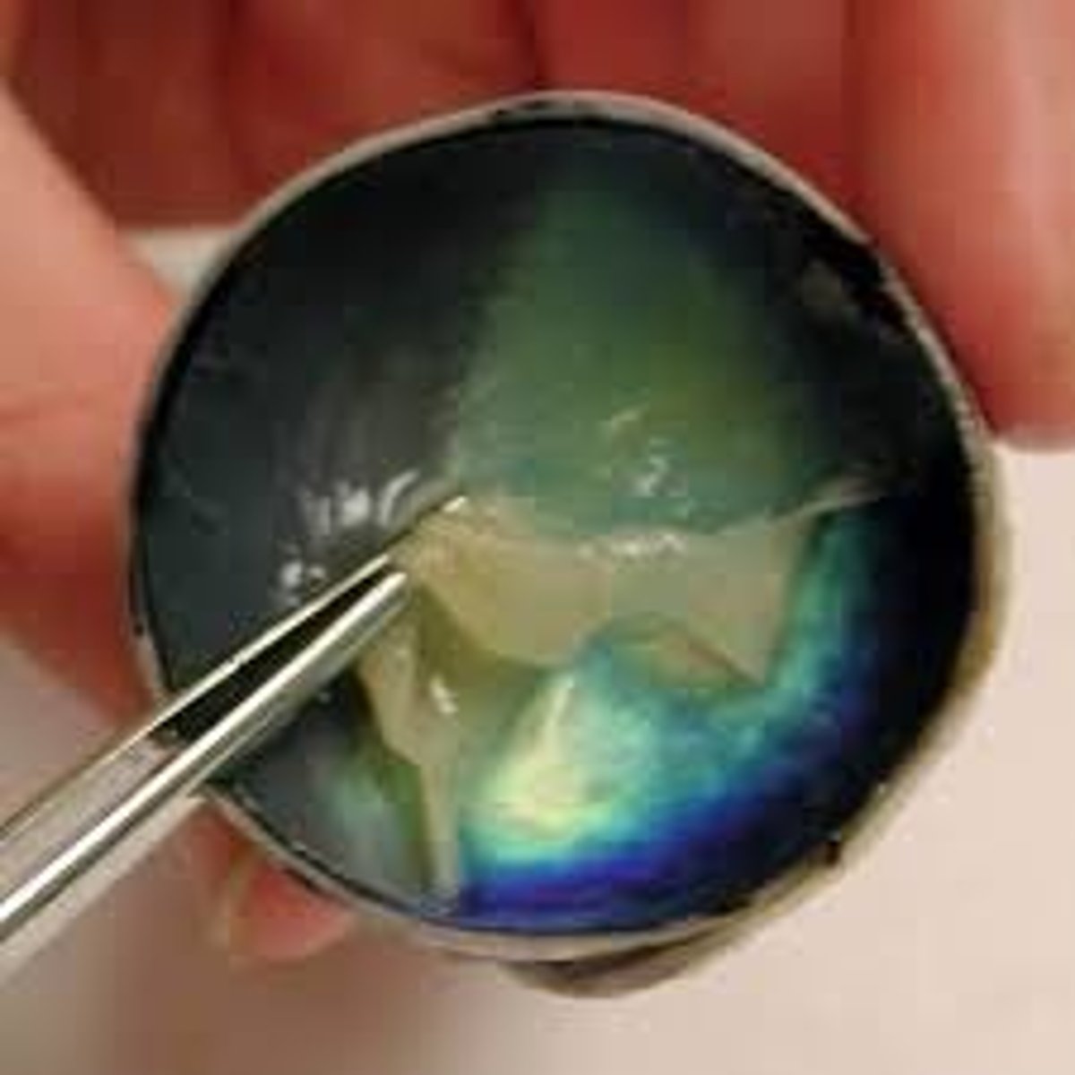
choroid
What is the black layer inside the eye called?
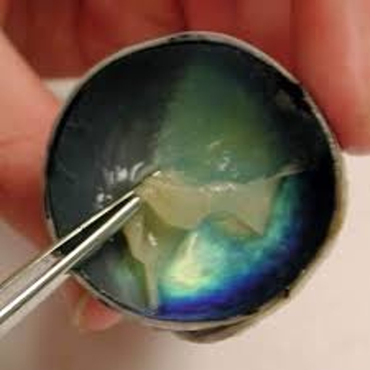
sclera, choroid, retina
what are the layers of the eye from top to bottom?
choroid
nourishes the other layers of the eye, and helps to absorb excess light
fovea centralis
What is I?
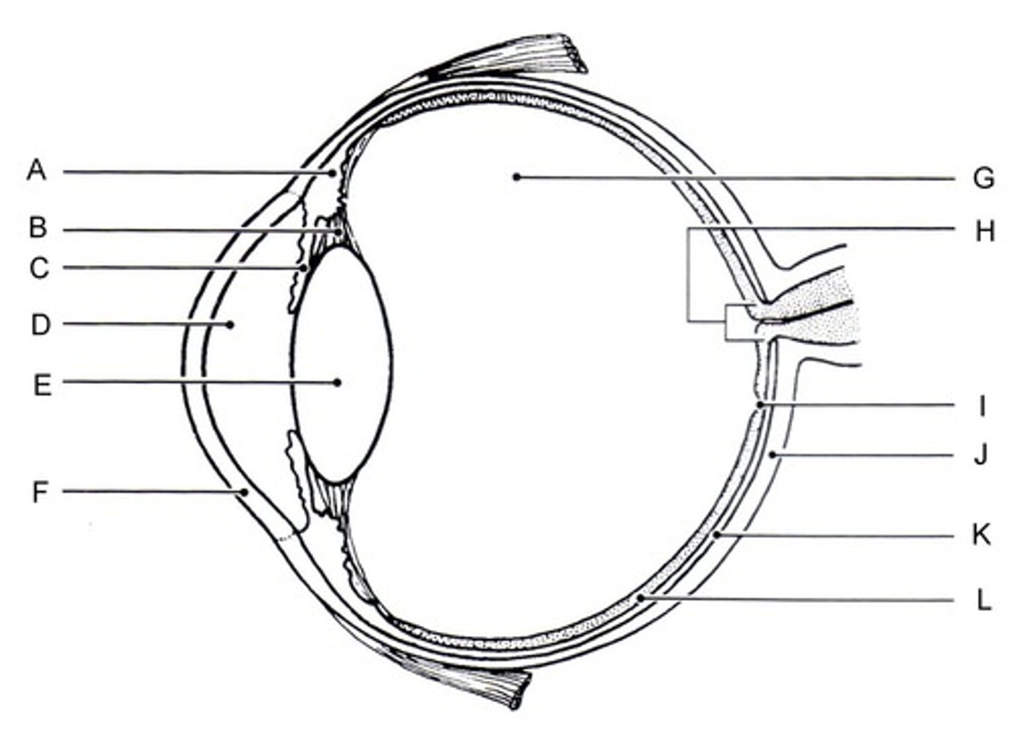
fovea centralis
tiny pit or depression in the retina that is the region of clearest vision
tapetum lucidum
What is the colored part of the eye?
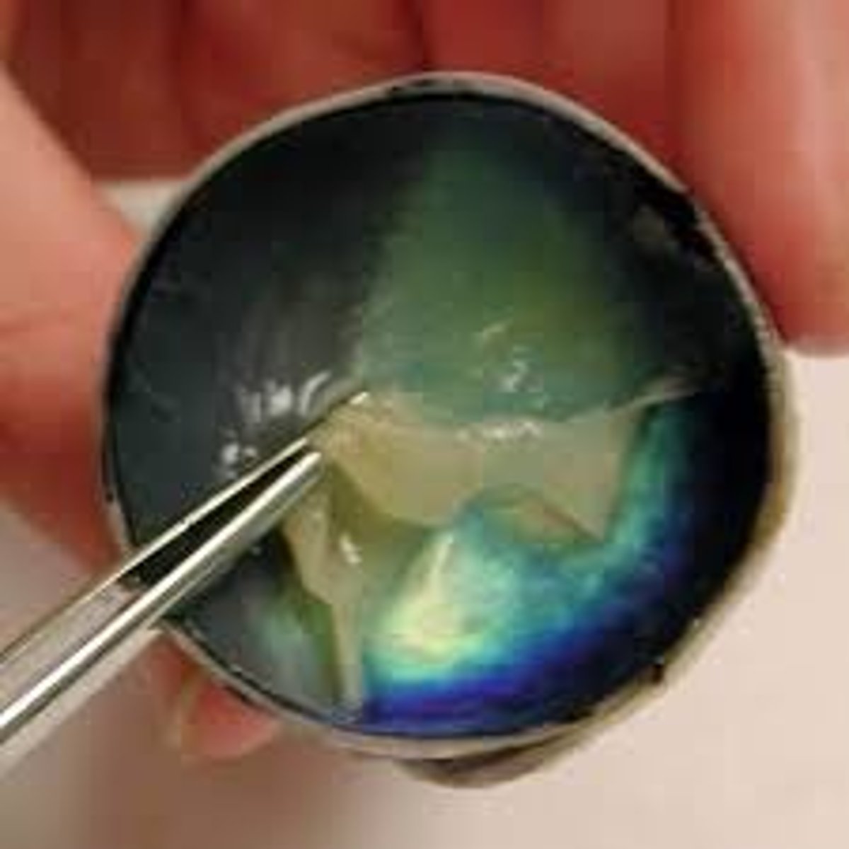
tapetum lucidum
Iridescent layer found in nocturnal animals for maximizing vision under low intensity light
rods
retinal receptors that detect black, white, and gray; necessary for peripheral and twilight vision.
cones
retinal receptor cells that are concentrated near the center of the retina and that function in daylight or in well-lit conditions, and give rise to color.
pinna
What is 1?
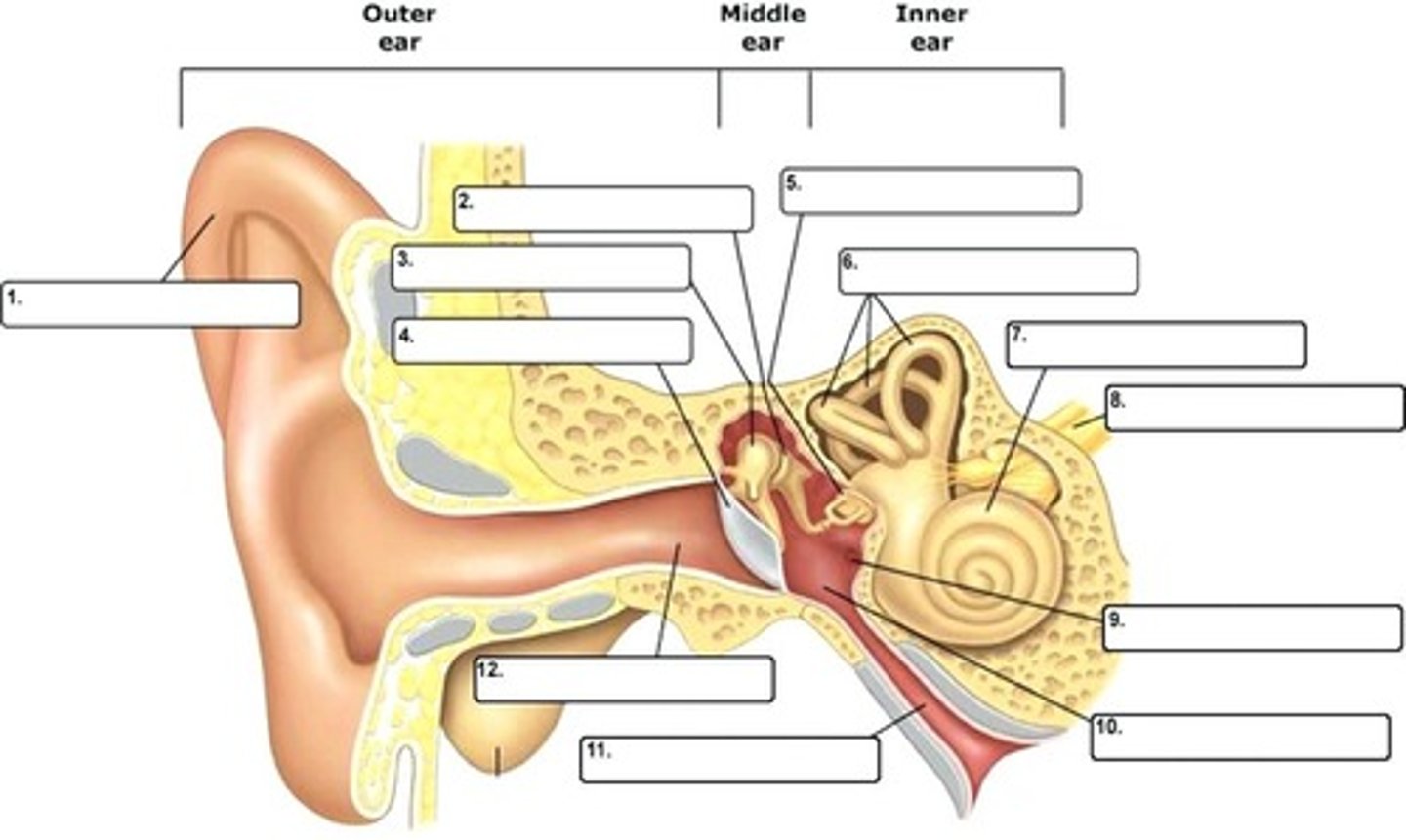
external auditory meatus
What is 12?
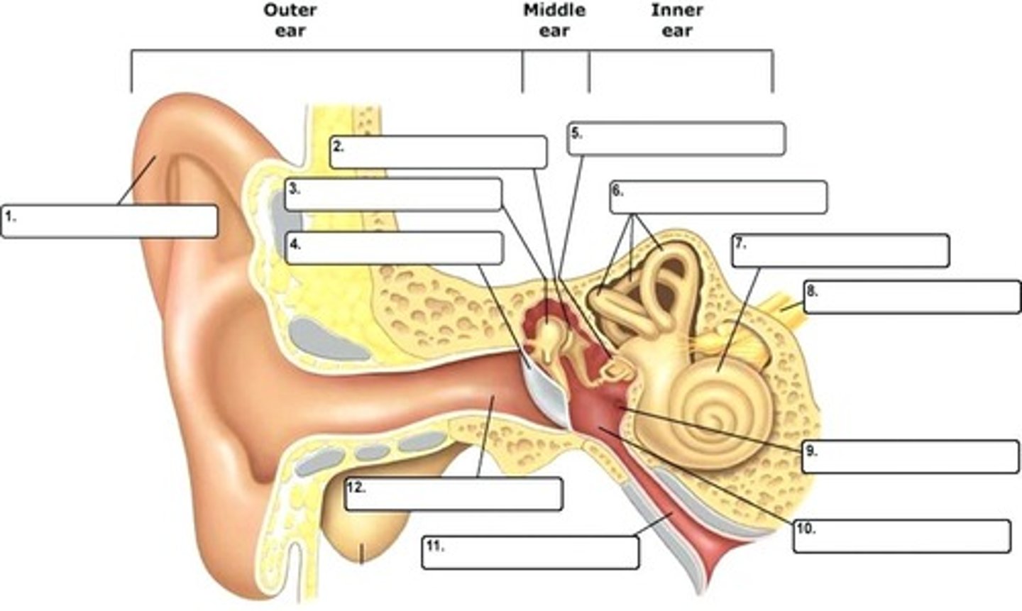
tympanum
What is 4?
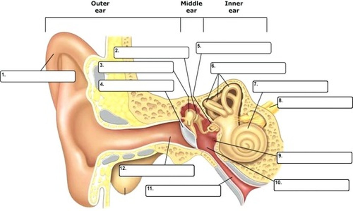
pinna
the visible part of the ear
external auditory meatus
the formal name for the ear canal
tympanum
picks up and amplifies sound waves
malleus
What is 3?
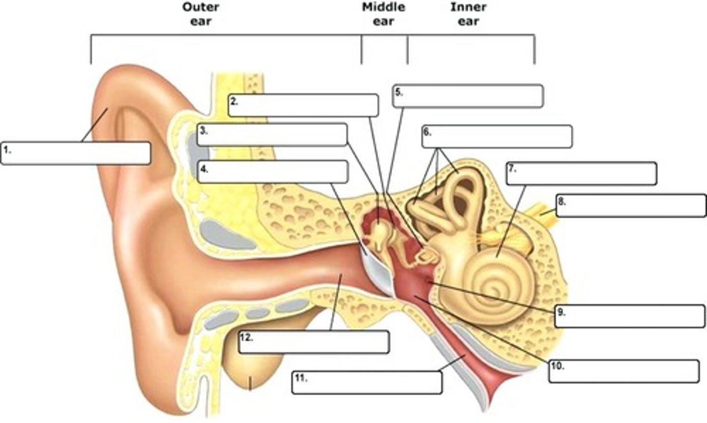
malleus
a small bone in the middle ear that transmits vibrations of the eardrum to the incus.
incus
What is 2?
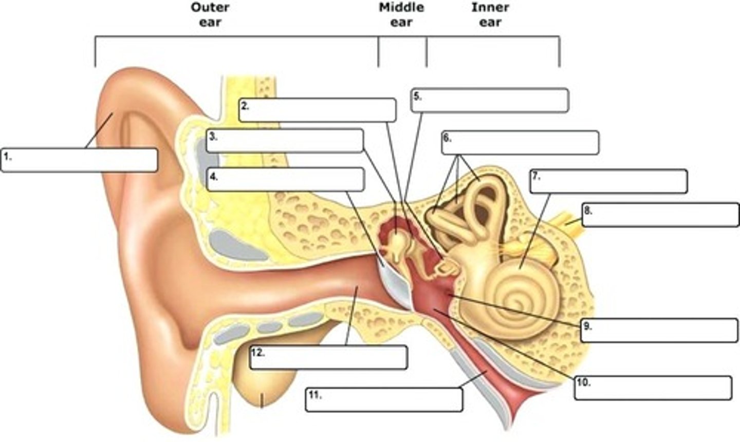
incus
a small anvil-shaped bone in the middle ear, transmitting vibrations between the malleus and stapes.
stapes
What is 5?
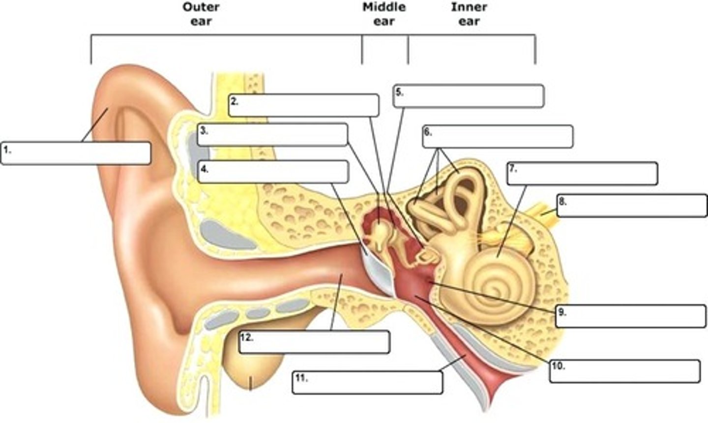
stapes
a small bone in the middle ear, transmitting vibrations from the incus to the inner ear.
round window
What is 9?
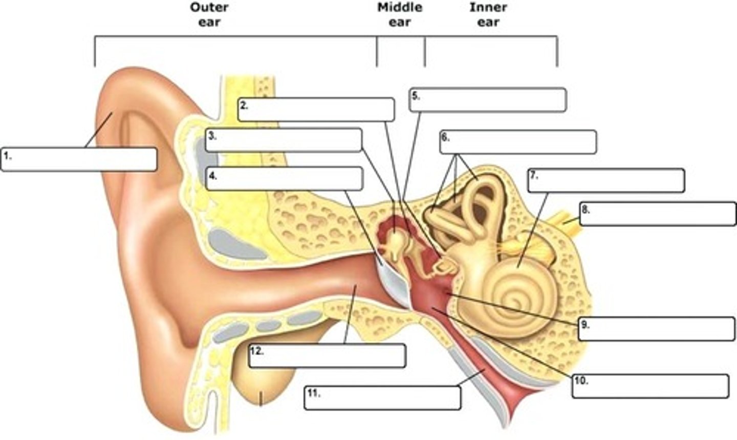
round window
A membrane-covered opening in the inner wall of the middle ear that compensates for changes in cochlear pressure
cochlea
What is 7?
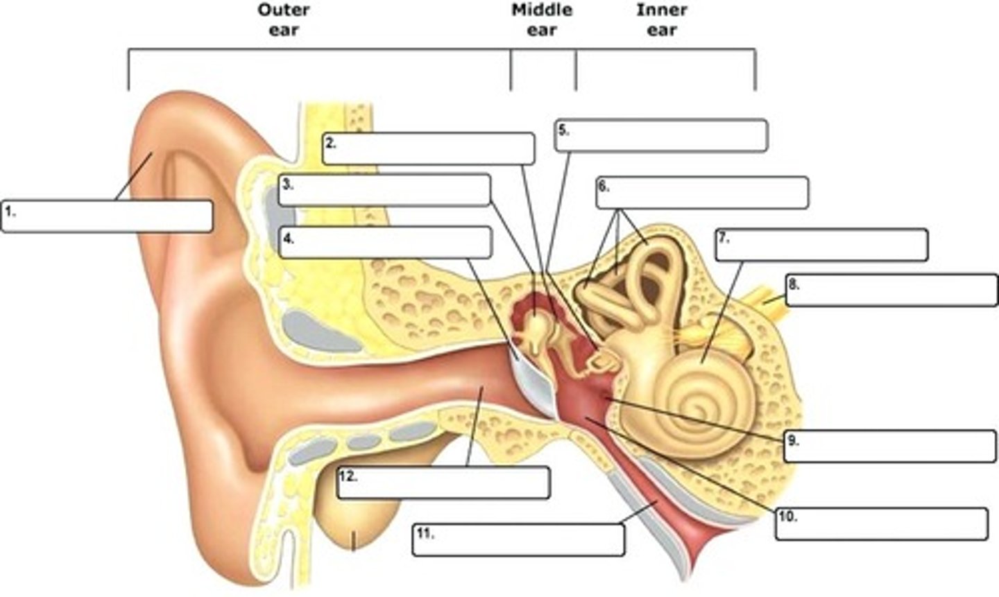
cochlea
a coiled, bony, fluid-filled tube in the inner ear through which sound waves trigger nerve impulses
scala vestibuli
What part is the red box pointing to?
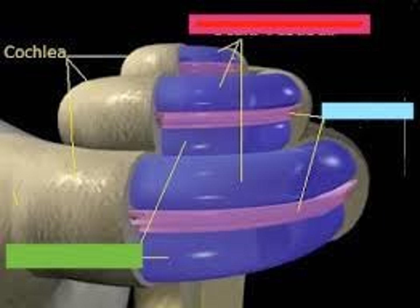
scala vestibuli
leads from oval window to apex of cochlea; contains perilymph
scala vestibuli
also known as vestibular canal
scala media
What part is the blue box pointing to?
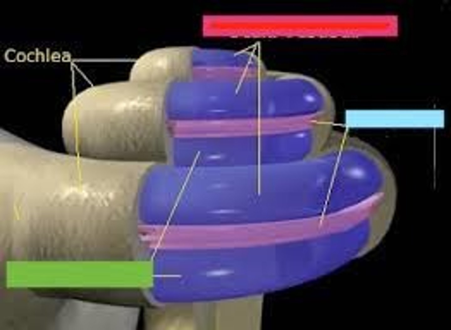
scala media
Middle chamber of the cochlea; filled with endolymph
scala media
also known as the middle canal
scala tympani
What part is the green box pointing to?
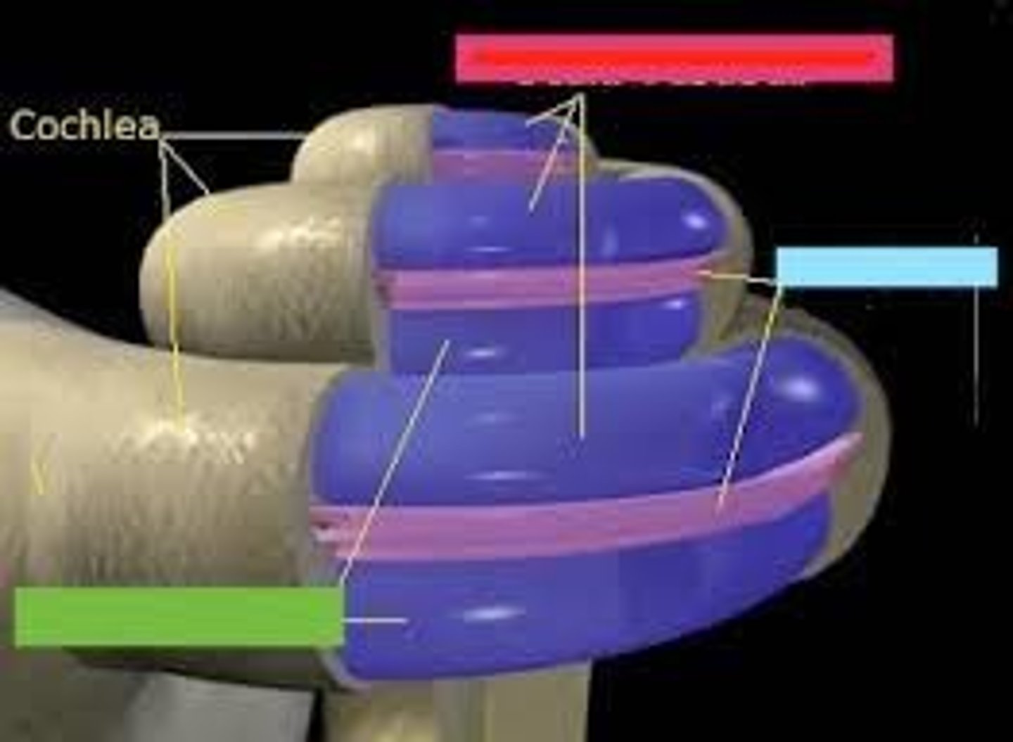
scala tympani
terminates at round window; contains perilymph
scala tympani
also known as the tympanic canal
organ of corti
tissue containing the hair cells necessary for hearing
organ of corti
What part of the ear is in the circle?
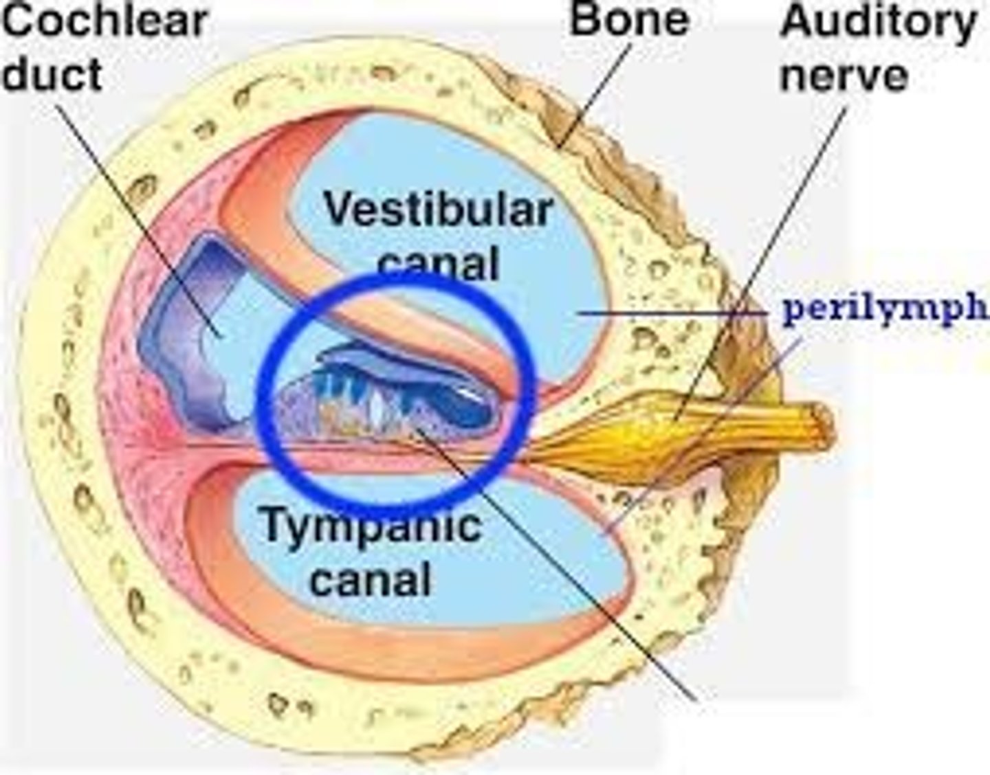
scala media
the organ of corti lies in what layer of the cochlea?
tectoral membrane
lies over organ of corti, distorts hair cells when displaced by sound waves in the cochlea
tectoral membrane
what is 3?
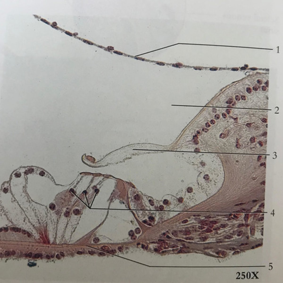
basilar membrane
A structure that runs the length of the cochlea in the inner ear and holds the auditory receptors, called hair cells.