Quiz 4 - Muscle cells, types
 Call Kai
 Learn
Learn Practice Test
 Spaced Repetition
 Match
Match Flashcards
 Knowt Play
1/58

There's no tags or description
Looks like no tags are added yet.
Name | Mastery | Learn | Test | Matching | Spaced | Call with Kai | Chat |
|---|
No analytics yet
Send a link to your students to track their progress
59 Terms
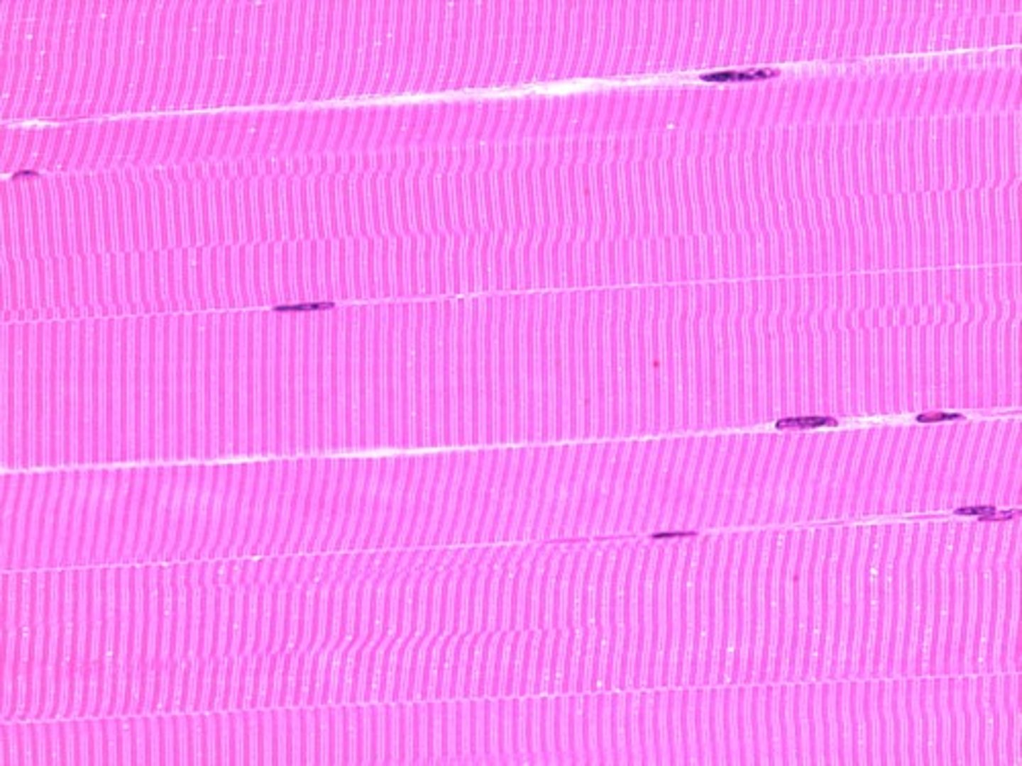
Skeletal Muscle
Striated, voluntary muscle that attaches to bones and produces body movement. Multinucleated
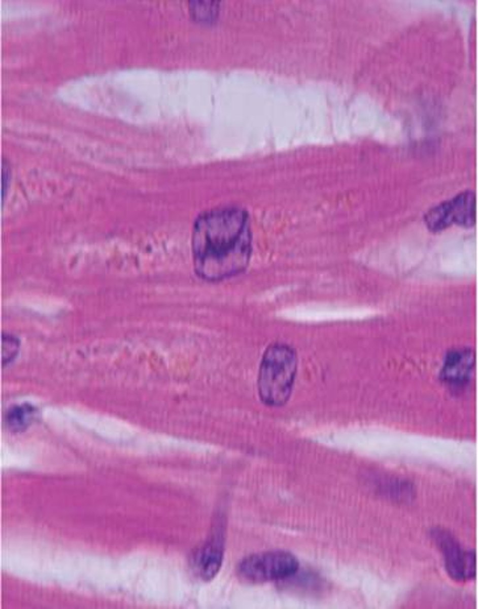
Cardiac Muscle
Striated, involuntary muscle found in the heart; cells branch and connect with intercalated discs.
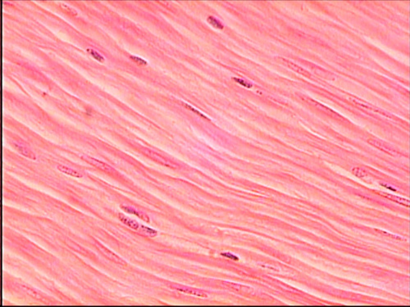
Smooth Muscle
Non-striated, involuntary muscle found in walls of organs and blood vessels.
Movement (muscle function)
Skeletal muscle contraction moves bones and body contents.
Stability
Muscles hold bones in place and resist gravity.
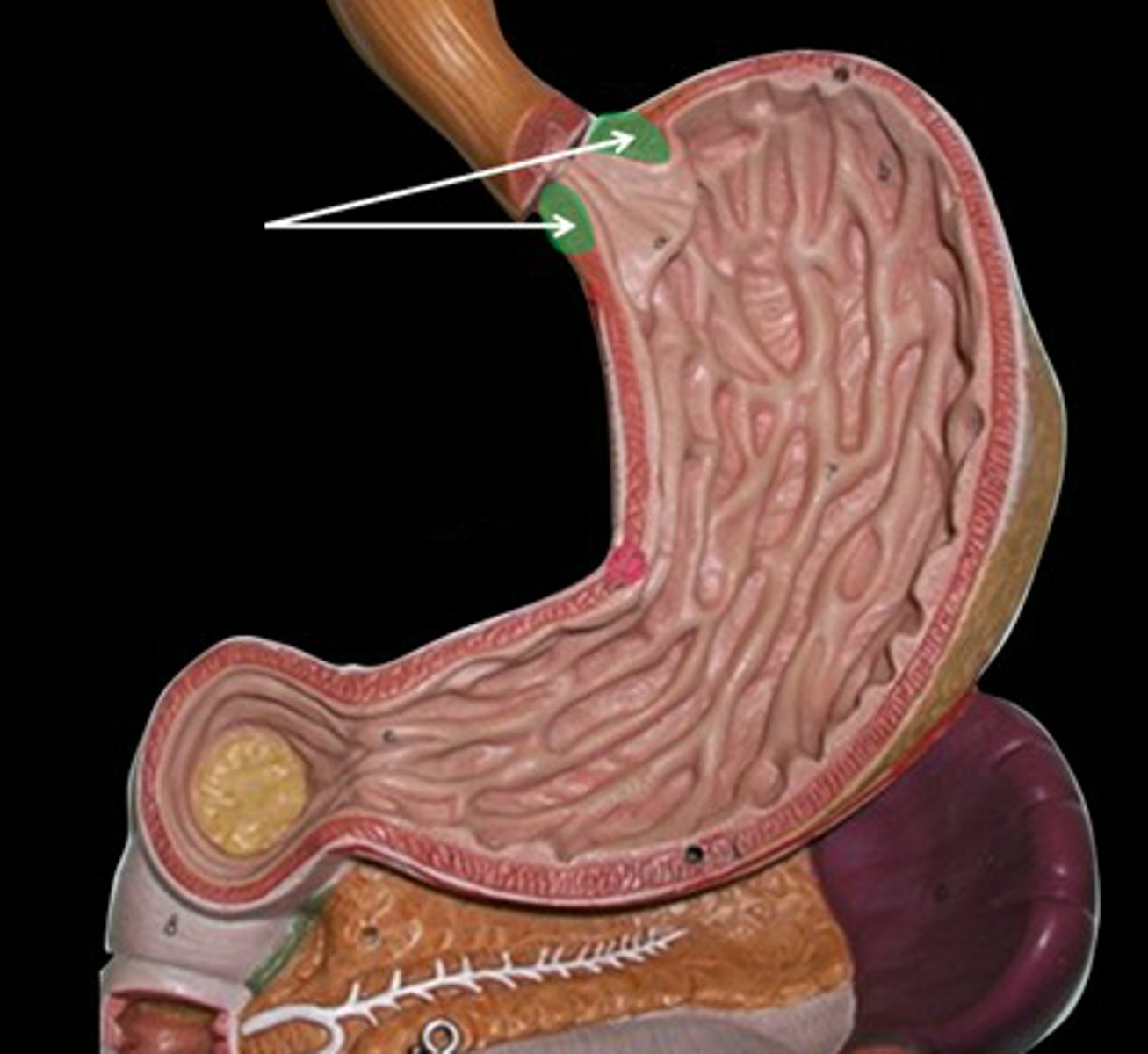
Sphincter Control
A ring-shaped muscle that contracts and relaxes to close or open body passages, acting as a valve to regulate the flow of substances. Muscles regulate openings such as eyelids, urethra, and anus.
Thermogenesis
Heat production by skeletal muscle.
Glycemic Control
Skeletal muscles absorb and use large amounts of glucose.
Epimysium
Connective tissue surrounding the entire muscle.
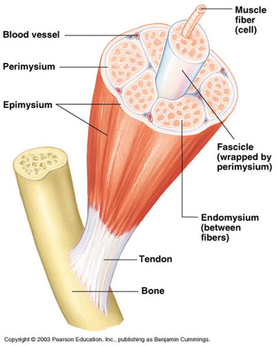
Perimysium
Connective tissue that surrounds bundles of muscle fibers (fascicles).
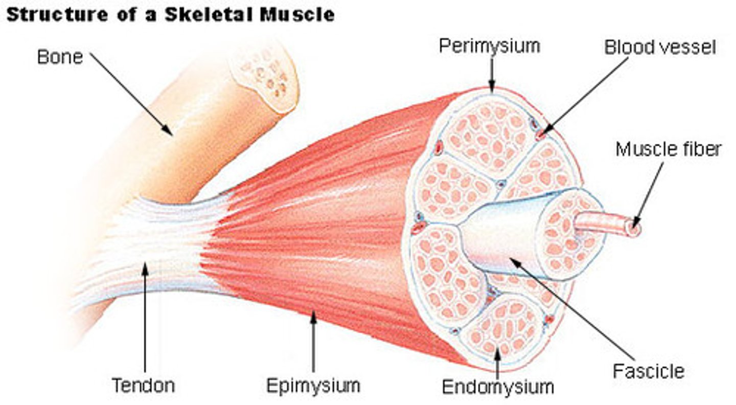
Endomysium
Thin connective tissue surrounding each individual muscle fiber.
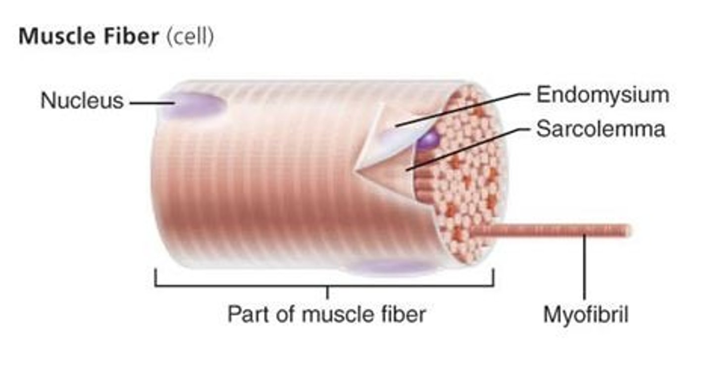
Fascicle
Bundle of muscle fibers wrapped by perimysium.
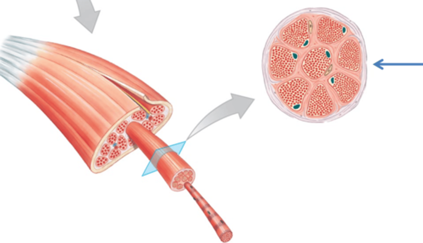
Muscle Fiber
A single skeletal muscle cell.
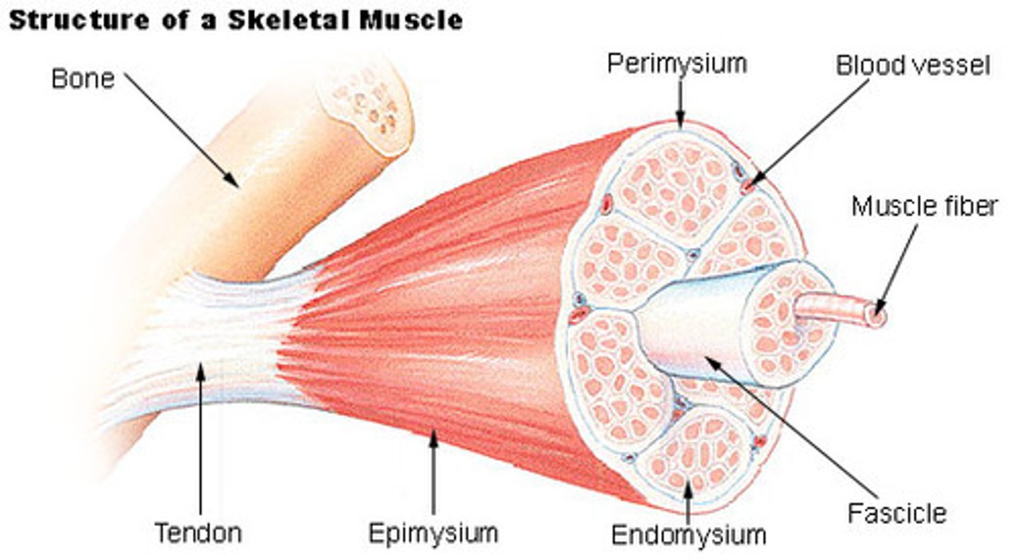
Sarcolemma
Plasma membrane of a muscle fiber.
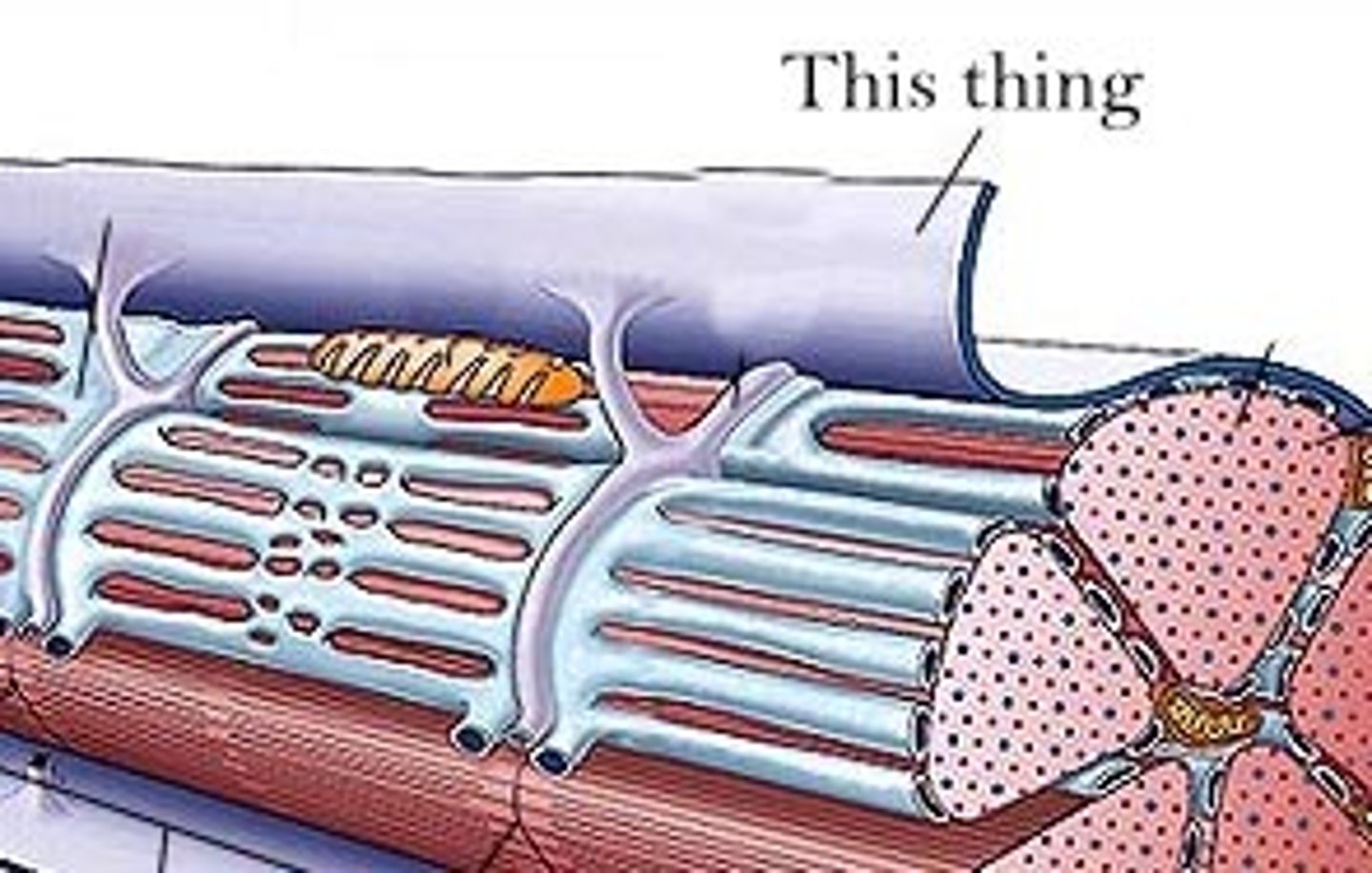
Nuclei (skeletal muscle)
Multiple nuclei located at the edge of muscle fibers.
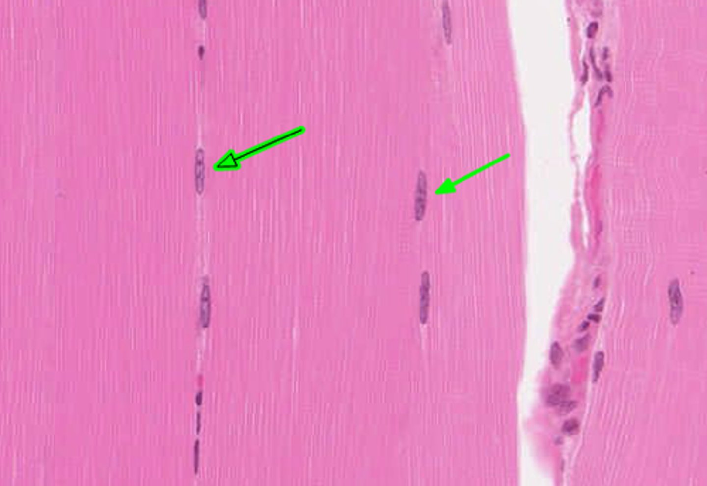
Sarcoplasm
Cytoplasm of a muscle fiber.
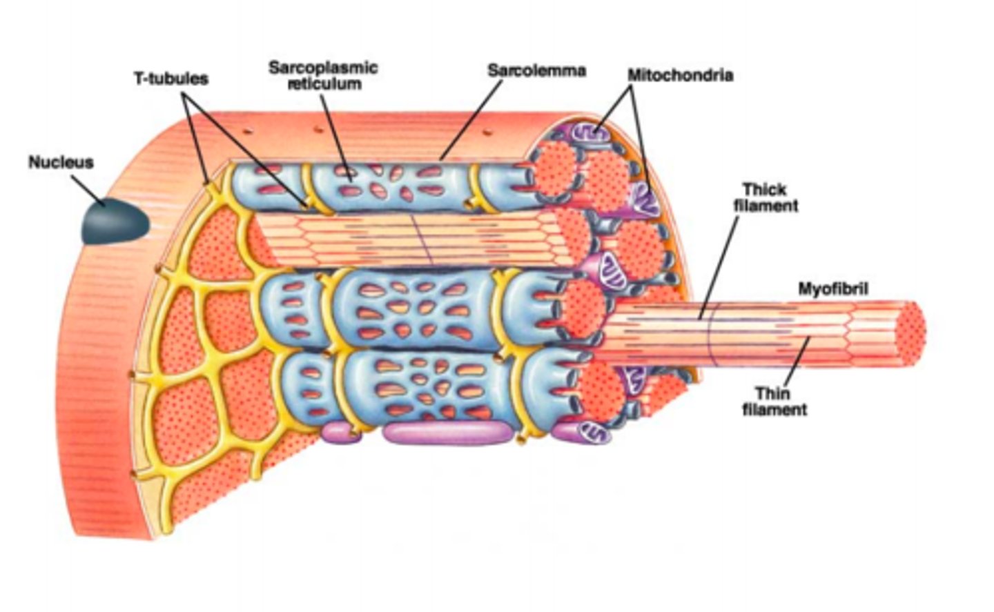
Myofibril
Long protein bundles inside muscle fibers responsible for contraction.
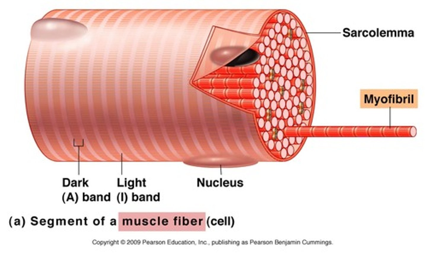
Myofilaments
Protein filaments inside myofibrils responsible for muscle contraction.
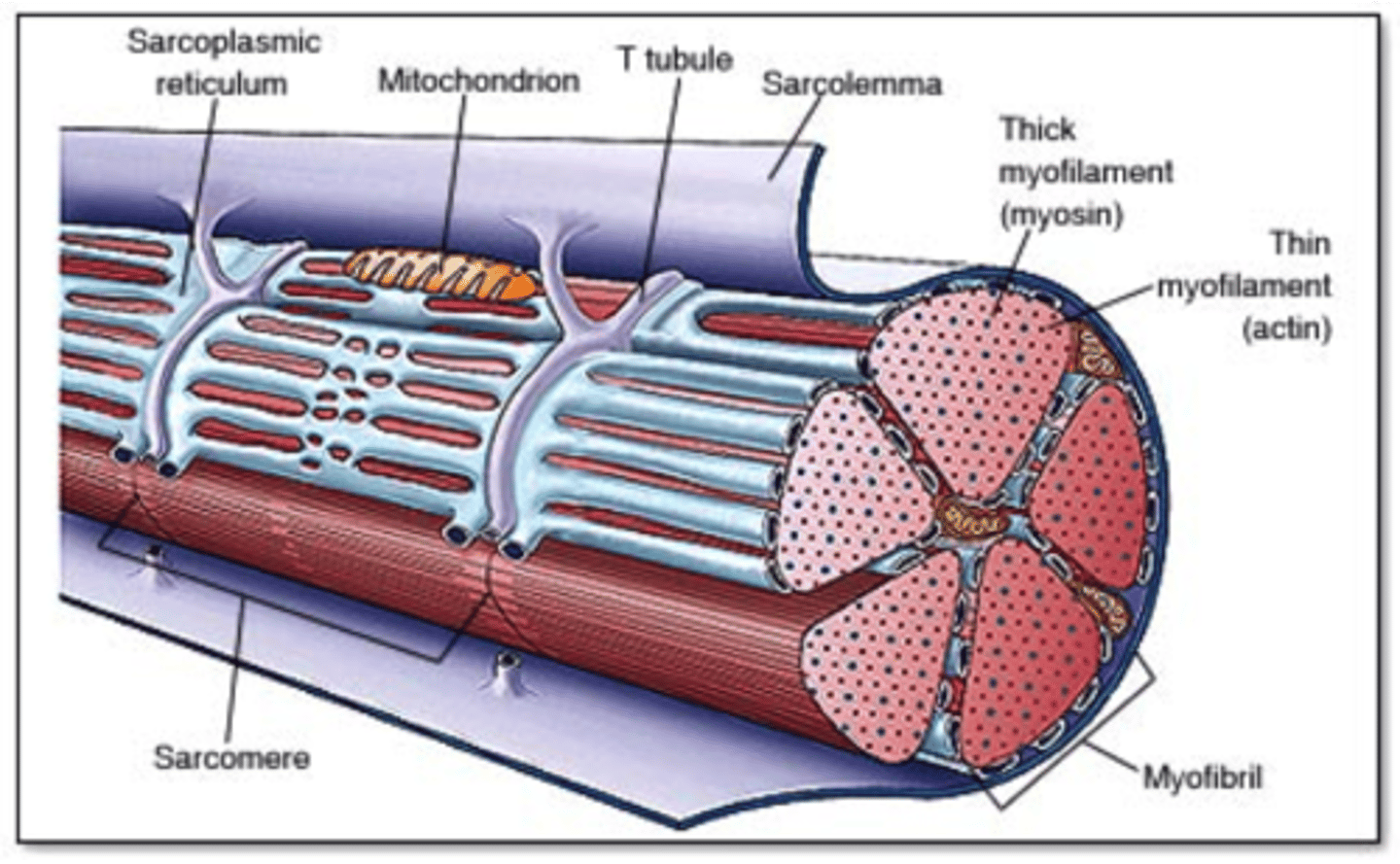
Thick Filament
Myosin protein filament that pulls actin during contraction
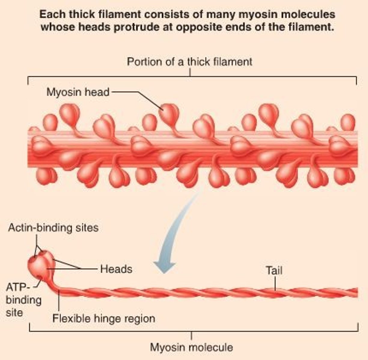
Thin Filament
Actin filament that interacts with myosin during contraction.
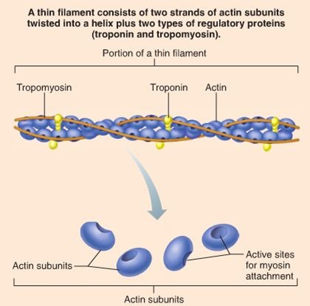
Myosin
Thick filament protein with heads that form cross bridges with actin.
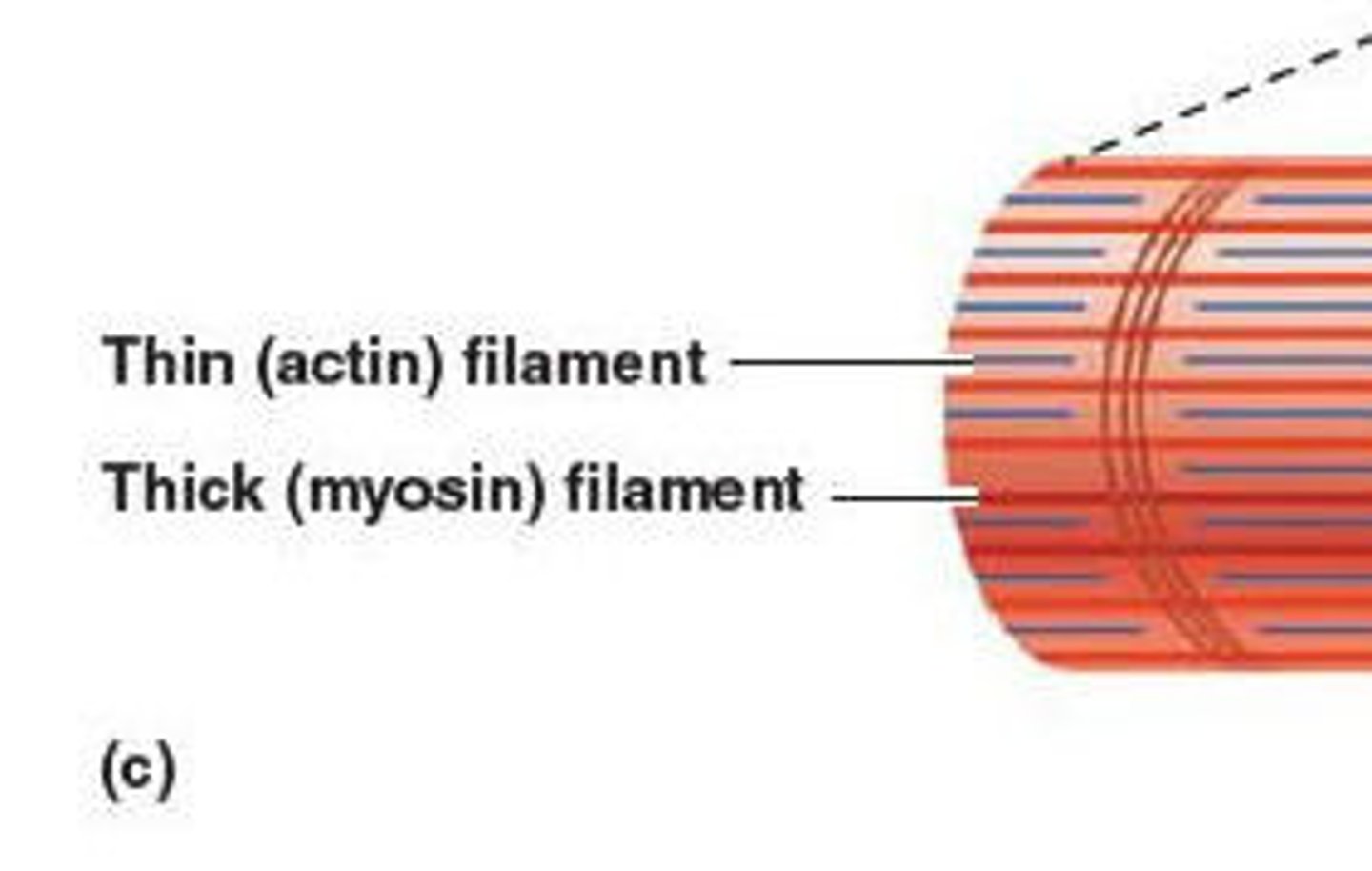
Actin
Thin filament protein that provides binding sites for myosin.
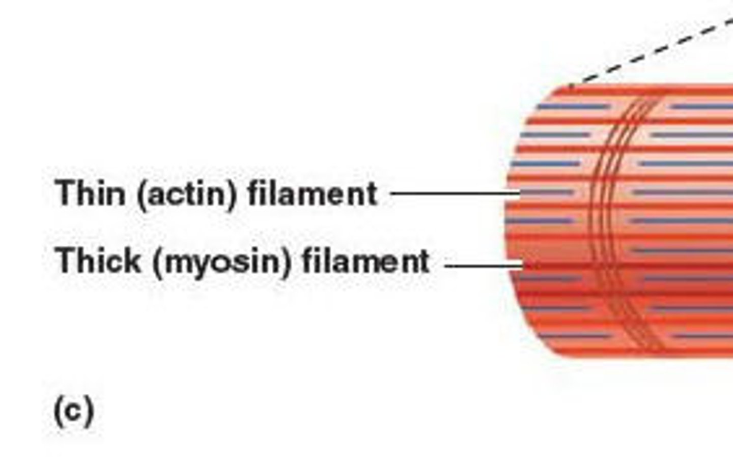
Tropomyosin
Protein that blocks myosin binding sites on actin when muscle is relaxed.
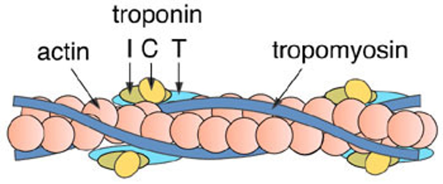
Troponin
Protein that binds calcium and moves tropomyosin to allow contraction.
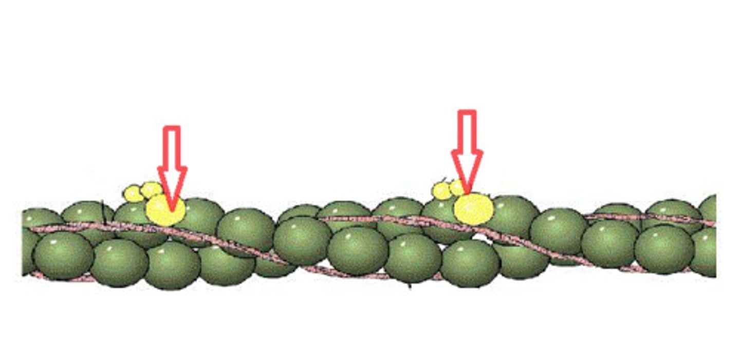
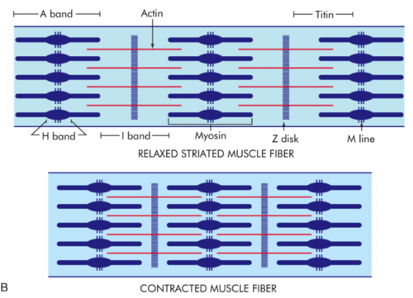
Titin
Elastic protein that stabilizes thick filaments and prevents overstretching.
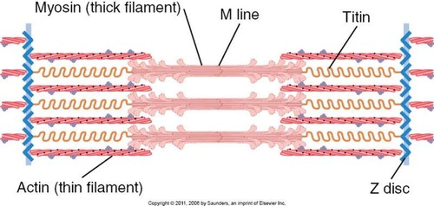
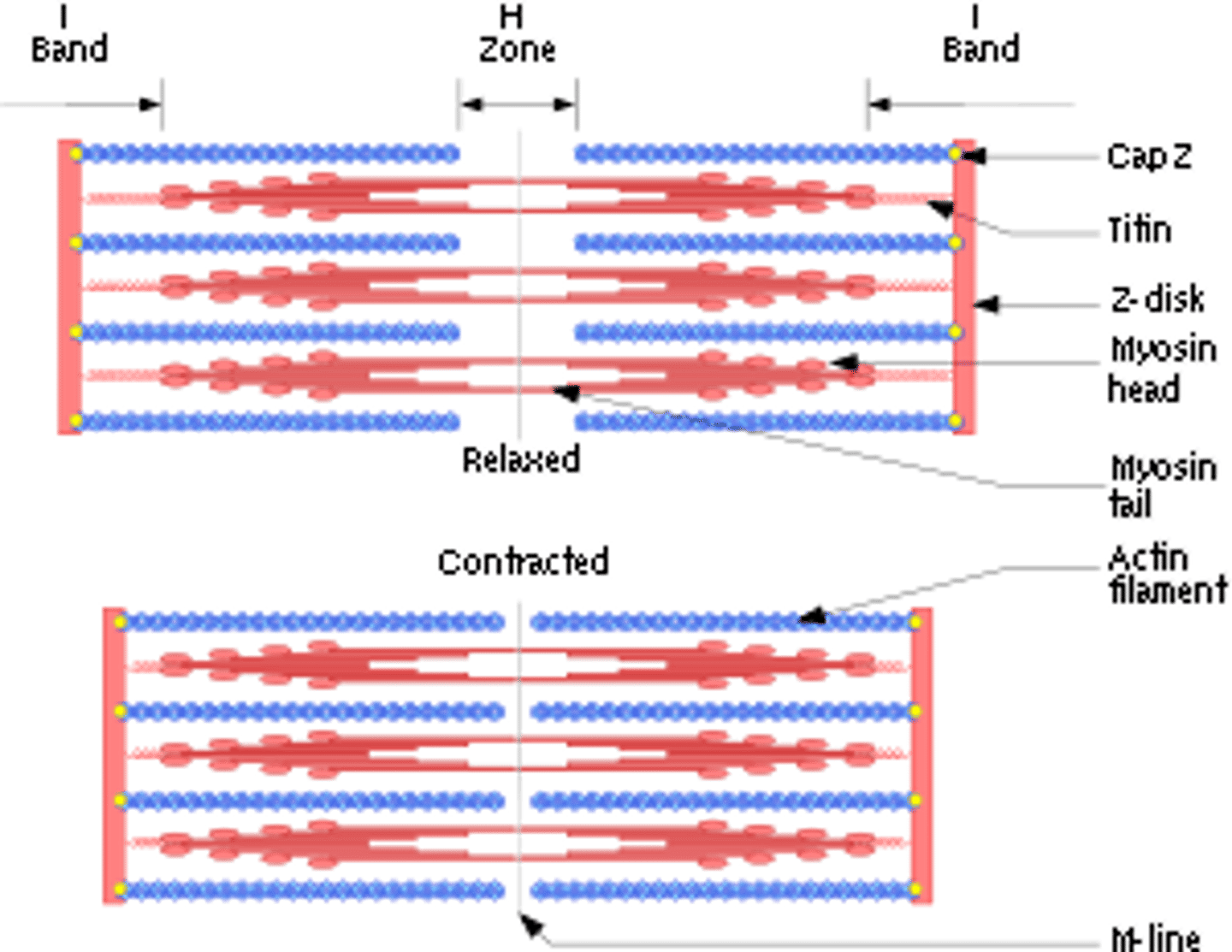
Sarcomere
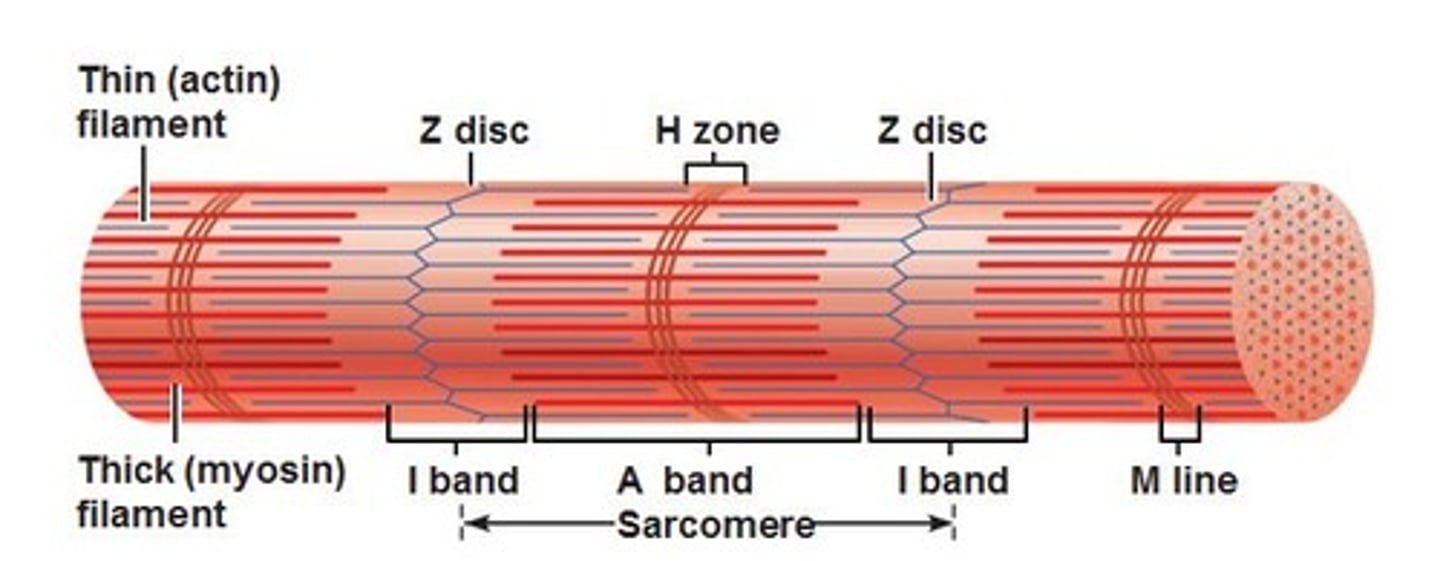
Functional contractile unit of muscle fiber (Z disc to Z disc).
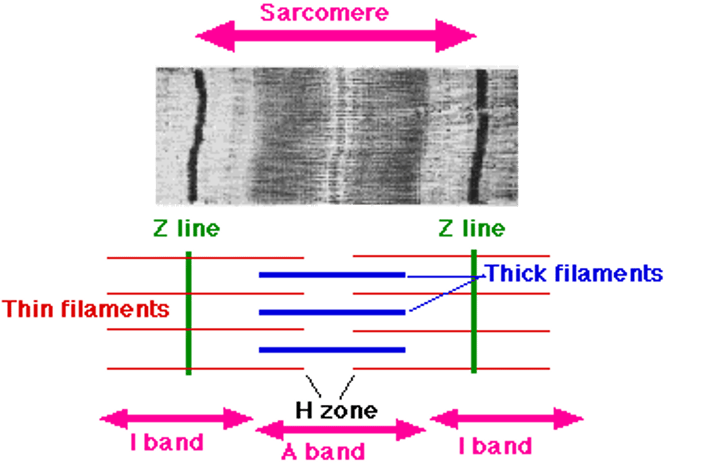
Z Disc
Boundary of a sarcomere; anchors thin filaments. On each sides
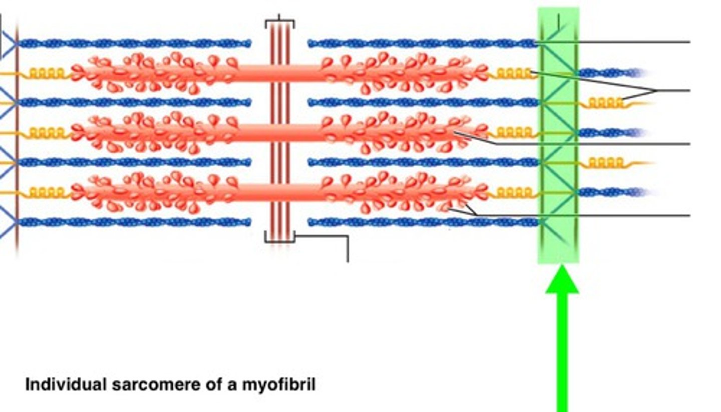
A Band
Dark region where thick and thin filaments overlap.
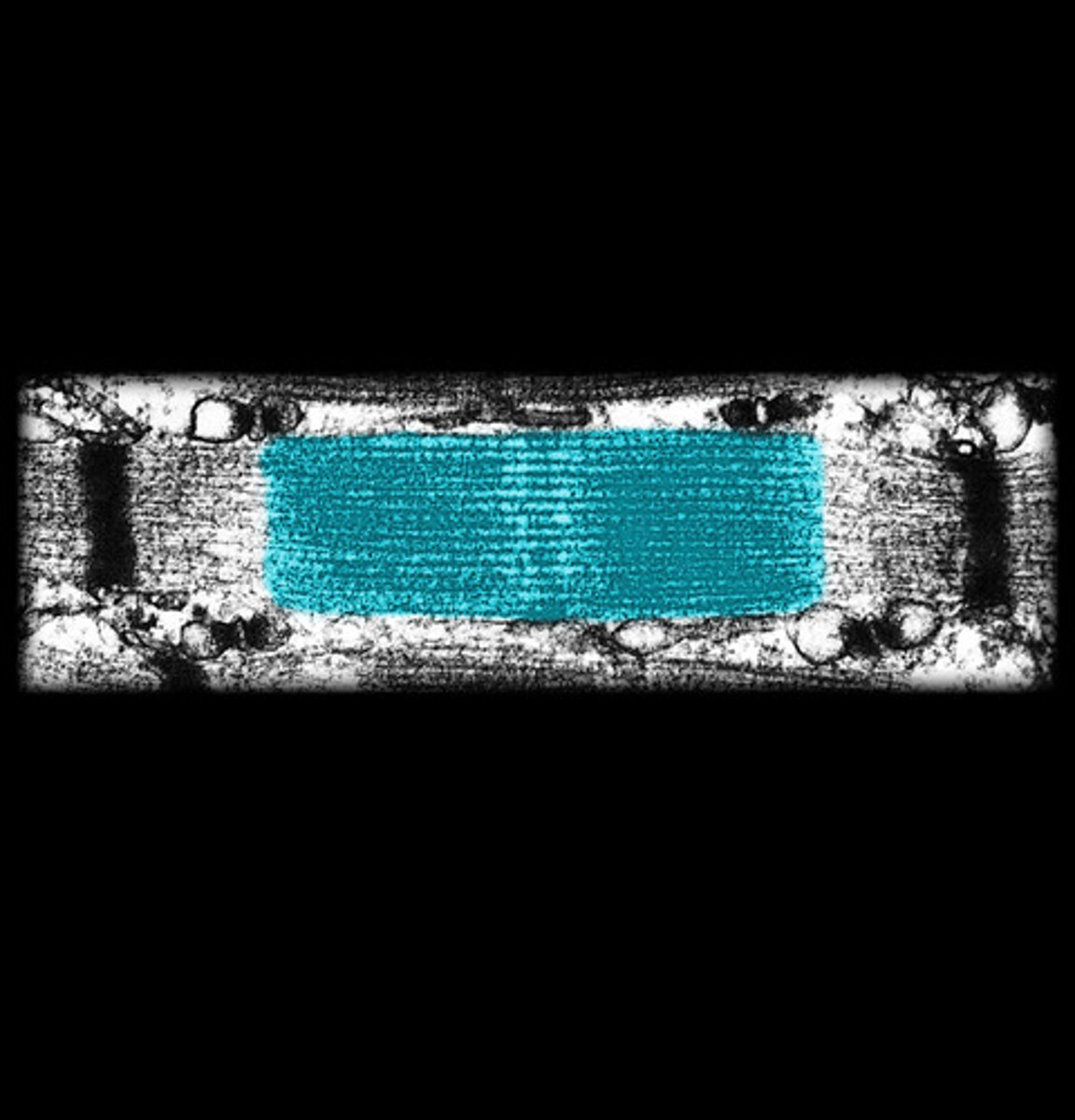
I Band
Light region containing only thin filaments.
H Zone
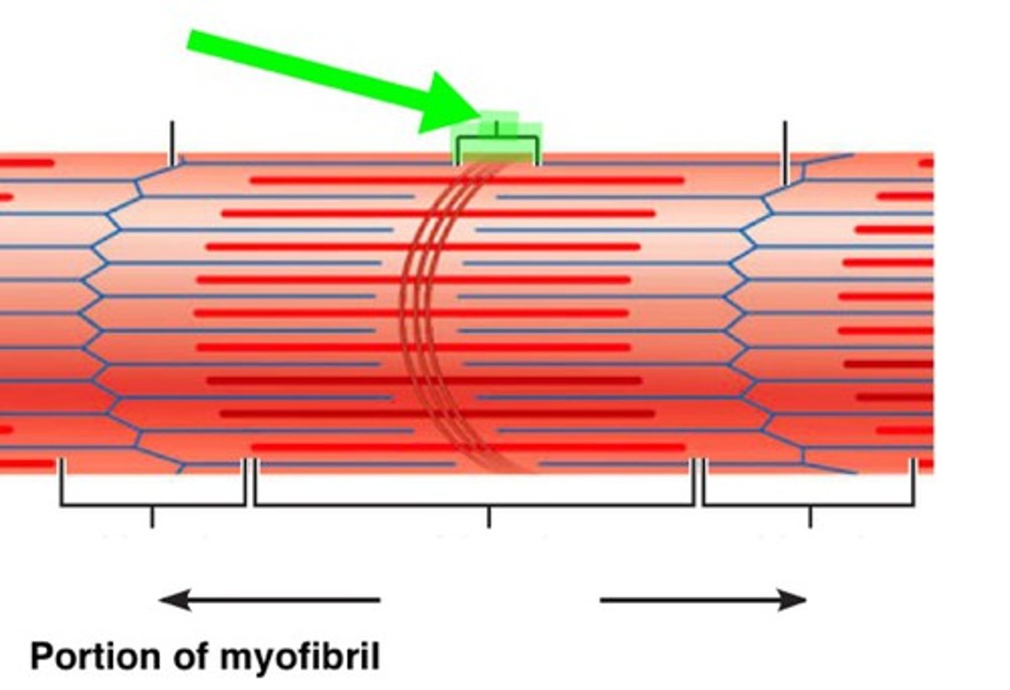
Region containing only thick filaments.
M Line
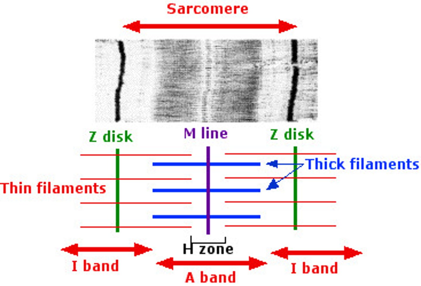
Center of sarcomere where thick filaments are anchored.
Direct (Fleshy) Attachment
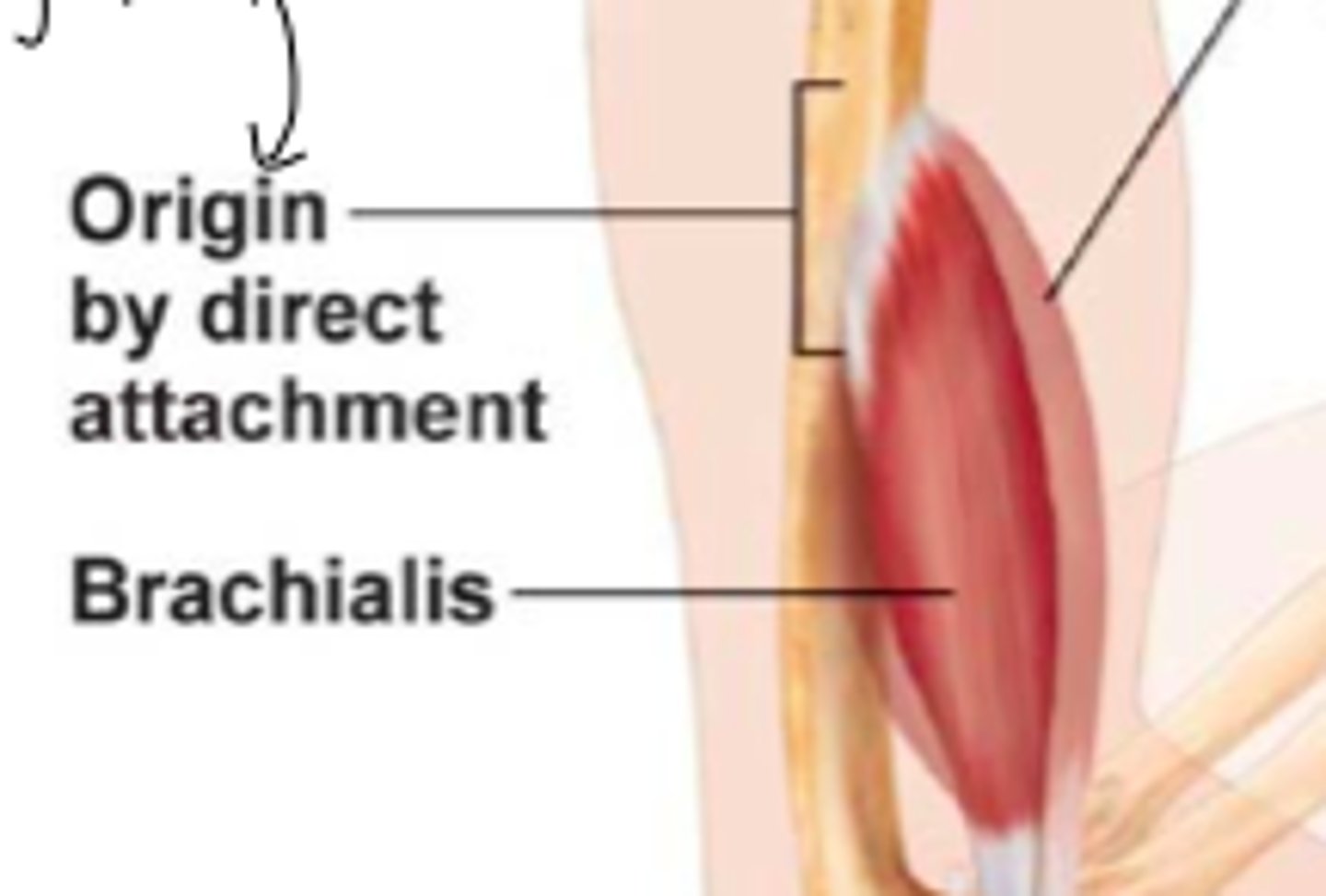
Muscle connects directly to bone via epimysium fibers.
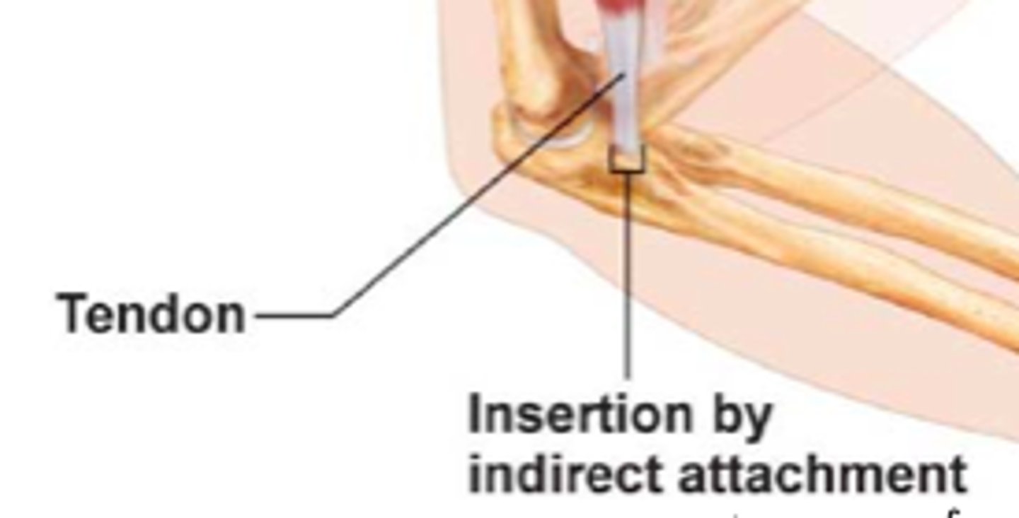
Indirect Attachment
Muscle connects to bone through a tendon.
Aponeurosis
Broad sheetlike tendon connecting muscle to bone.
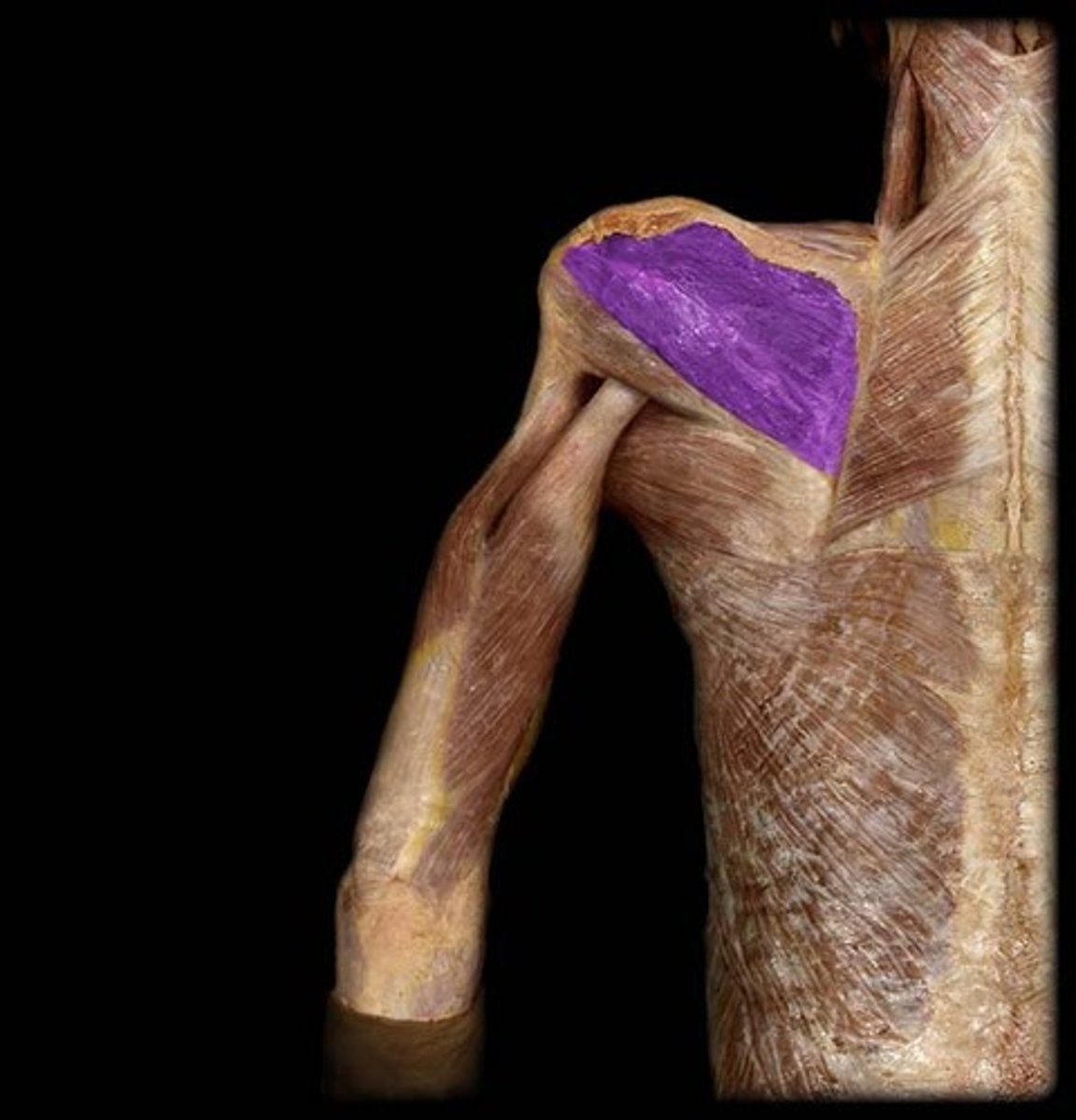
Infraspinatus
Example of muscle with direct attachment.
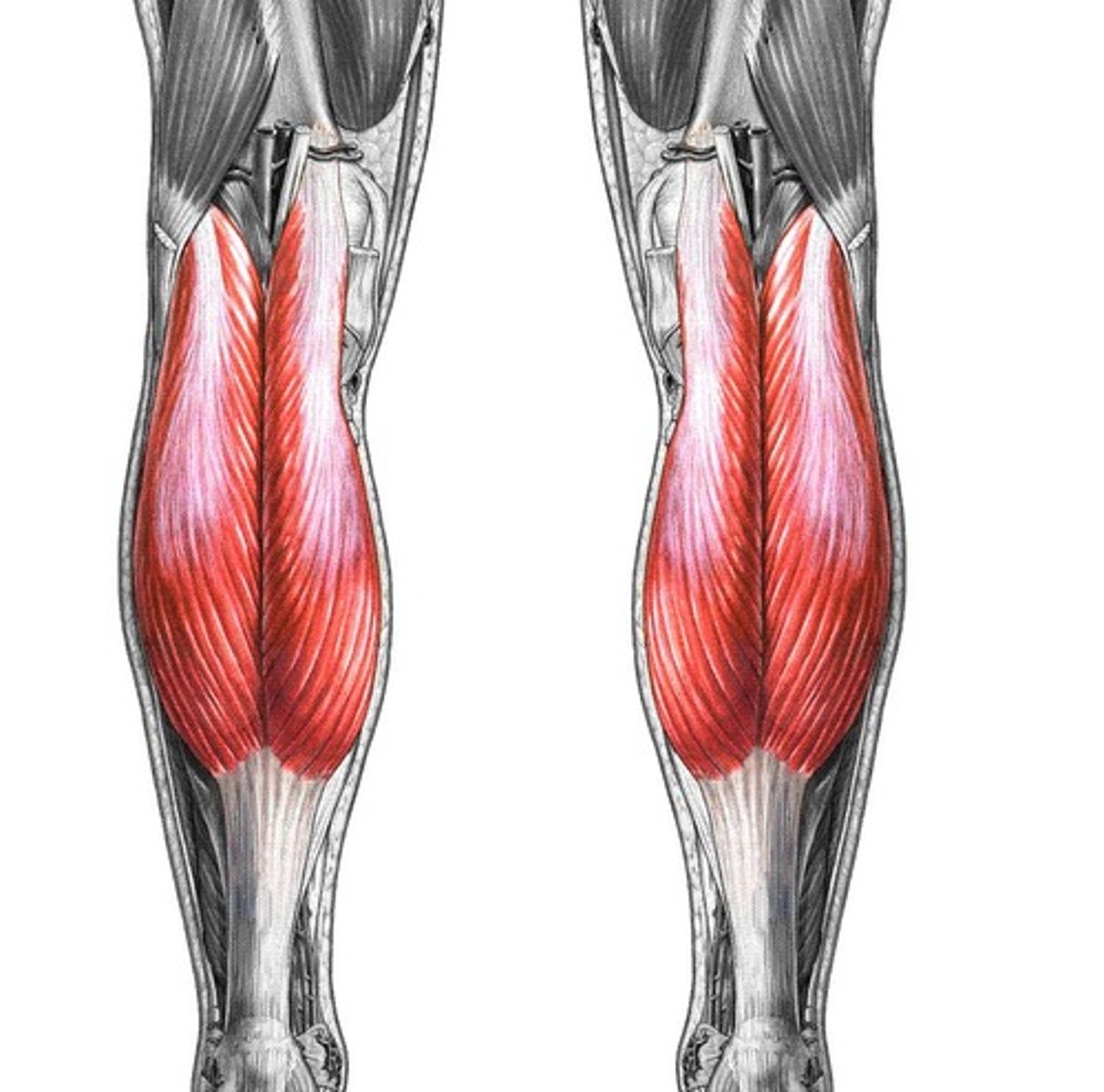
Gastrocnemius
Calf muscle attached indirectly via the calcaneal tendon.
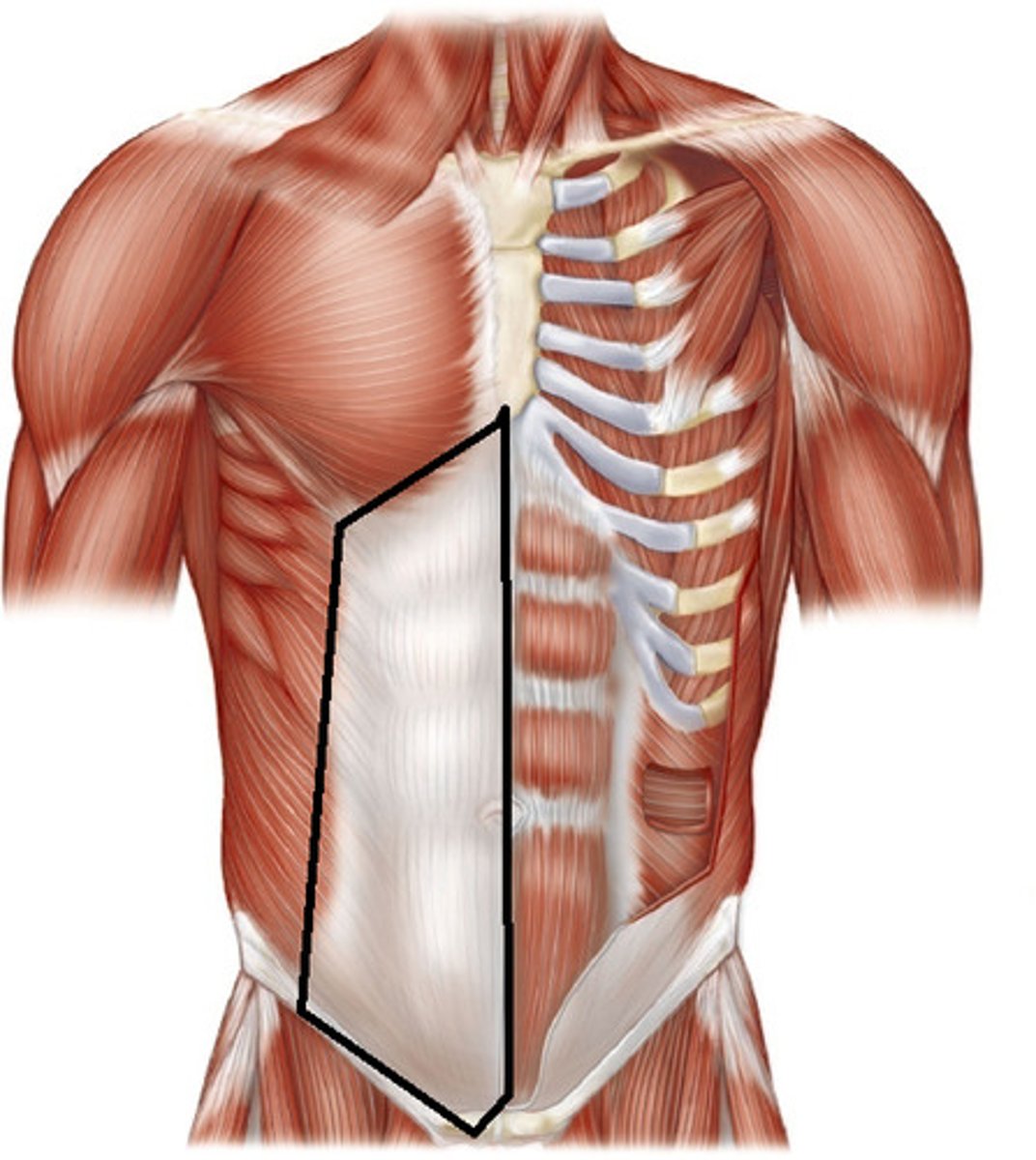
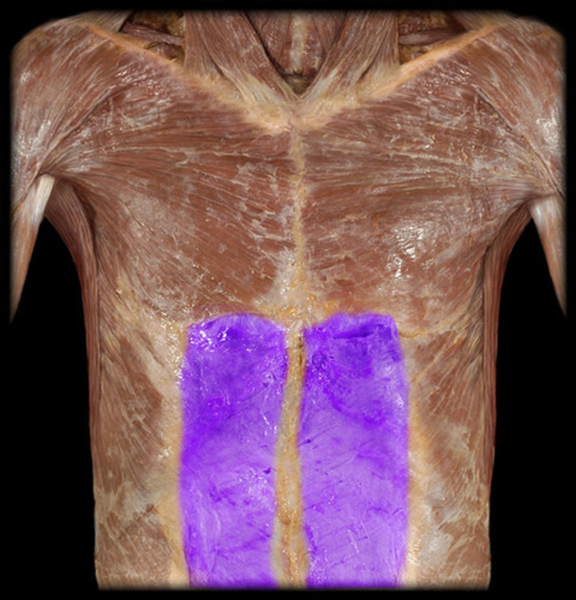
Rectus Sheath
Aponeurotic sheath surrounding rectus abdominis.
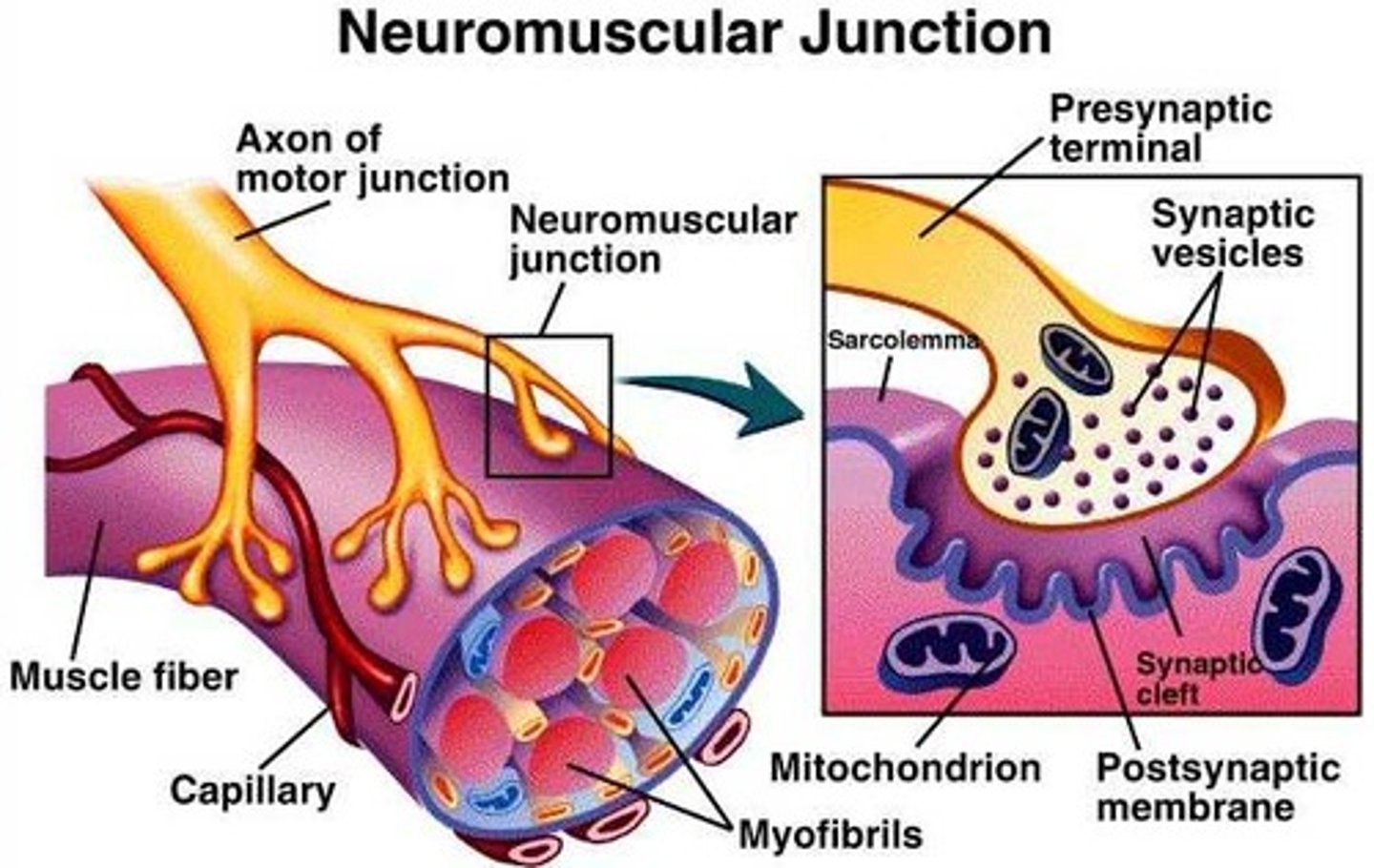
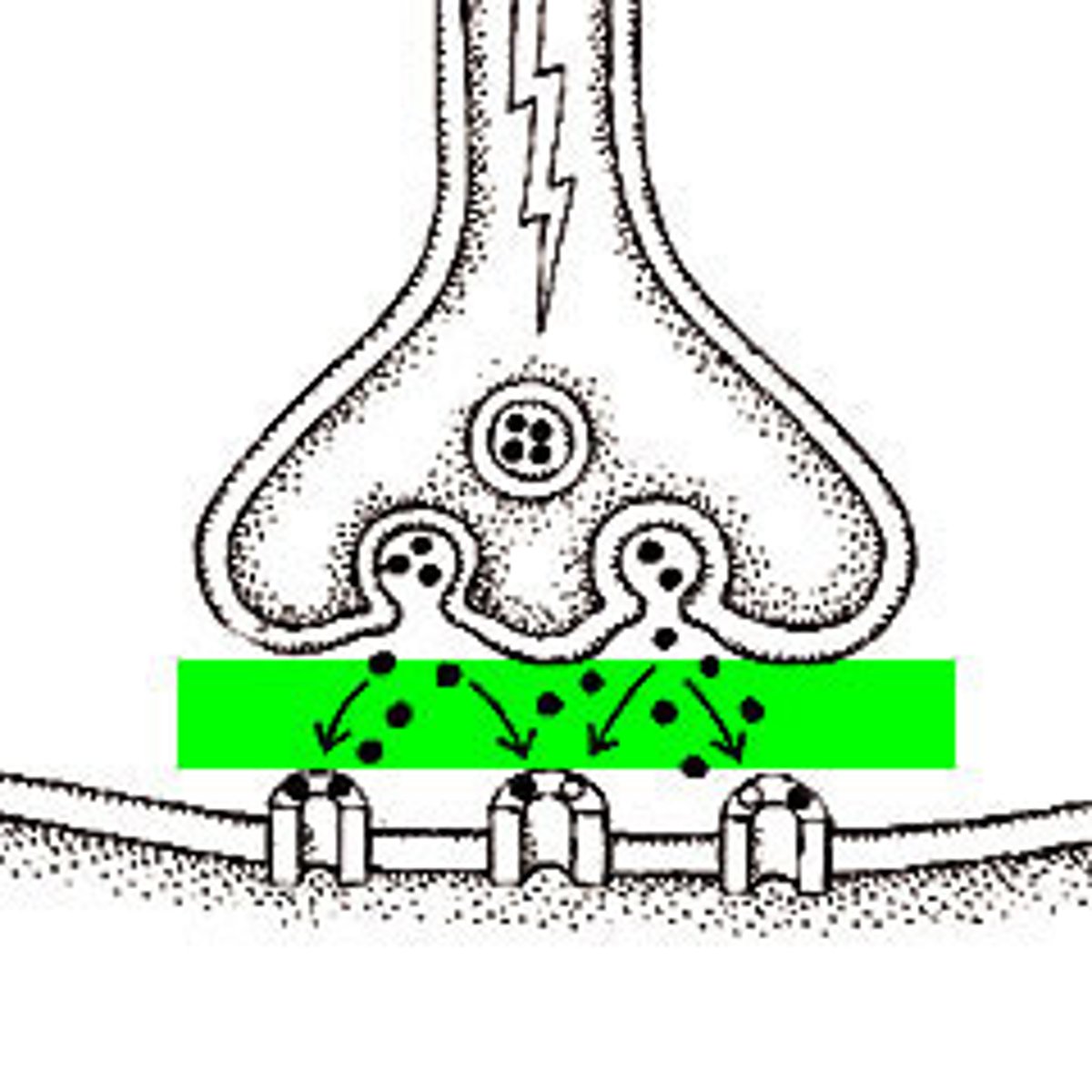
Neuromuscular Junction
Synapse between motor neuron and muscle fiber.
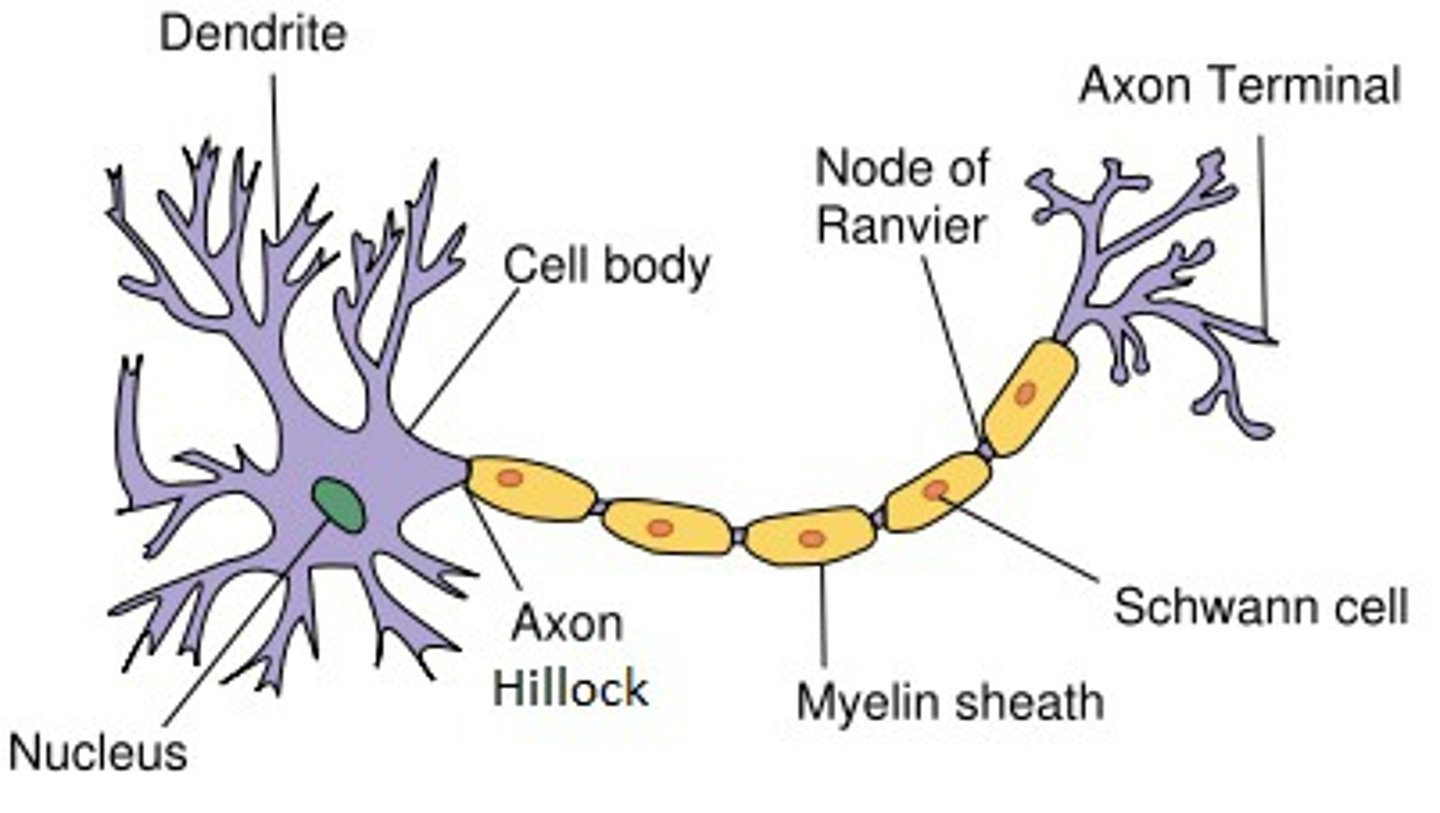
Motor Nerve Fiber
Neuron that stimulates muscle contraction.
Presynaptic Terminal / Axon Terminal
End of neuron that releases neurotransmitter.
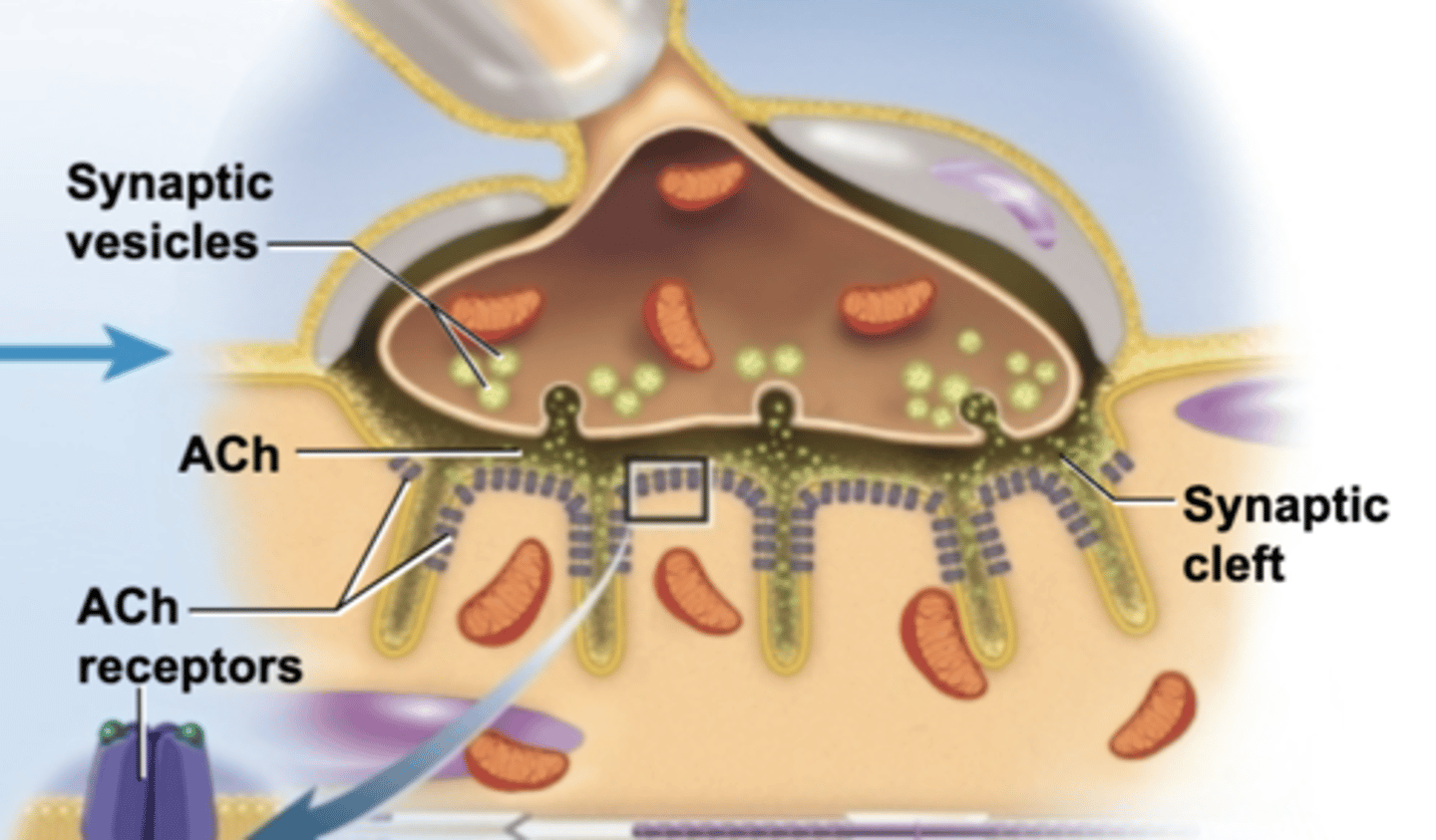
Synaptic Vesicles
Vesicles containing acetylcholine.
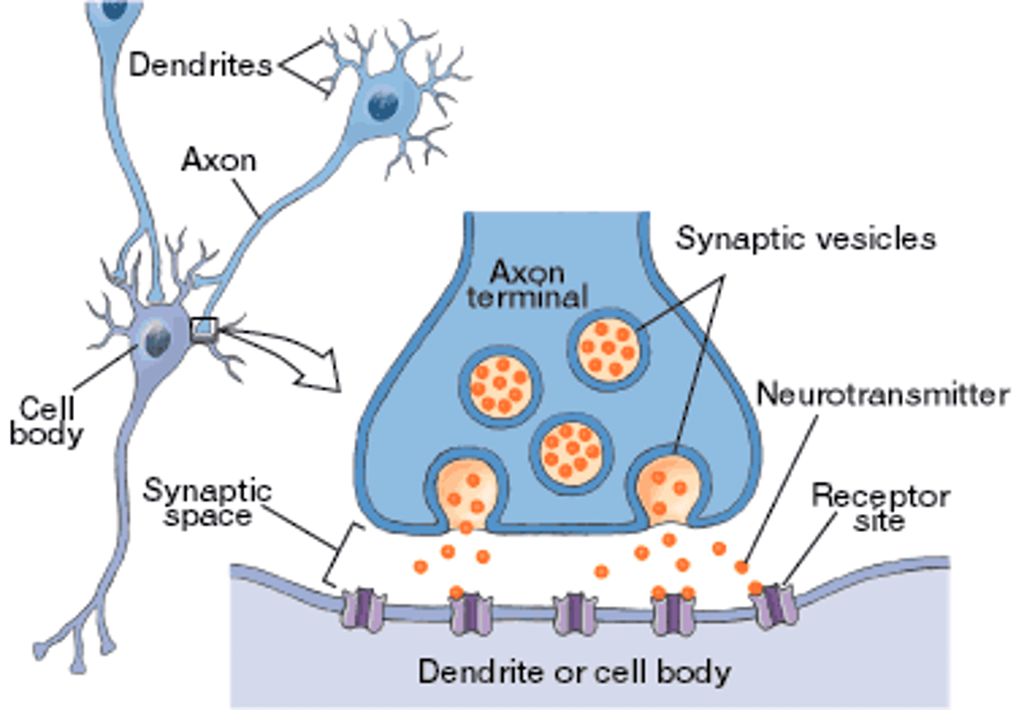
Synaptic Cleft
Gap between neuron and muscle fiber.
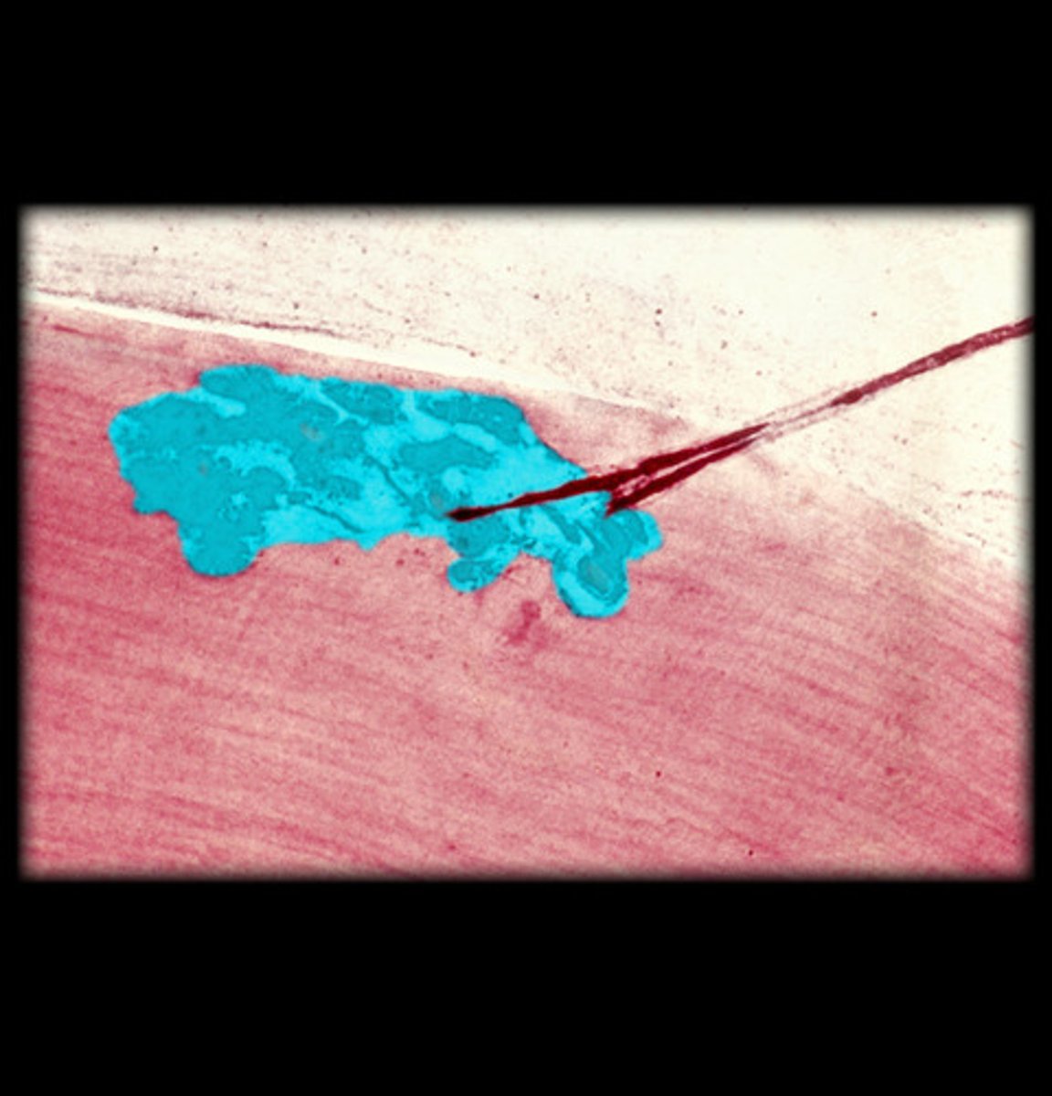
motor end plate
Region of sarcolemma containing ACh receptors.
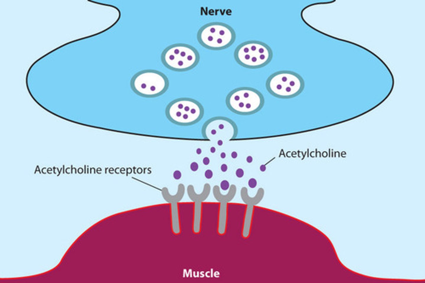
Acetylcholine (ACh)
Neurotransmitter that stimulates muscle contraction.
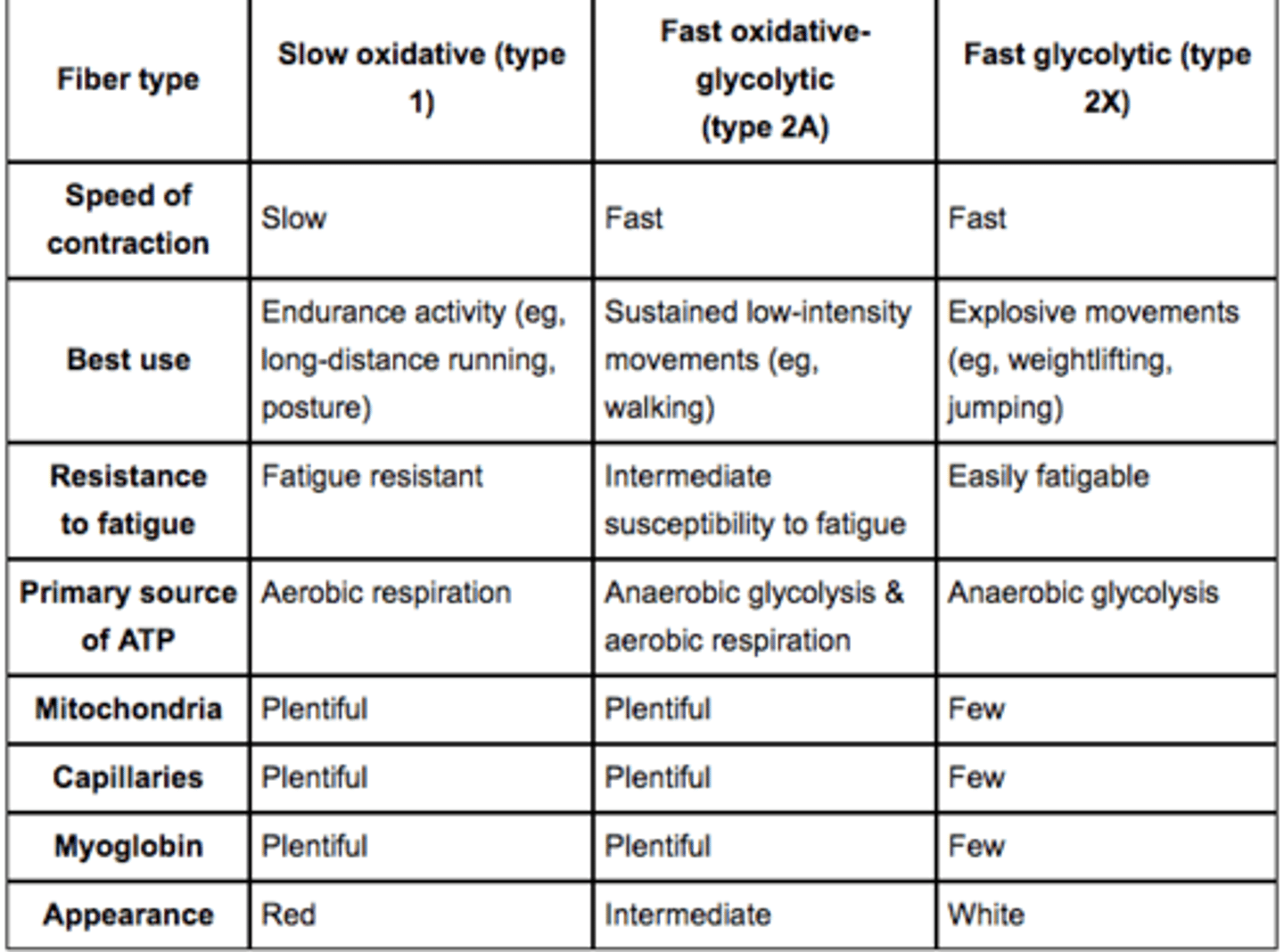
Slow Oxidative Fibers (Type I)
Red, fatigue-resistant fibers using aerobic respiration.
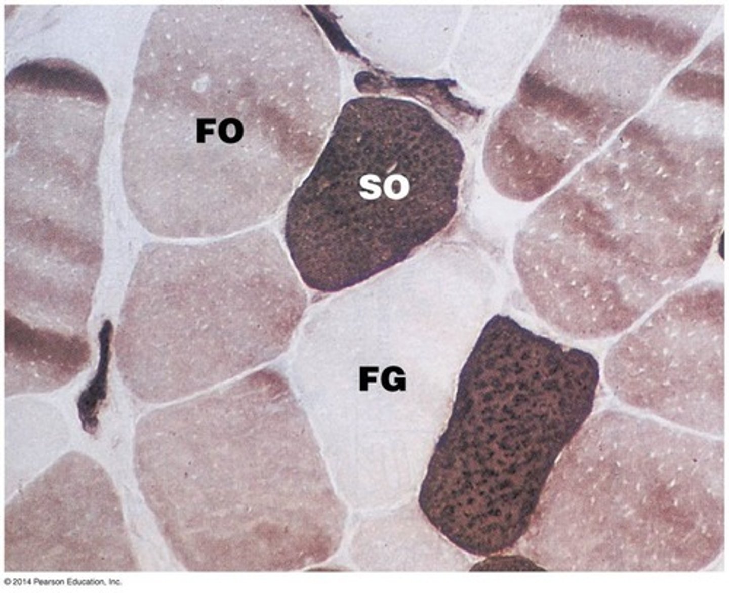
Fast Glycolytic Fibers (Type II)
White fibers that contract quickly but fatigue rapidly.
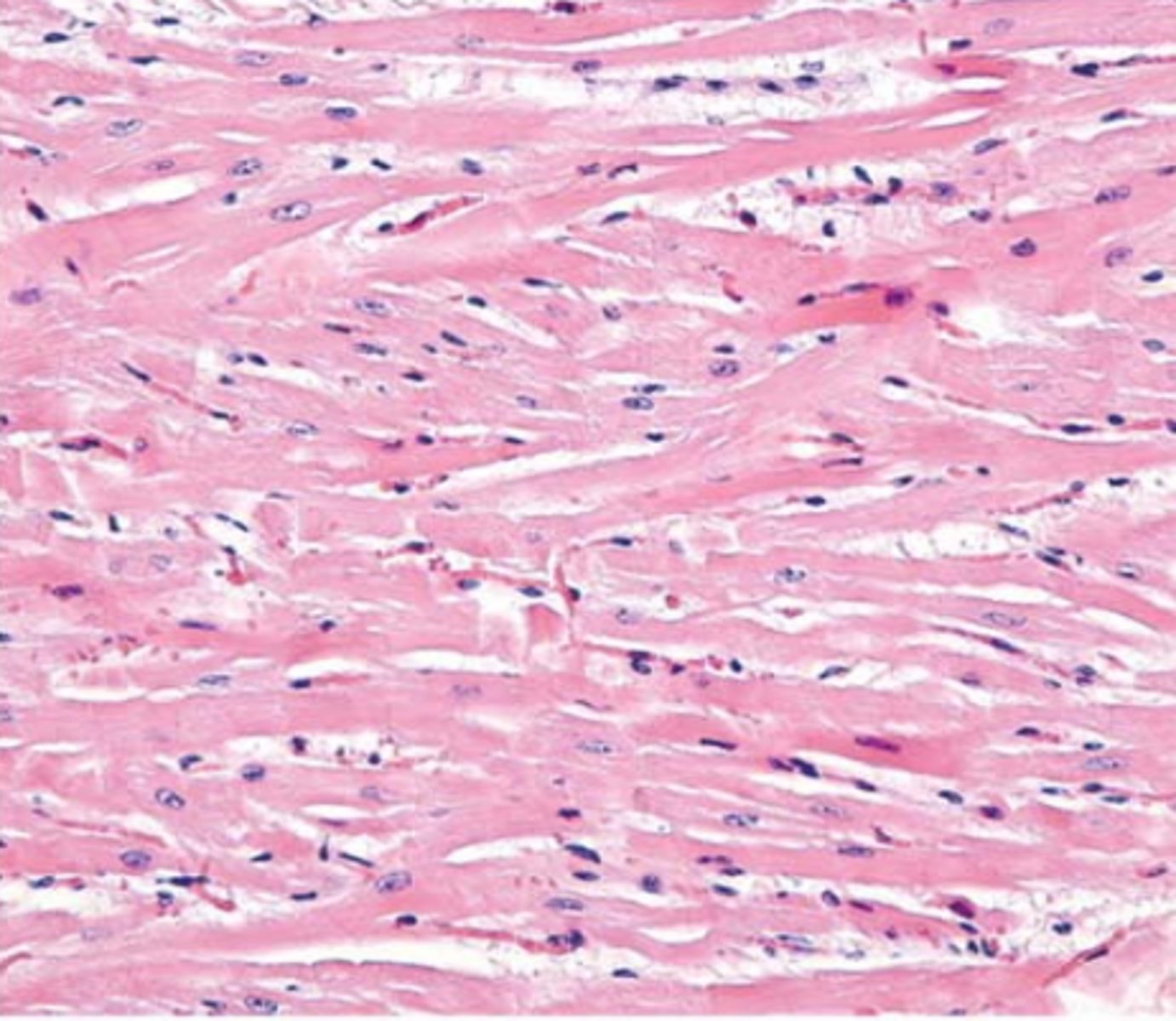
Cardiomyocytes
Cardiac muscle cells.
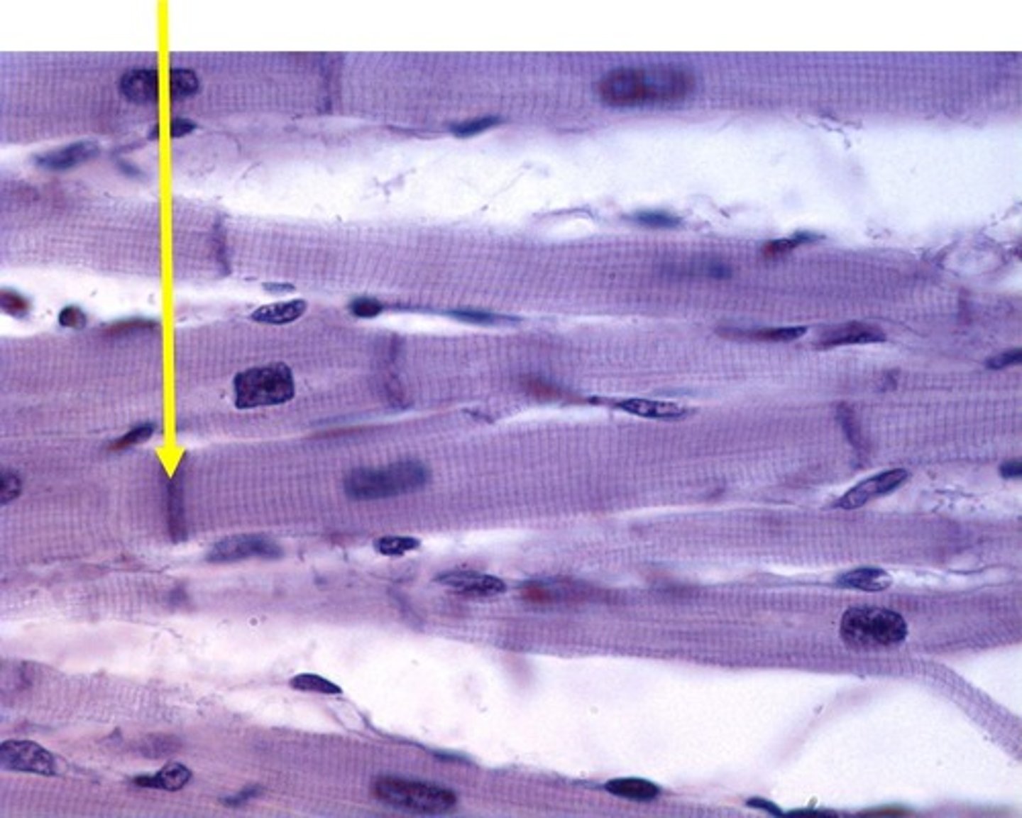
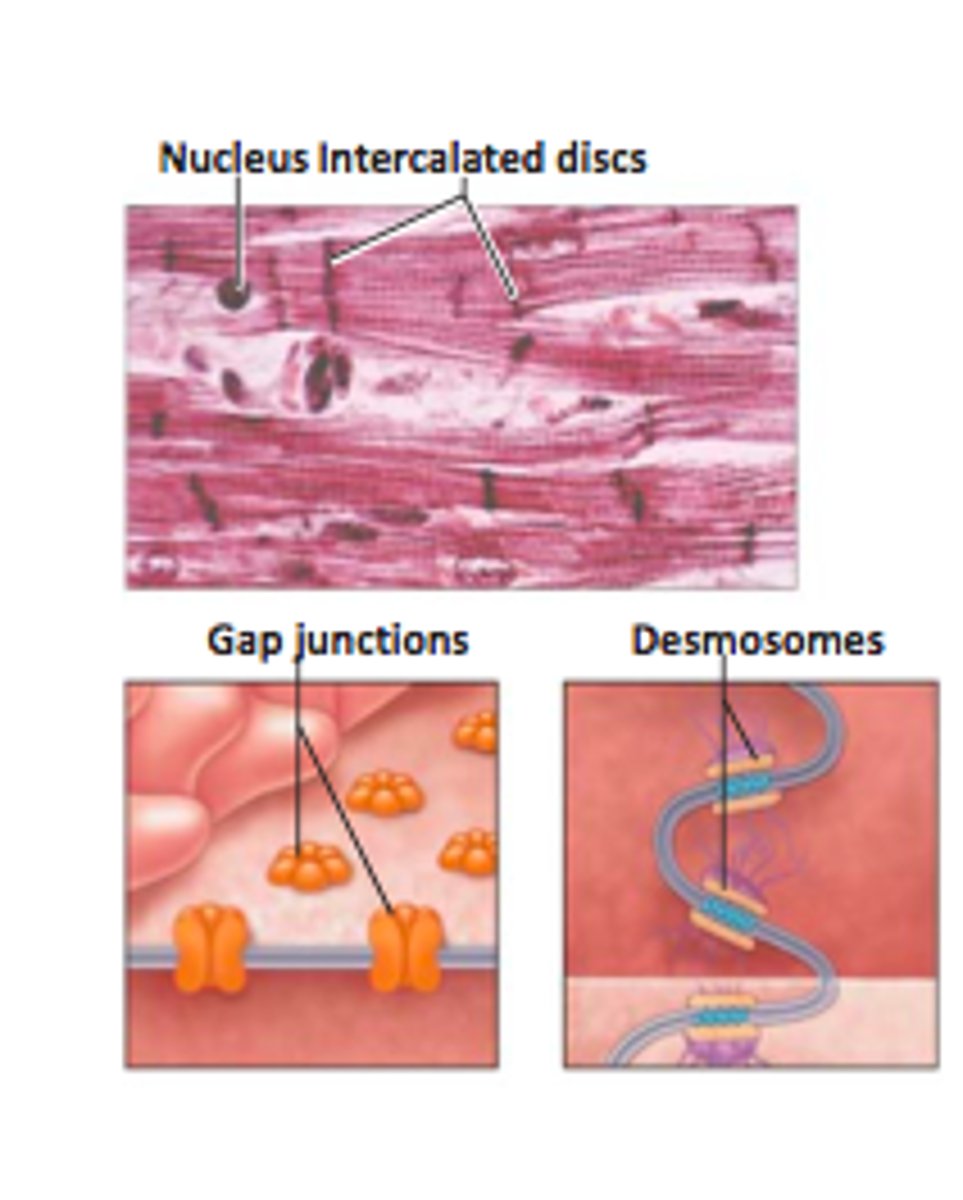
Intercalated Discs
Specialized junctions connecting cardiac cells.
Gap Junctions
Channels allowing electrical signals to pass between cardiac cells.
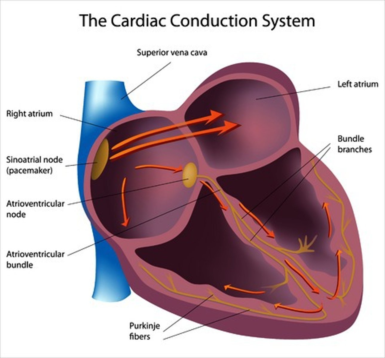
Sinoatrial Node
Natural pacemaker that sets heart rhythm.
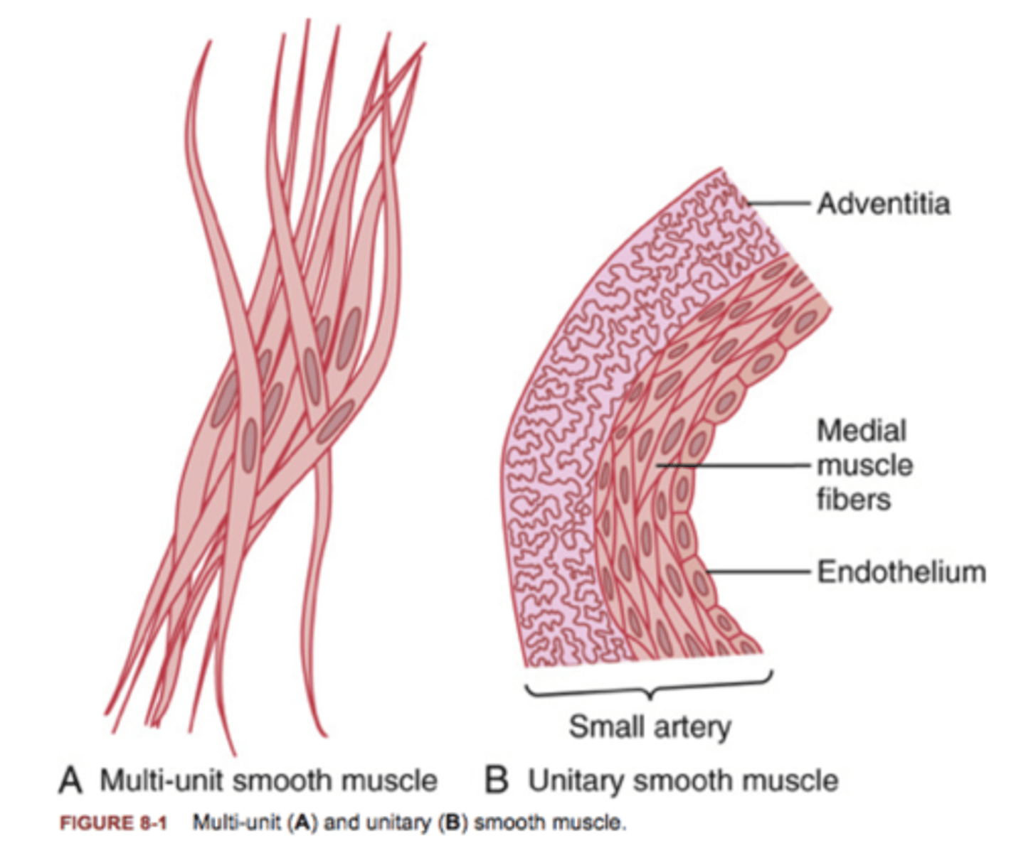
Multiunit Smooth Muscle
Smooth muscle where fibers contract independently.
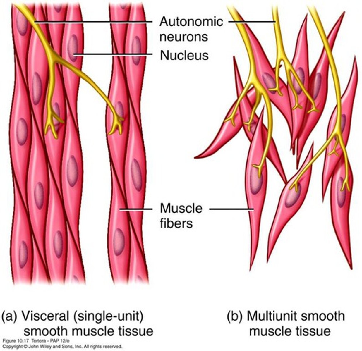
Unitary Smooth Muscle
Smooth muscle where many cells contract together.
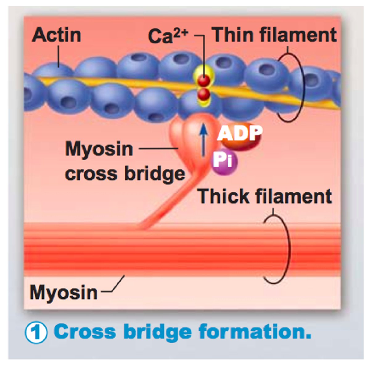
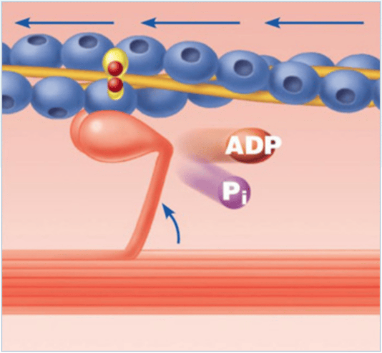
Cross Bridge
Connection formed when myosin head binds actin.
Power Stroke
Myosin head pulls actin toward center of sarcomere.
Sliding Filament Theory
Actin slides over myosin causing sarcomere shortening.
Excitation
Nerve signal releases ACh and stimulates muscle fiber.
Excitation-Contraction Coupling
Electrical signal triggers calcium release.
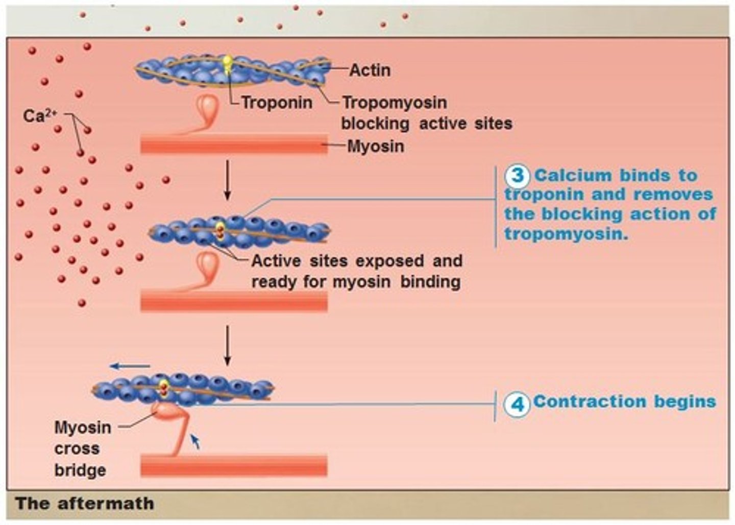
Contraction
Myosin cross bridges pull actin and shorten sarcomere.
Relaxation
Calcium removed and muscle returns to resting state.