Scrub Tech
 Call Kai
 Learn
Learn Practice Test
 Spaced Repetition
 Match
Match Flashcards
 Knowt Play
1/159

There's no tags or description
Looks like no tags are added yet.
Name | Mastery | Learn | Test | Matching | Spaced | Call with Kai | Chat |
|---|
No analytics yet
Send a link to your students to track their progress
160 Terms
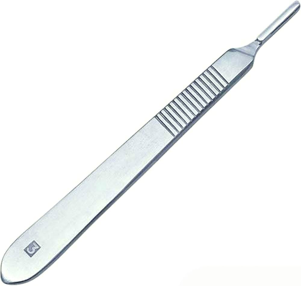
Knife (#10 blade)
Bard-Parker handle with a #10 blade; short knife for skin, long or deep knife for fascia and capsule.
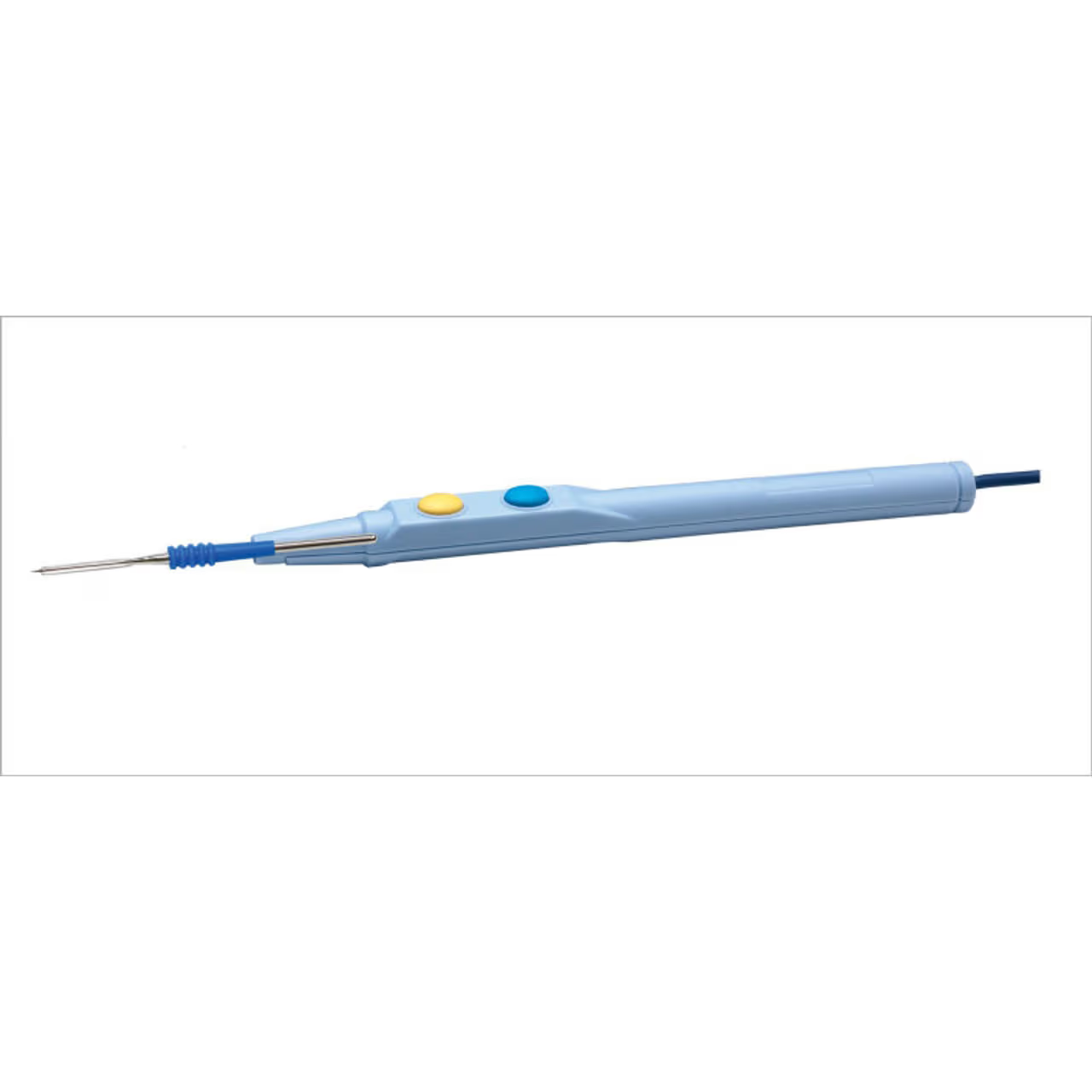
Bovie (electrocautery)
Cuts and coagulates tissue; also used to mark Whiteside's line on the femur.
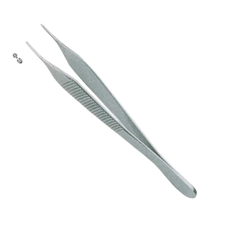
Pickups (Adson / heavy)
Toothed forceps for grasping tissue; the heavy pickup debrides the fat pad.
Adson clamp
Fine clamp for vessels and tissue; an Adson tonsil clamp places the hip liner (Gupta).
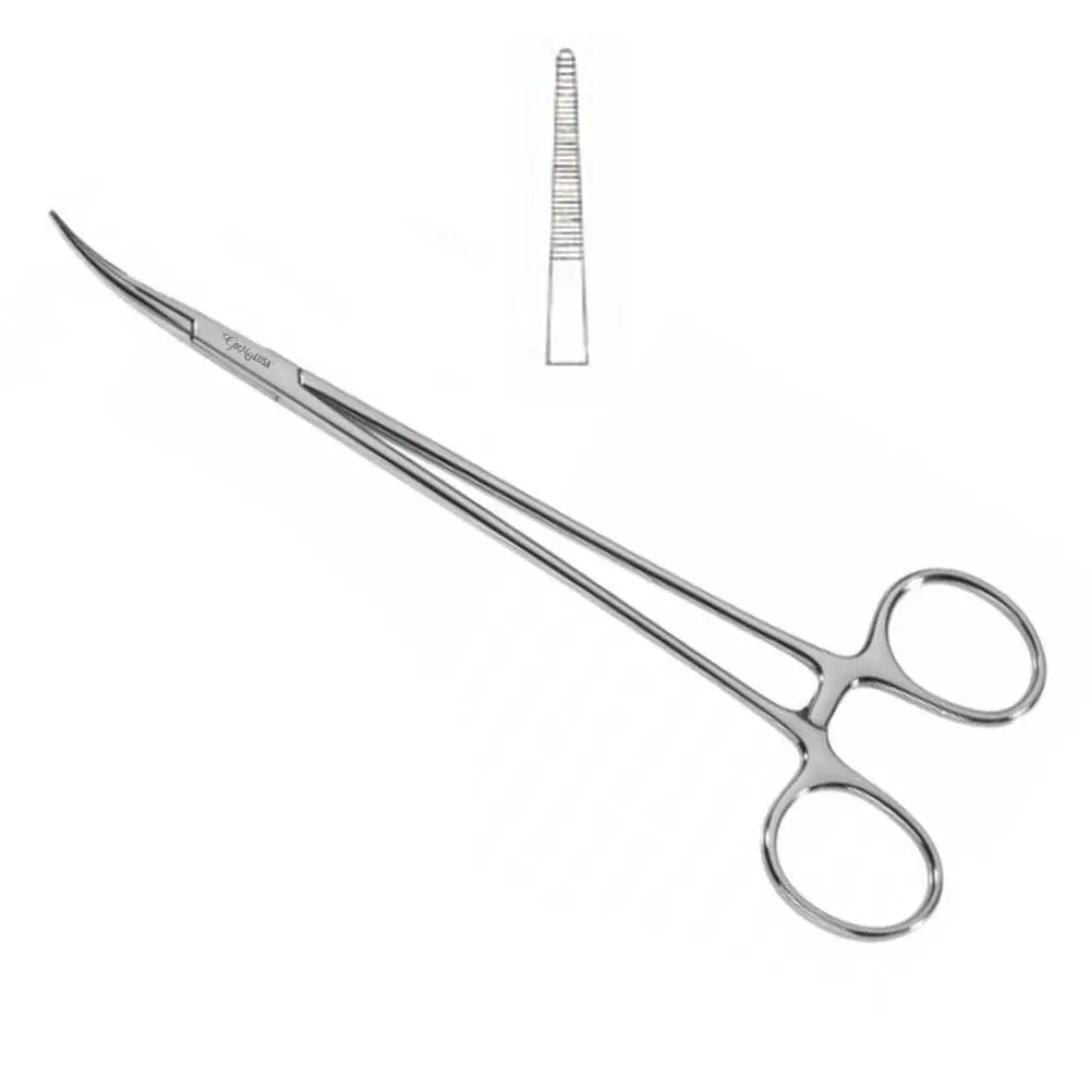
Kocher clamp
Heavy toothed clamp; grasps tissue, pulls out bone cuts, separates the TFL, and removes trials.
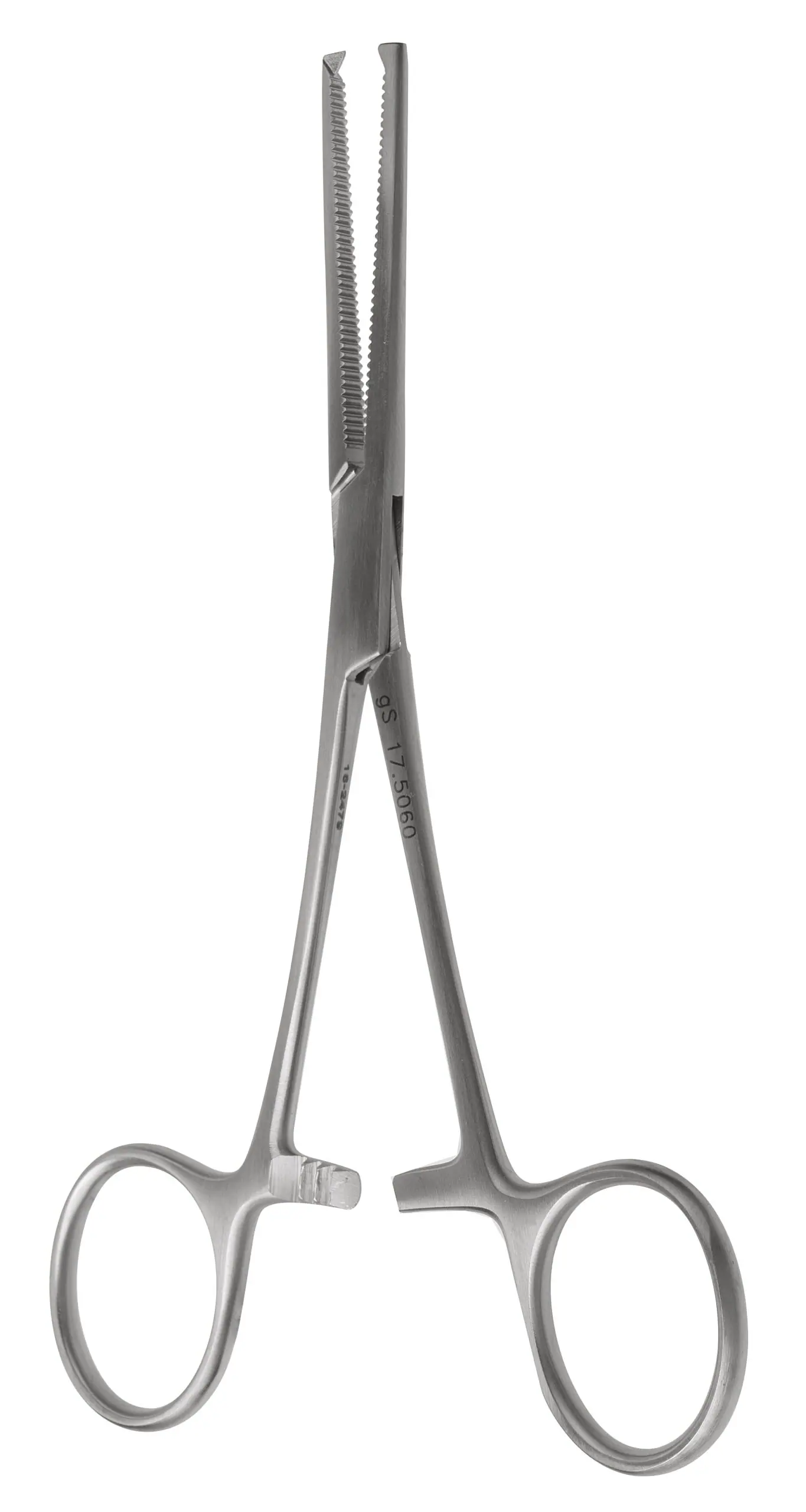
Allis clamp
Atraumatic grasping clamp used for blunt dissection (Gupta hip).
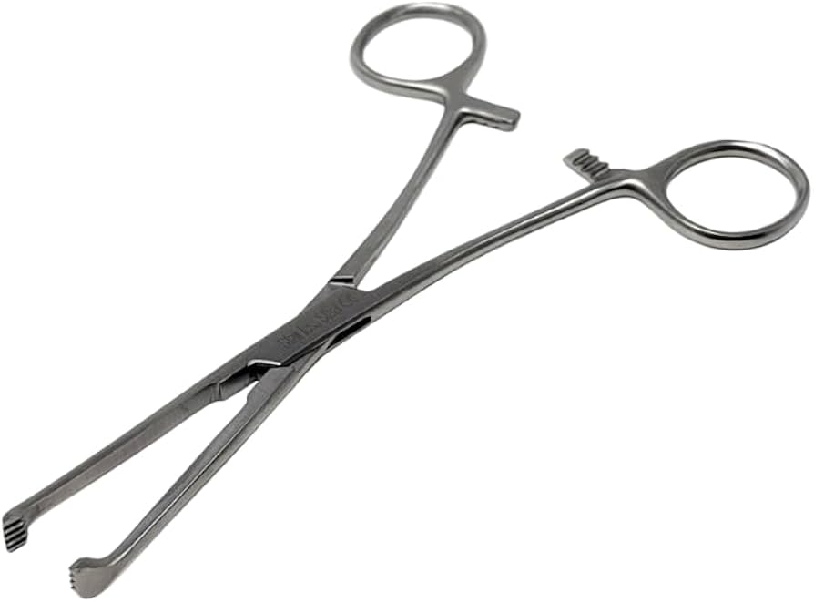
Towel clamp
Clamp used to help prep the patella (Seng knee).
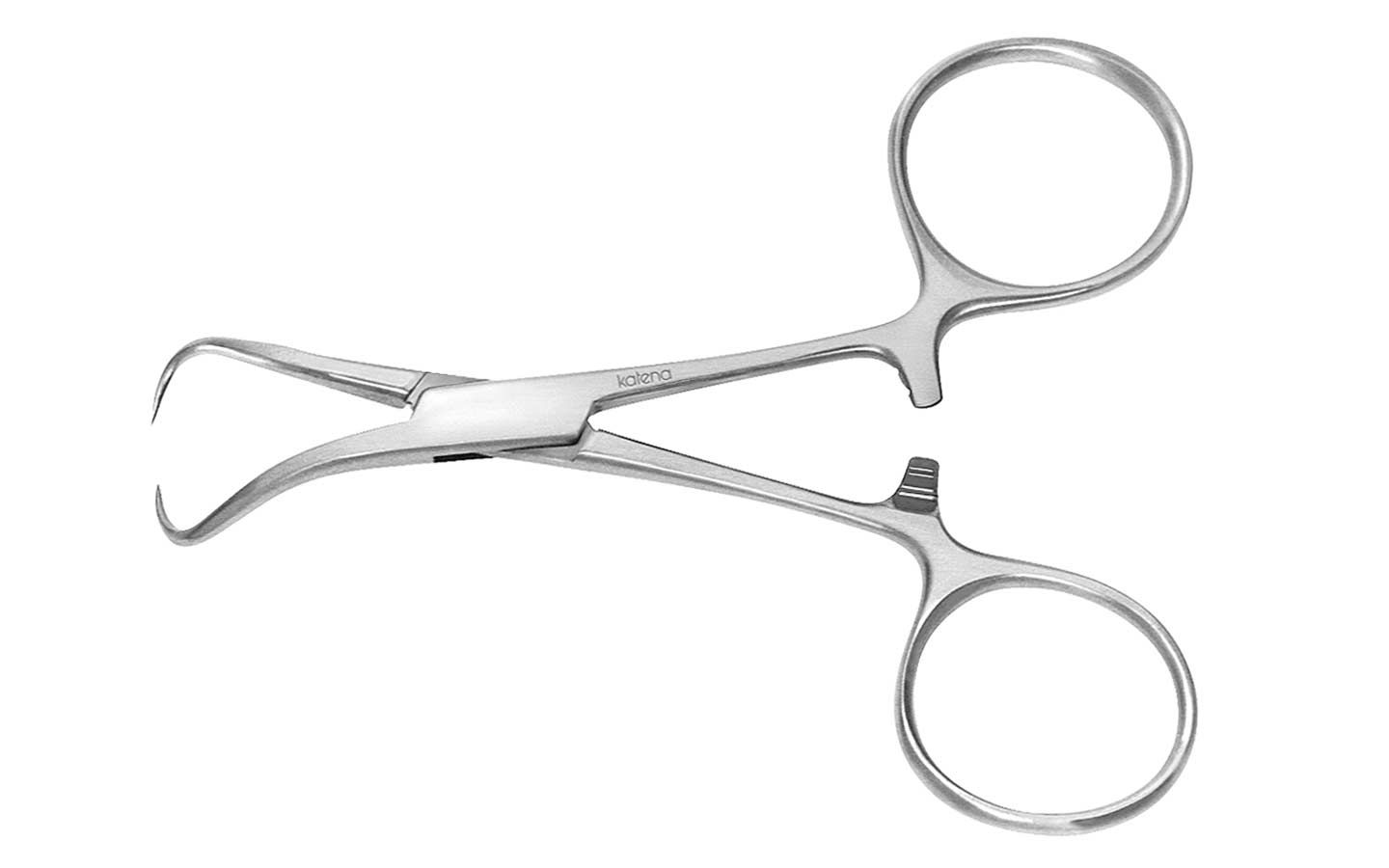
Cobb elevator
Periosteal elevator for dissection, capsule release, and removing the napkin-ring bone in hips.
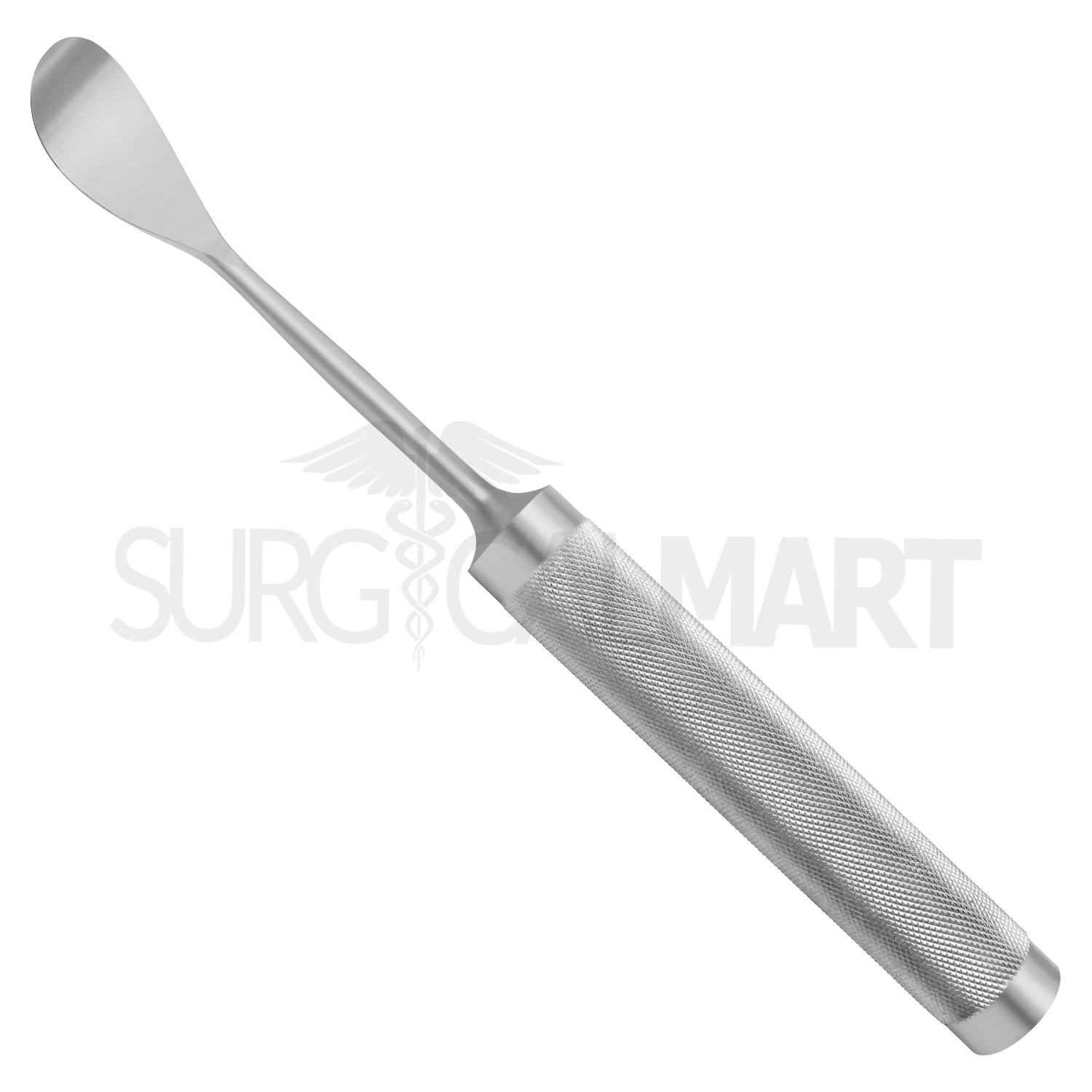
Key elevator
Heavy periosteal elevator for exposure (Seng knee).
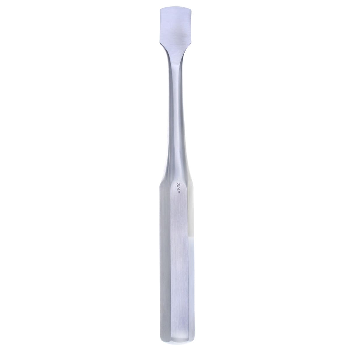
Freer elevator
Fine elevator; also clears excess cement during cementing (Gupta knee).
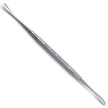
Rongeur (double-action)
Bites bone, trims osteophytes, cleans the canal, and can make a bone plug.
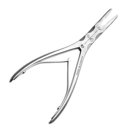
Pituitary rongeur
Narrow rongeur to clean the back of the knee and release the hip capsule.
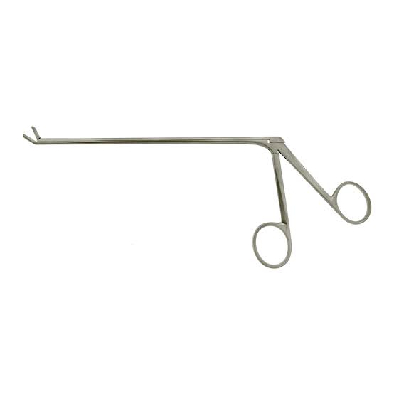
Curved curette
Scrapes soft tissue and bone from the back of the knee (Seng).
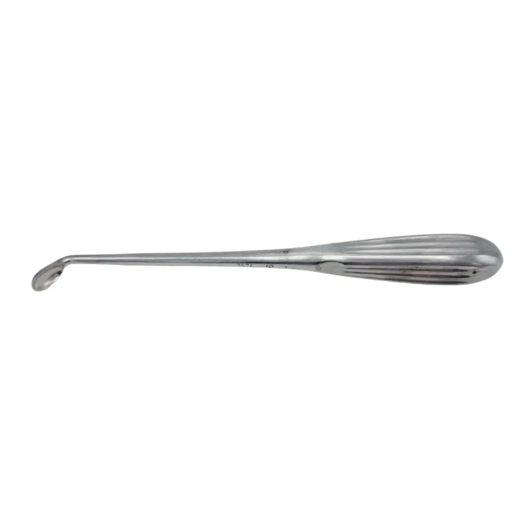
Curved osteotome
Pries up the bone cut and cleans the back of the knee.
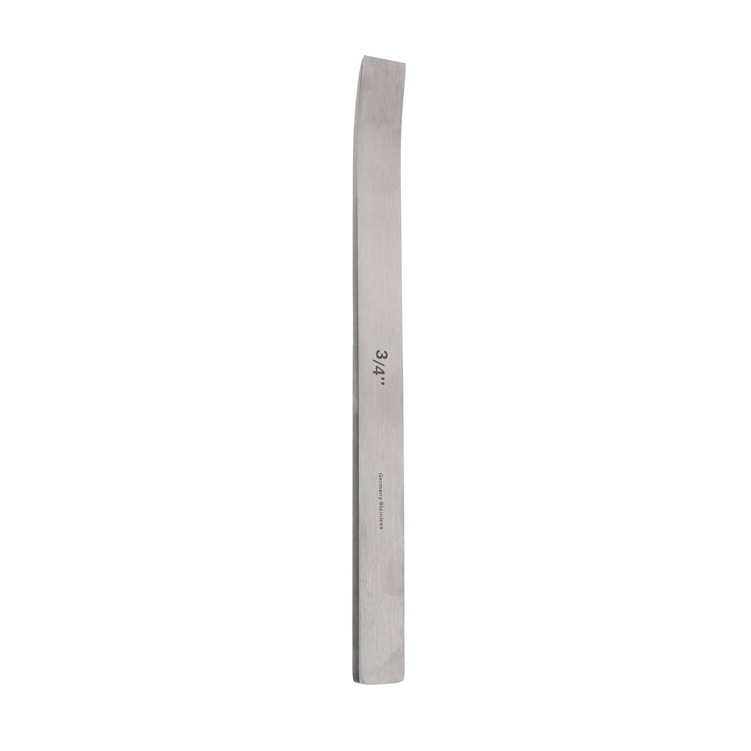
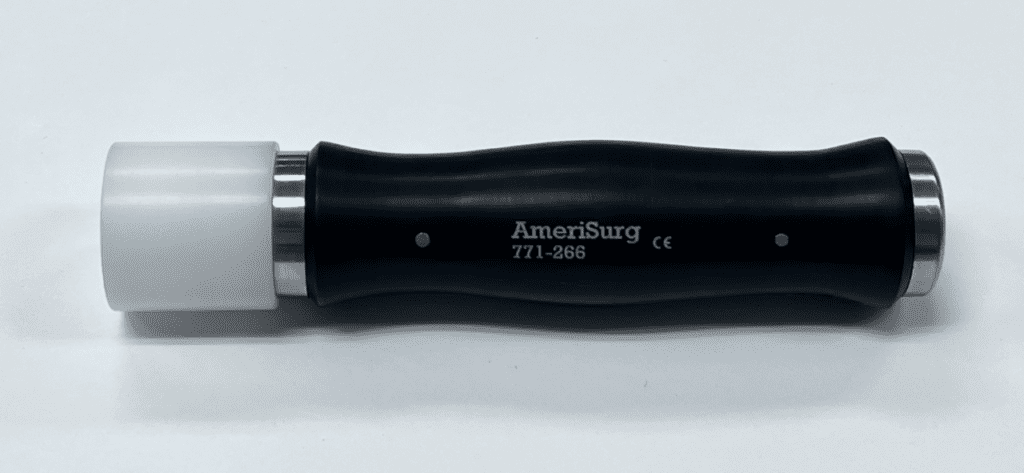
Mallet
Drives osteotomes, impactors, and broaches.
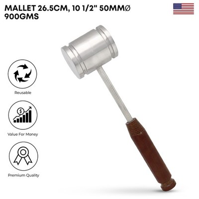
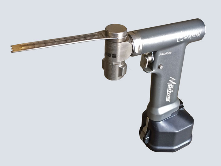
Saw (sagittal)
Makes the bone resections and the femoral neck cut.
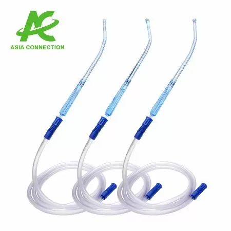
Yankauer suction
Clears blood and irrigation from the field.
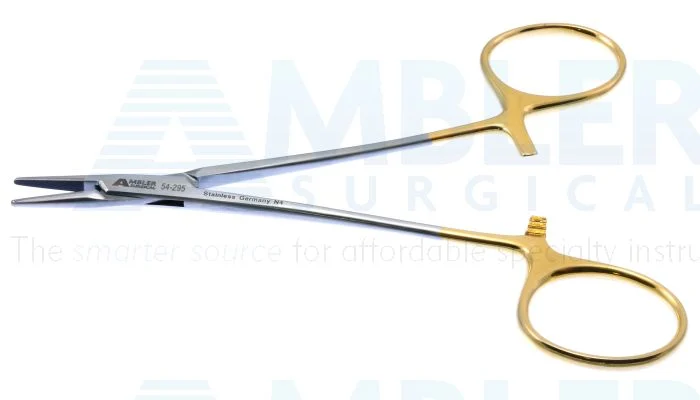
Needle driver
Holds the suture needle during closing.
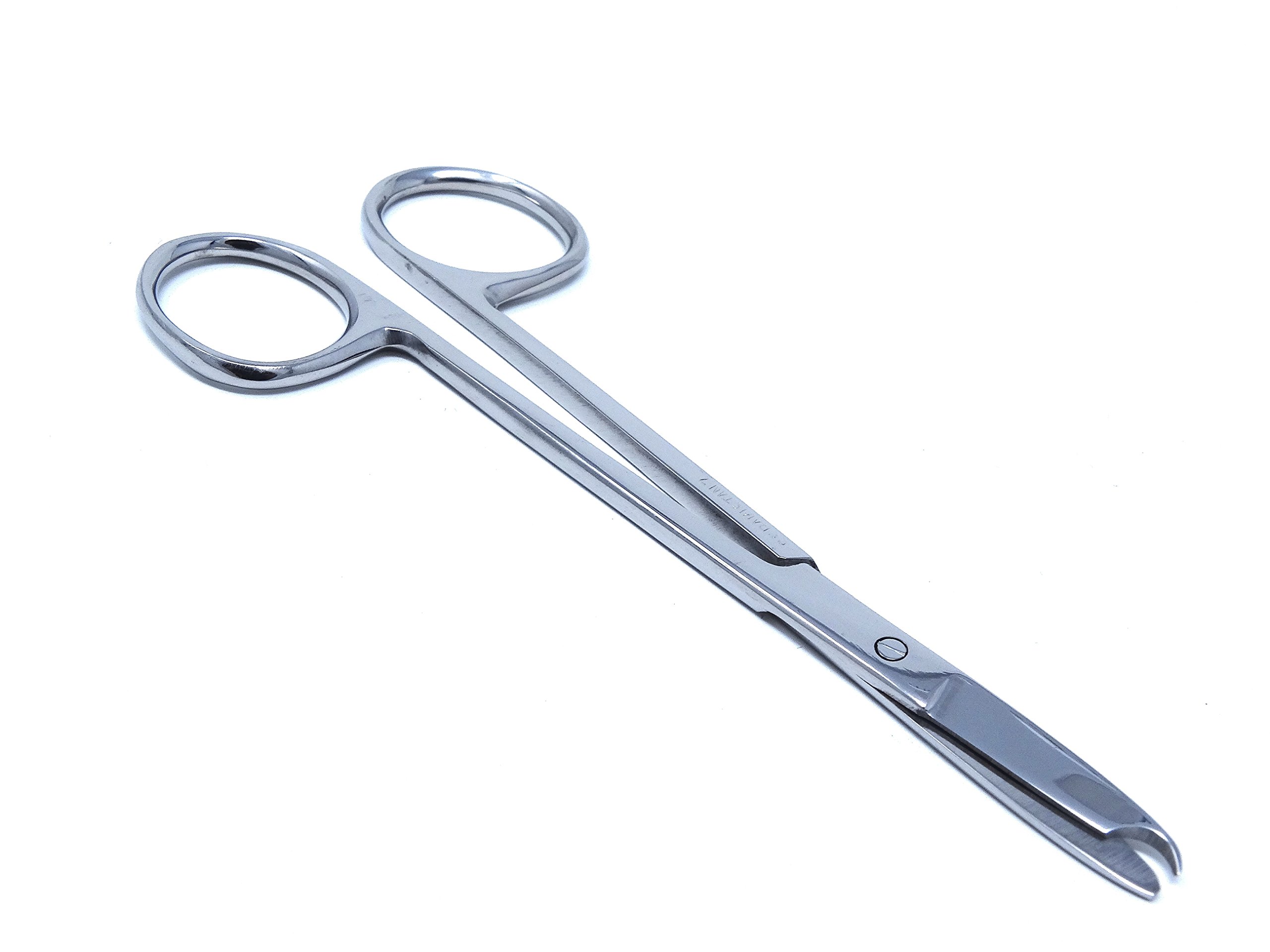
Suture scissors (Mayo)
Cut suture and dense tissue during closing.
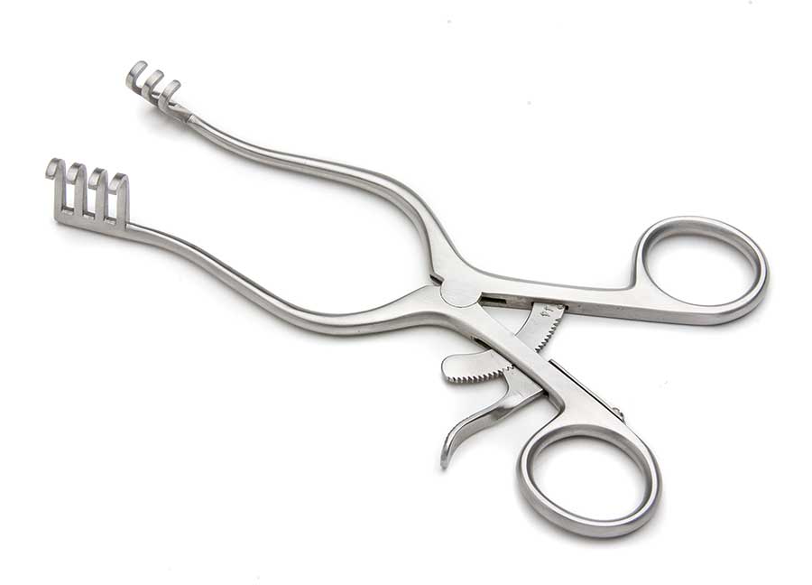
Weitlaner retractor (MDS1867016)
Small self-retaining retractor with rake tips; used at the opening (Gupta hip).
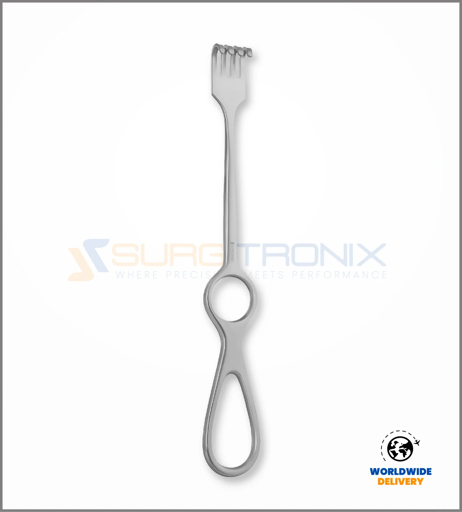
Rake retractor
Sharp handheld retractor for medial knee exposure (Land, Gupta).
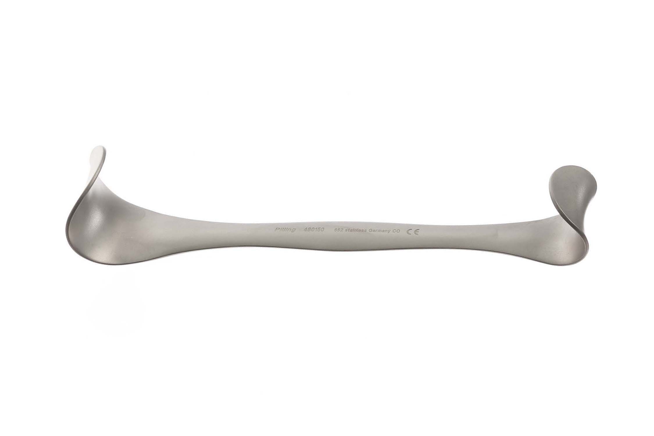
Goelet retractor
Double-ended knee retractor for exposure and femoral sizing (Land).
Smillie retractor
Knee retractor for tibial exposure and after the first femoral cut (Land, Gupta).
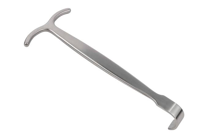
PCL retractor (pickle)
Protects the PCL and posterior tibia during the tibial cut (Land, Gupta).
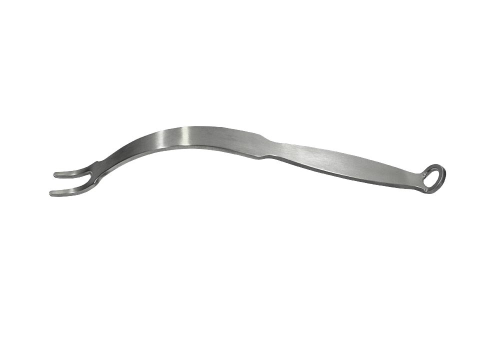
90-degree Hohmann
Right-angle bone lever for knee exposure (Land).
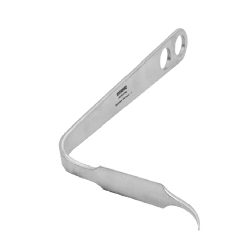
Bent Hohmann
Angled bone lever for tibial exposure (Gupta knee).
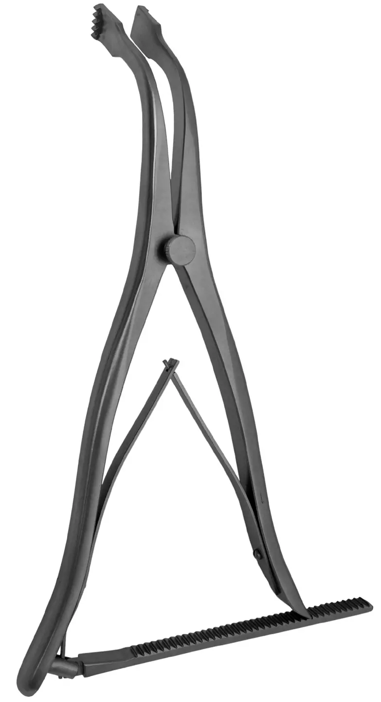
Lamina spreader
Opens the joint to clean out the back of the knee (Seng, Land).
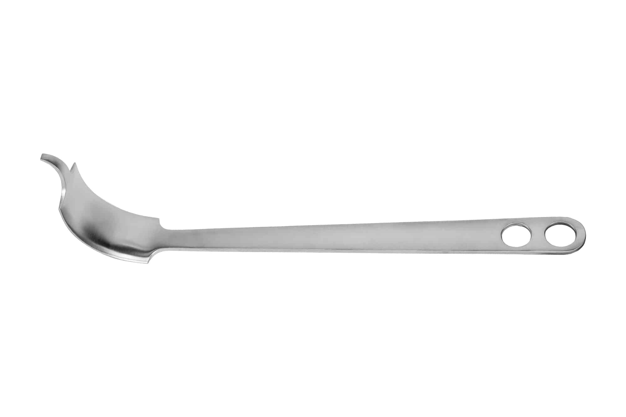
Cobra retractor (MDS3298002)
Broad curved hip retractor over the acetabular rim and femur (Land, Gupta).
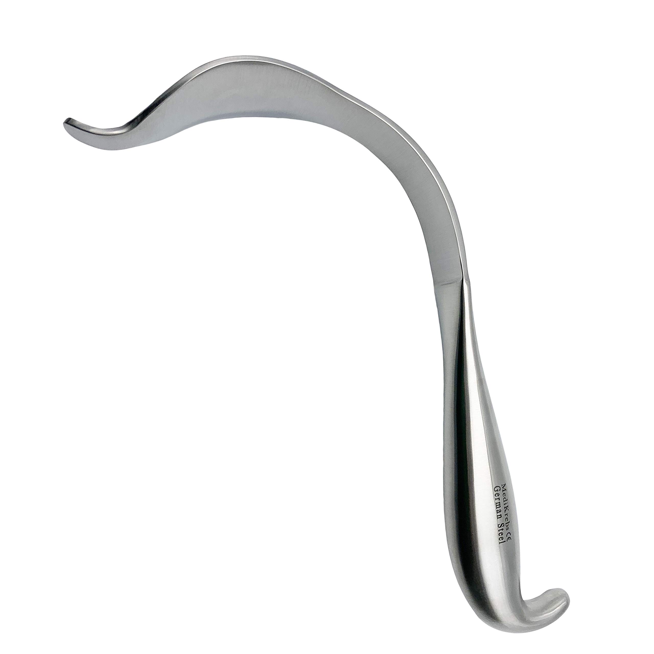
Meyerding retractor
Bladed hip retractor for socket and femoral visualization (all hips).
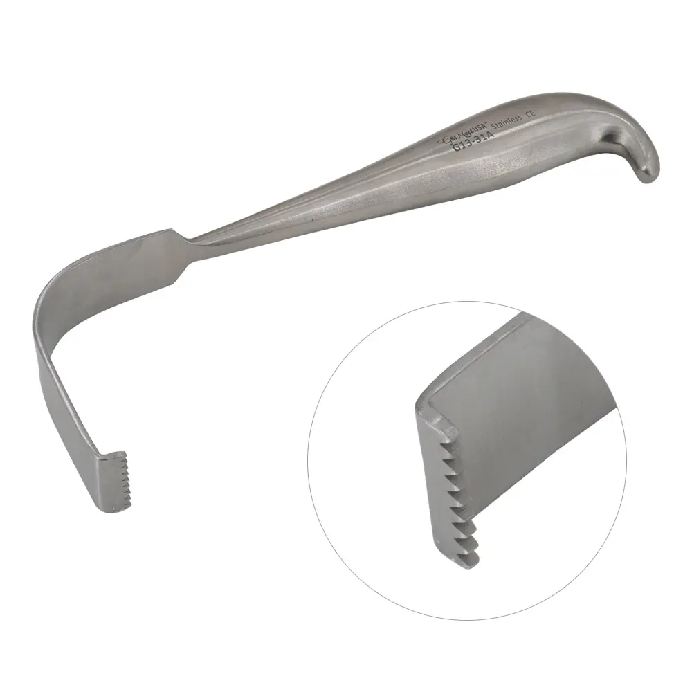
Mueller retractor (Woody)
Forked-tip femoral retractor with a blue handle; femoral exposure (Land, Gupta).
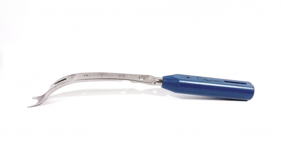
Big J retractor (77-1188)
Long retractor with a deep hooked J tip for deep hip exposure (Land).
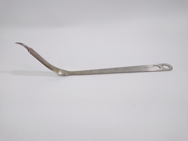
Cerebellar retractor (24-2810)
Self-retaining retractor with rake tips; placed during the neck cut (Gupta hip).
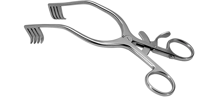
1 retractor ('big ugly')
The #1 hip retractor, called the big ugly; goes in for femoral exposure (Land, Gupta).
Femoral hook (6850-140 L / 6850-144 R)
Side-specific curved hook that elevates and delivers the femur; also helps dislocate the hip.
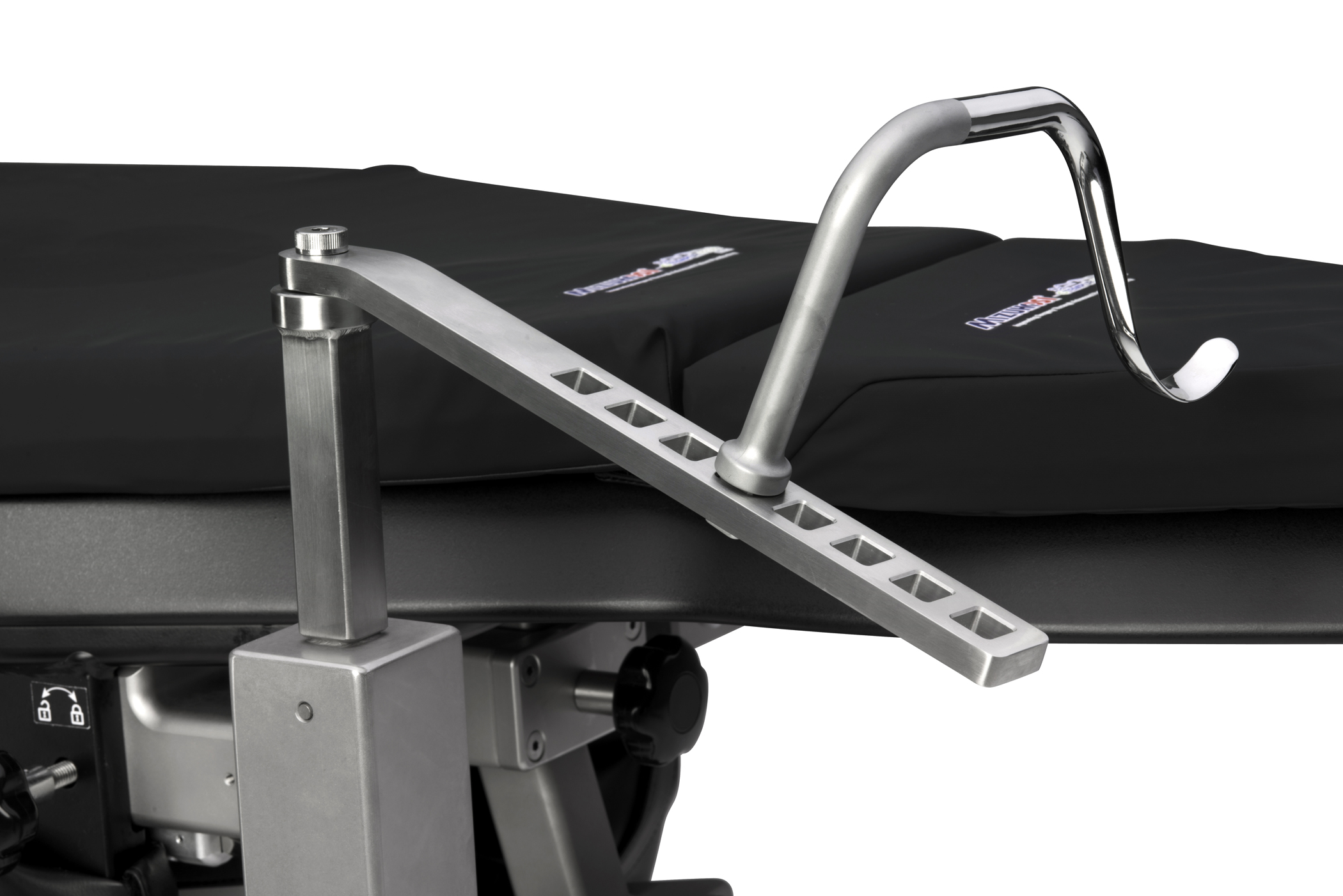
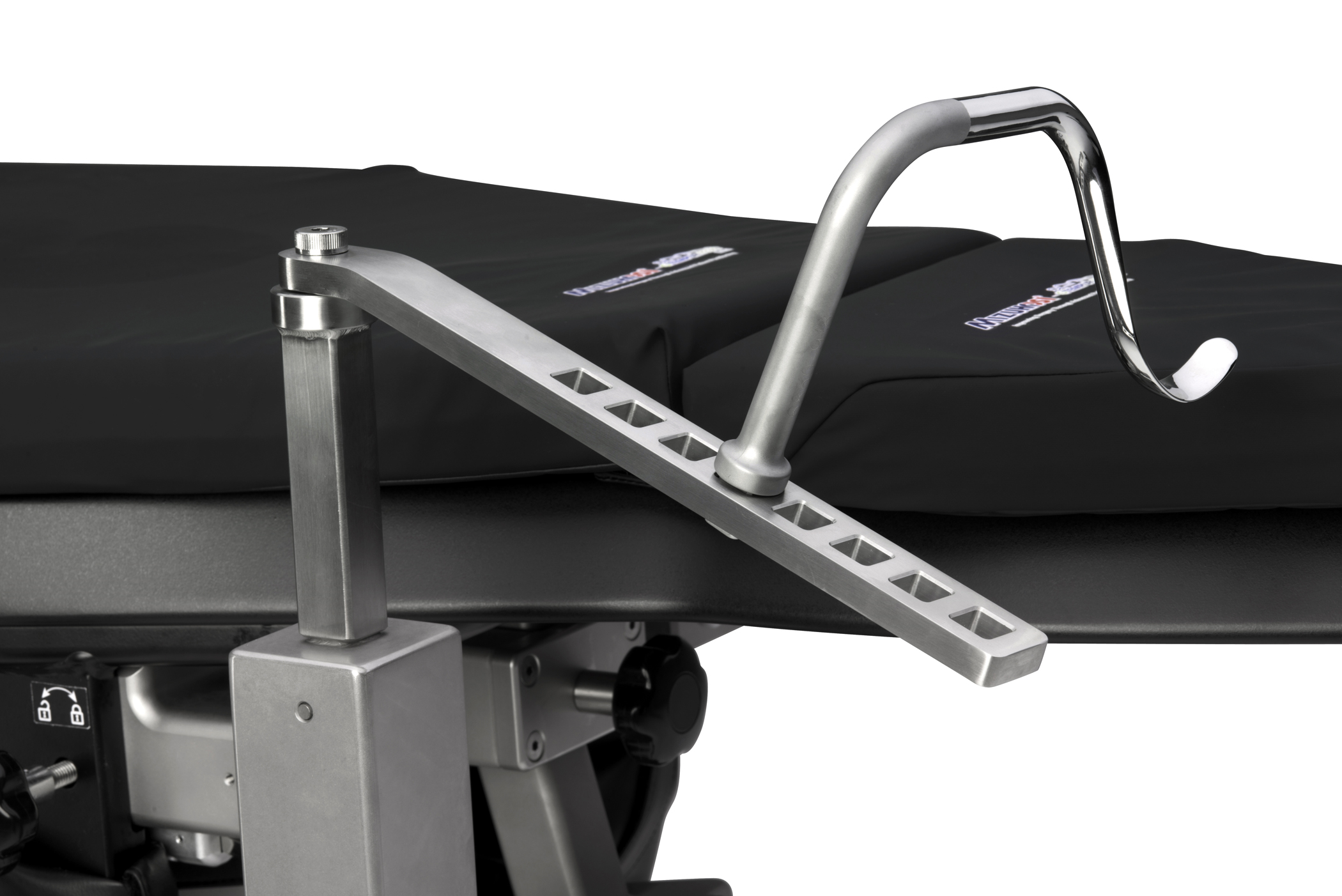
Hana bar (6850-110)
Right-angle slotted bar that the femoral hook mounts to for femoral elevation.
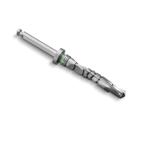
Starter drill
Opens the femoral canal to start the distal femoral cut.
T-handle
Handle on the starter drill / IM rod; comes out before the saw cut (Land).
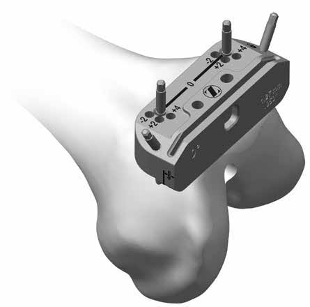
Distal femoral cutting guide
Guides the distal femur resection.
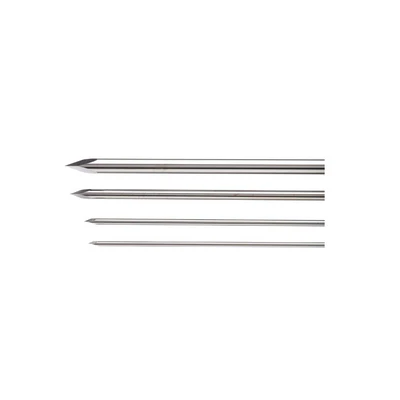
Smooth pins
Non-threaded pins that hold the cutting guides to bone.
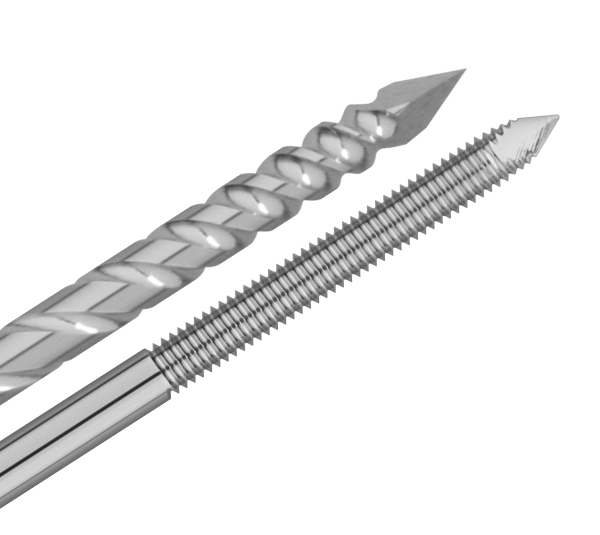
Threaded pin
Threaded pin that secures the 4-in-1 block.
Pin driver
Inserts and removes the pins (Seng).
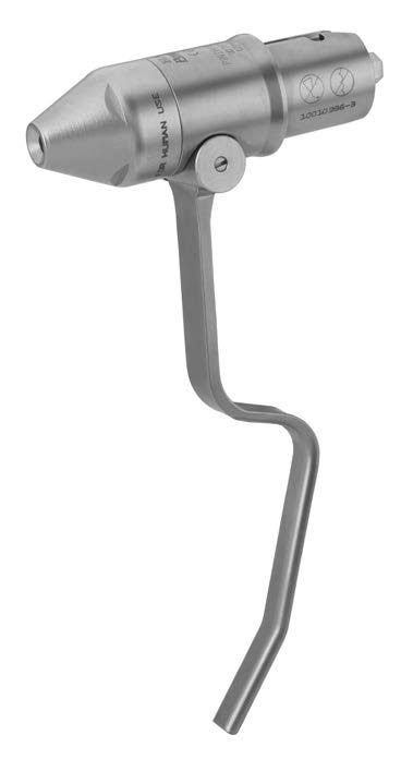
Slap hammer
Removes the 4-in-1 block and guides off their pins.
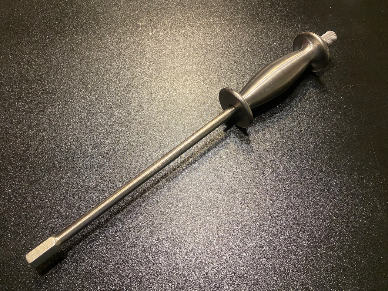
Tibial cutting guide
Guides the proximal tibial cut.
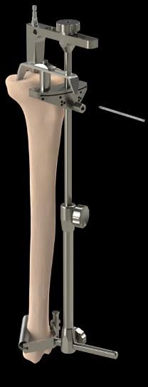
Stylus
Sets the depth of the tibial cut.
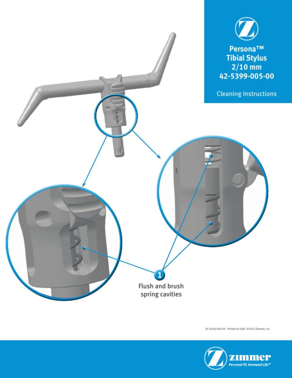
Spacer block
Checks the flexion/extension gap after the cuts.
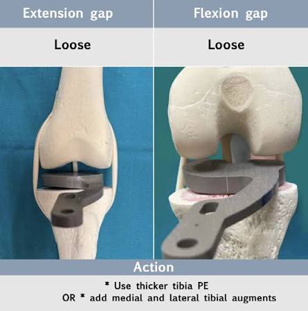
Femoral sizing guide (fusion jig)
Sizes the femur and sets rotation; Seng's has a magnetic sizing piece and is adjusted with a screwdriver.
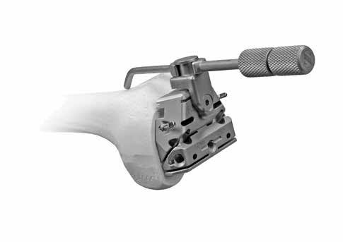
4-in-1 cutting block
Guides the anterior, posterior, and chamfer femoral cuts.
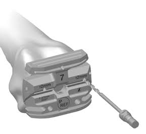
Angel wing
Thin blade that checks the 4-in-1 block is the right size and position before cutting.
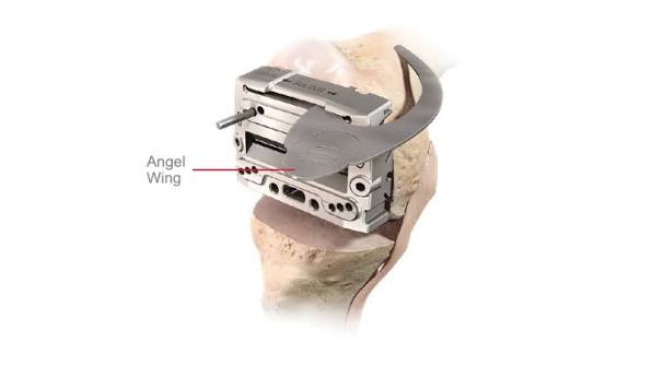
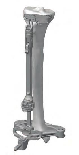
Tibial drill tower
Guides drilling the tibia for the keel/stem (Gupta 'tower'; Seng 'tibia drill').
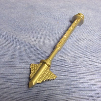
Tibial punch
Punches the tibial keel or stem slot.
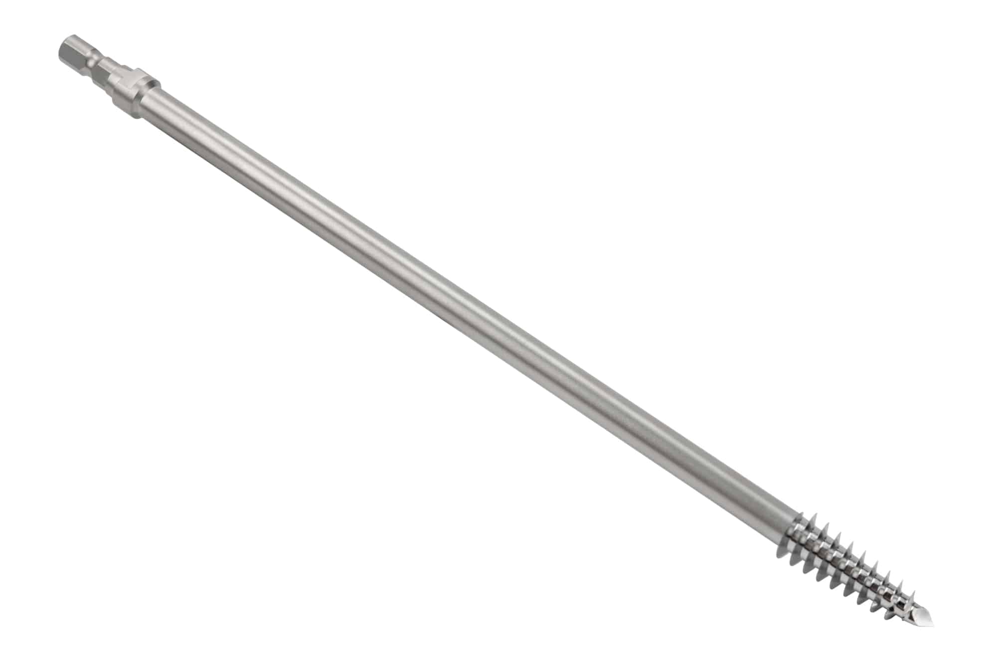
Femoral peg drill
Drills the femoral fixation peg/lug holes (Seng, Land).
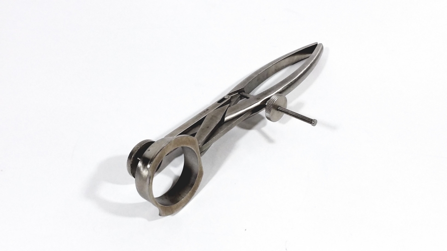
Patella clamp
Everts and holds the patella and guides drilling the patella holes (Seng).
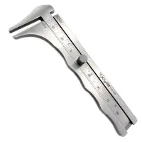
Patella caliper
Measures patellar thickness before and after the cut (Seng).
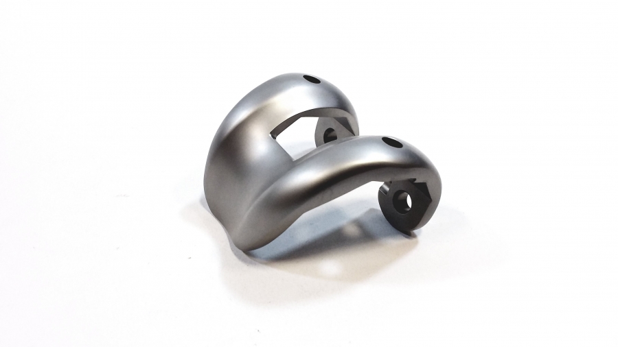
Femoral trial
Provisional femoral component to check fit and tracking.
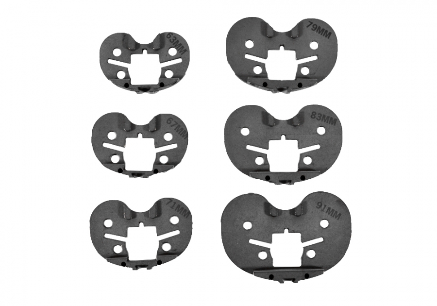
Tibial baseplate trial
Provisional tibial tray for sizing and position.
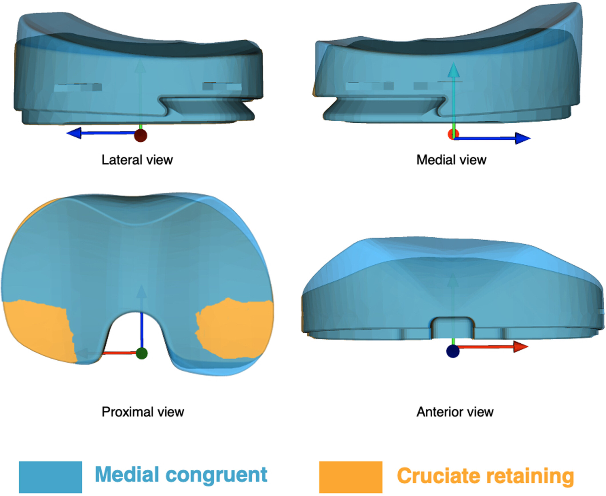
Poly trial (MC)
Provisional polyethylene insert (medial congruent) trialed on the tibia to set stability.
Patella trial
Provisional patellar button to check thickness and tracking.
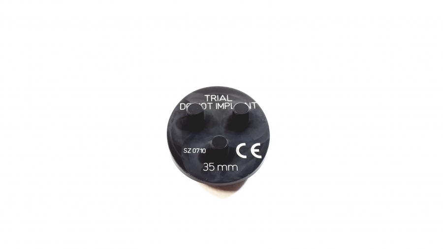
Pulse lavage
Pressurized irrigation that cleans the bone before cementing.
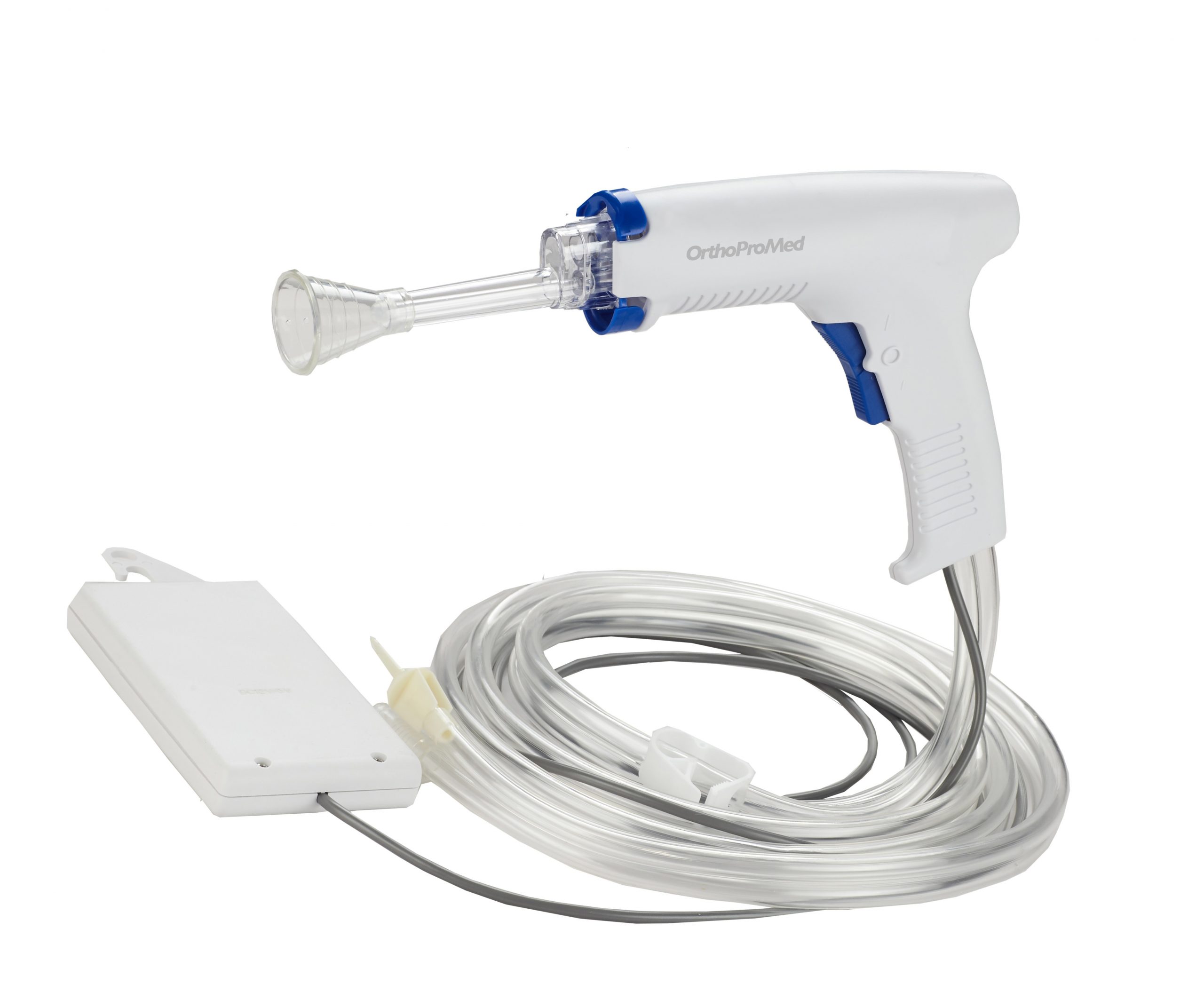
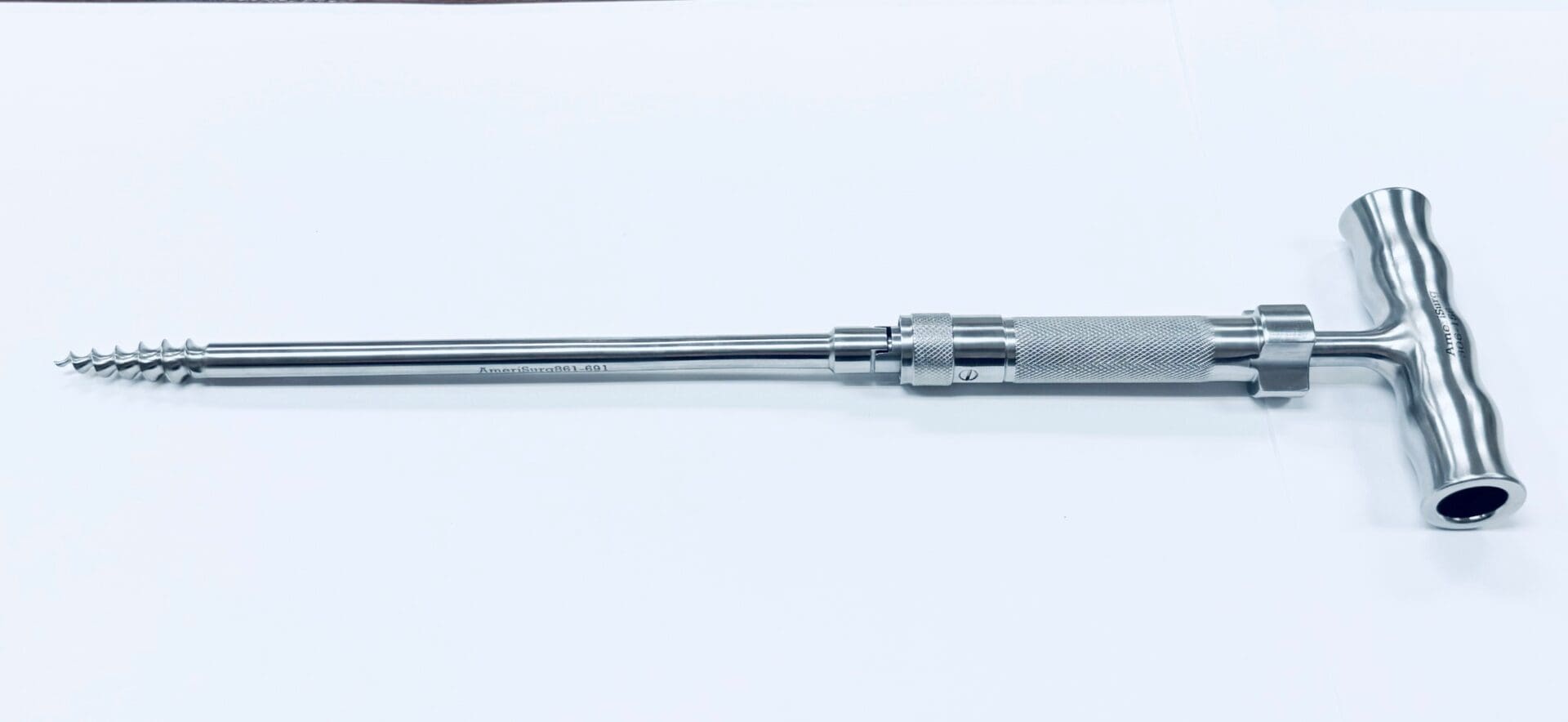
Corkscrew
Threads into the femoral head to remove it after the neck cut (all hips).
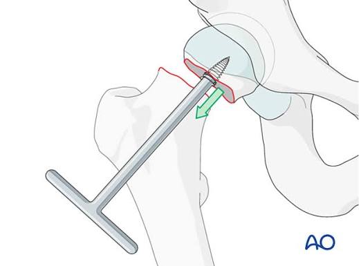
Acetabular reamer
Reams the socket to size.
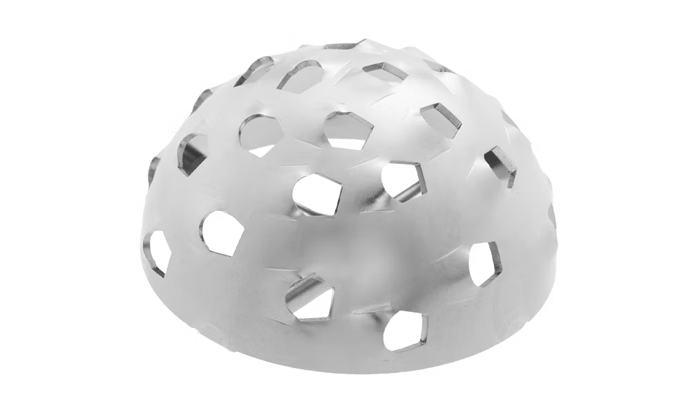
Curved impactor
Inserts and impacts the acetabular cup (Land).
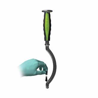
Straight impactor
Impacts the liner (a liner impactor head attaches); Gupta also seats the cup with it.
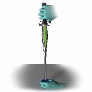
Rat tail
Curved hand rasp; first pass to open and shape the femoral canal before broaching.
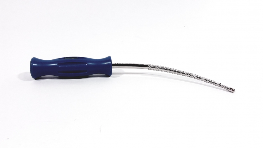
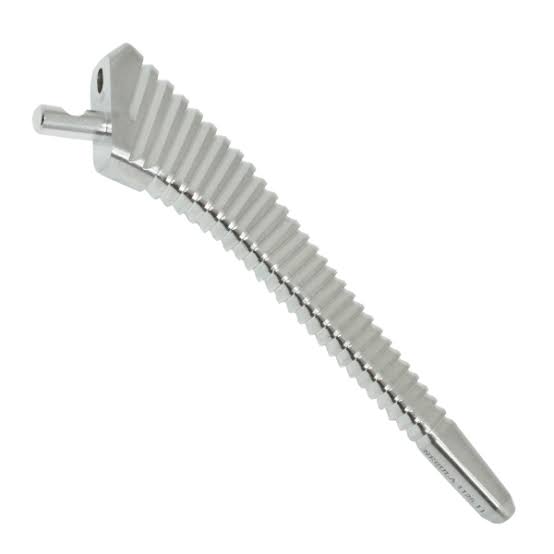
Starter broach / broaches
Sequential rasps that shape the femoral canal; the final broach serves as the stem trial.
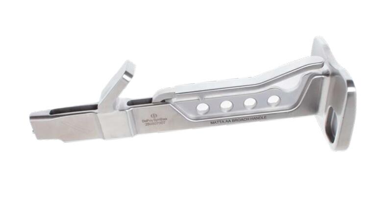
Straight broach handle
Holds the broach and the cookie cutter.
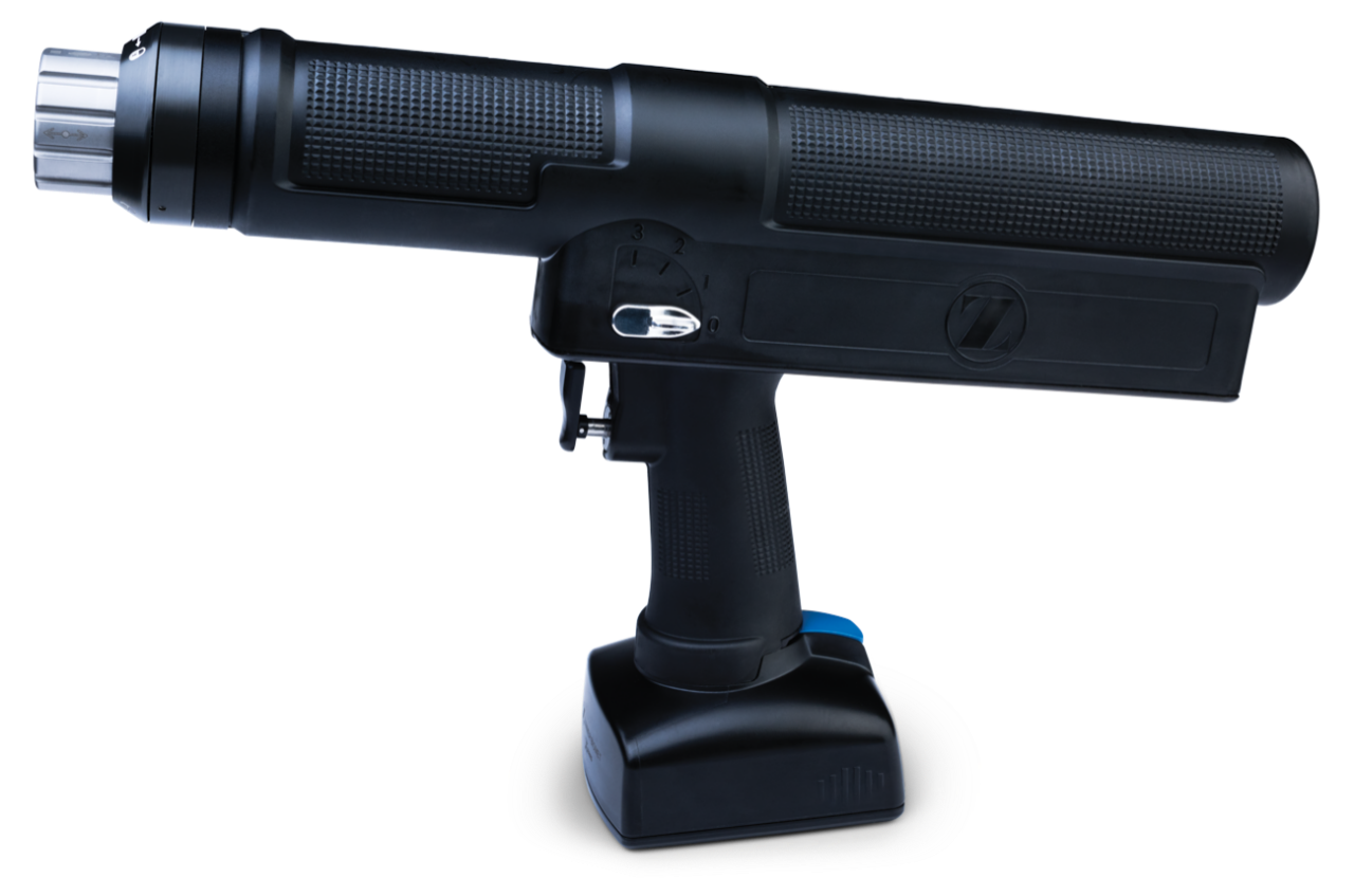
HAMMR
Powered impaction handle used to broach the femur (Land).
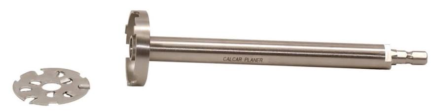
Calcar planer (Mill)
Flattens the calcar; surgeons alternate broaching and calcar planing.
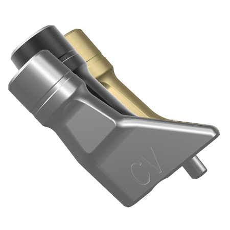
Trial neck
Provisional neck to set offset and version.
Trial head (head ball)
Provisional head to set leg length and stability (e.g., standard neck, +0 head).
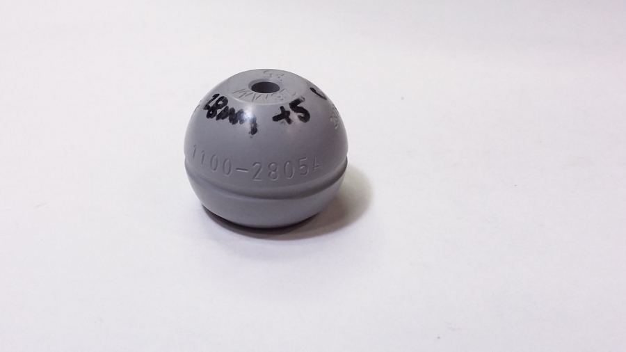
Stem impactor
Impacts the real femoral stem with the mallet.
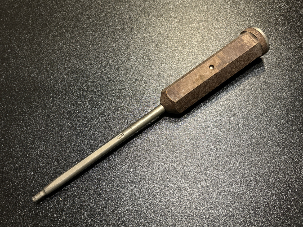
Femoral head ball pusher/impactor
Seats the real head on the stem trunnion; also pushes/reduces the hip.
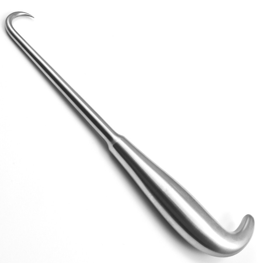
Bone hook
Dislocates the hip and helps deliver the femur.
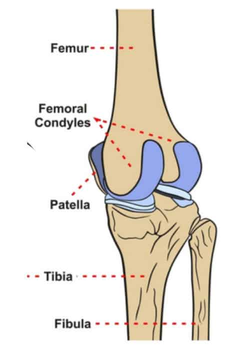
Distal femoral condyles (medial & lateral)
Resurfaced by the femoral component; removed by the distal and posterior cuts.
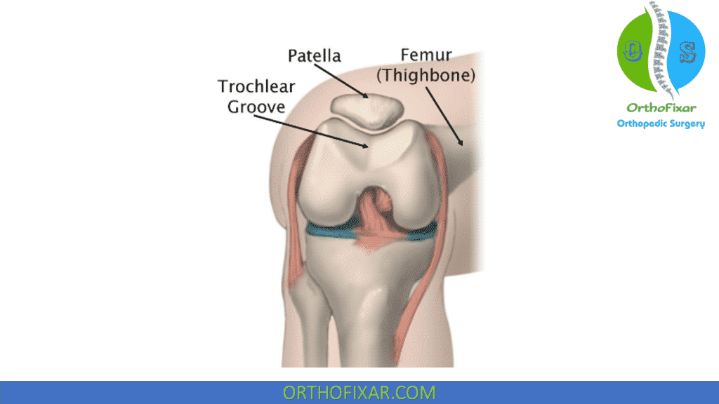
Trochlear groove (femoral sulcus)
Patellofemoral tracking surface; replaced by the anterior flange of the femoral component.
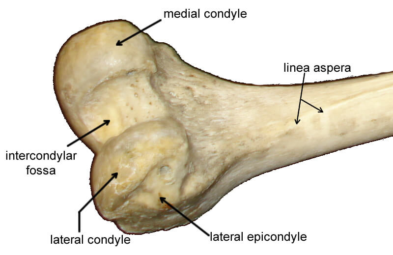
Intercondylar notch
Houses the cruciates; the box cut is made here for a posterior-stabilized component.
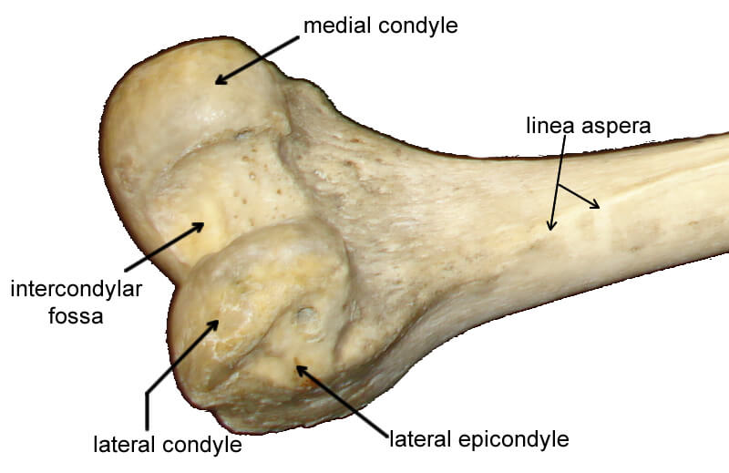
Medial and lateral epicondyles
Define the transepicondylar axis, the primary reference for setting femoral component rotation.
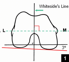
Whiteside's line (AP axis)
Anteroposterior trochlear axis used as a secondary check for femoral rotation.
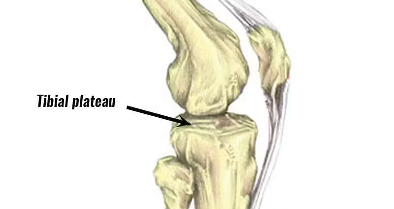
Tibial plateau (medial & lateral)
Resected flat to seat the tibial baseplate.
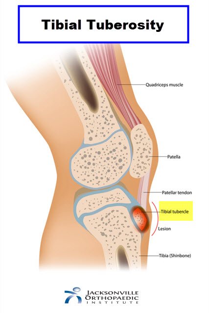
Tibial tubercle (tuberosity)
Patellar tendon insertion; its medial third is the rotational landmark for tibial component alignment.
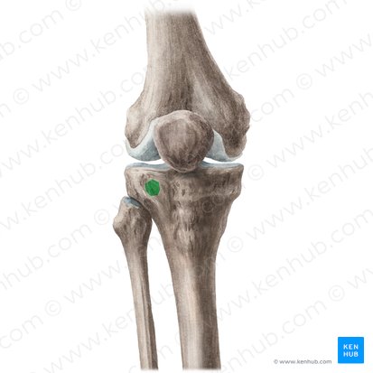
Gerdy's tubercle
IT band insertion; lateral exposure and retractor landmark.
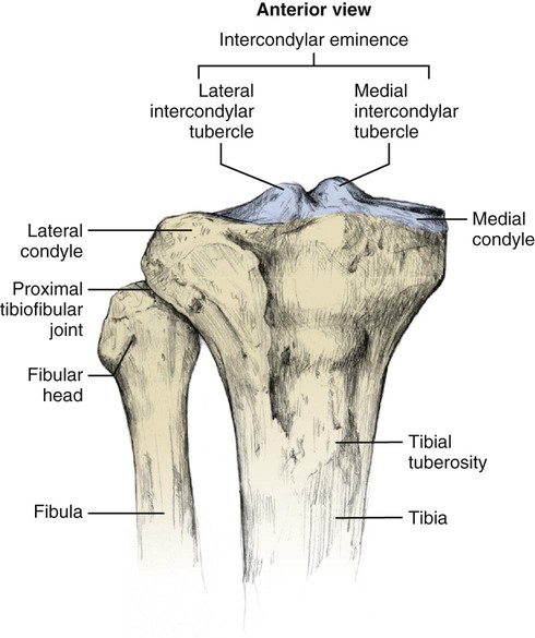
Intercondylar eminence (tibial spines)
ACL attachment; removed with the tibial resection.
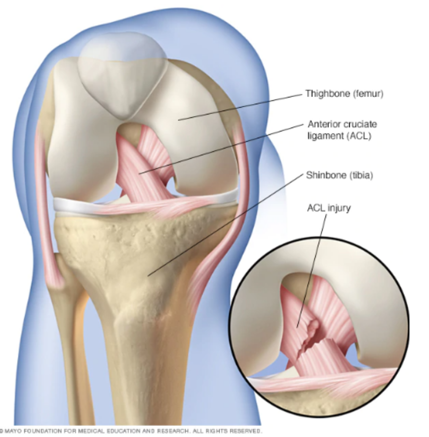
Anterior cruciate ligament (ACL)
Central stabilizer routinely resected during TKA.
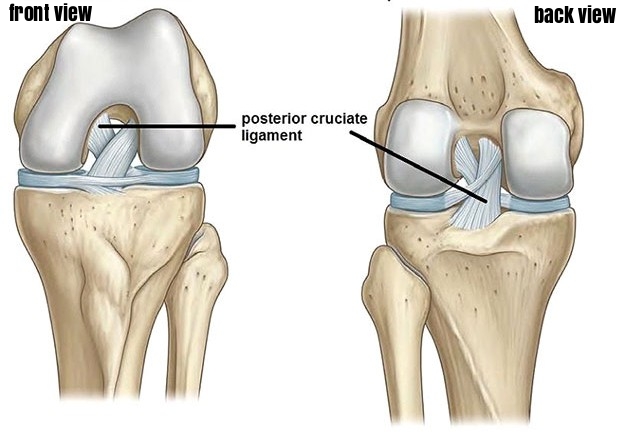
Posterior cruciate ligament (PCL)
Retained in cruciate-retaining designs, resected in posterior-stabilized; governs flexion stability and rollback.
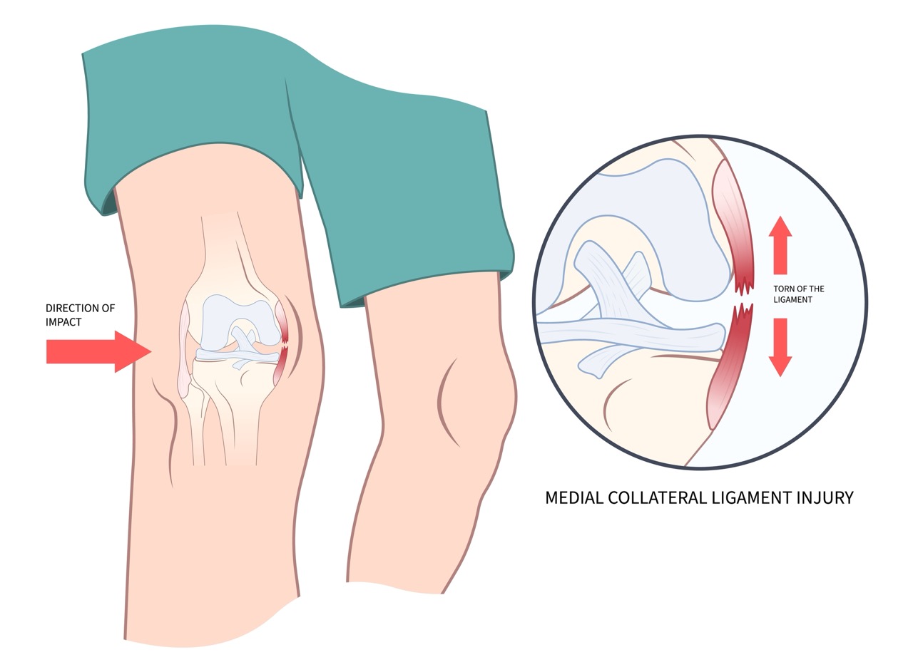
Medial collateral ligament (MCL)
Primary medial stabilizer; protected during exposure and selectively released to balance a tight medial gap.
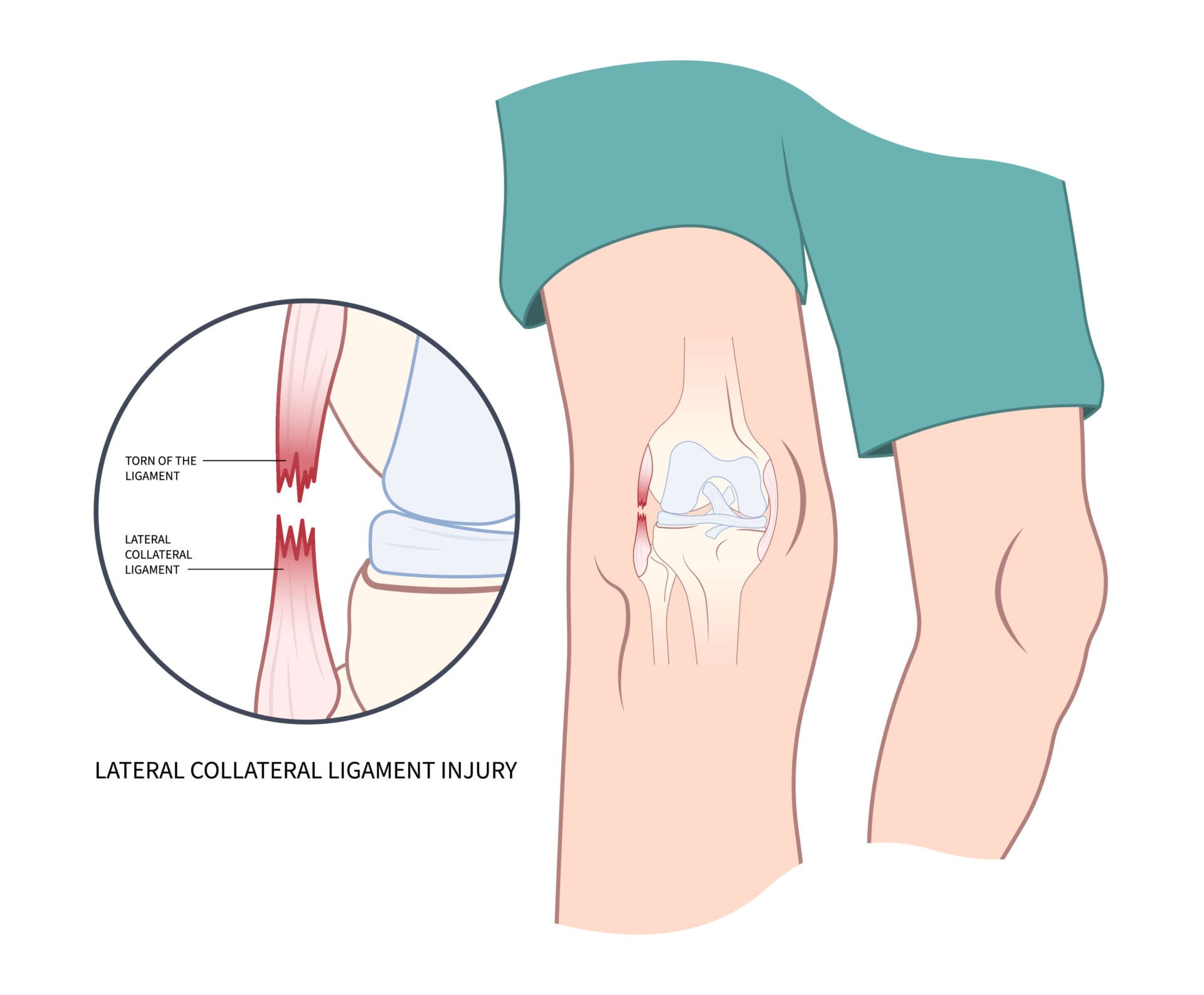
Lateral collateral ligament (LCL)
Primary lateral stabilizer; part of the lateral release sequence in a valgus knee.
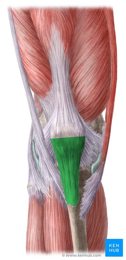
Patellar tendon (ligament)
Avulsion risk with aggressive eversion, so protected during exposure.
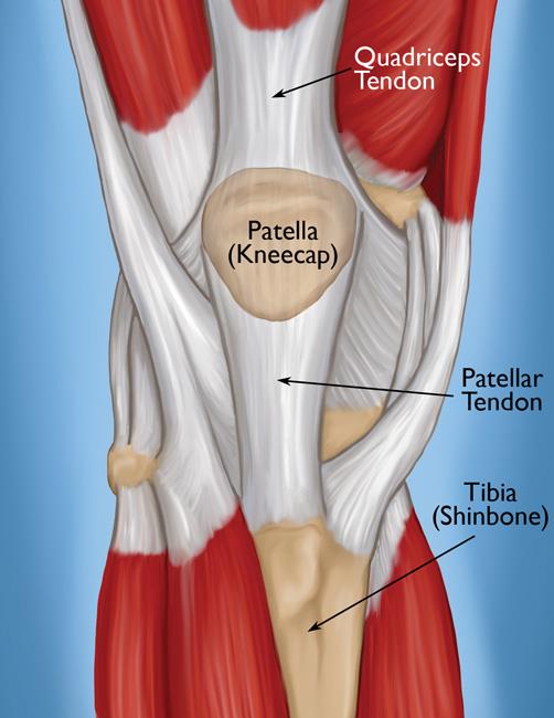
Quadriceps tendon
Incised in the arthrotomy; component of the extensor mechanism.
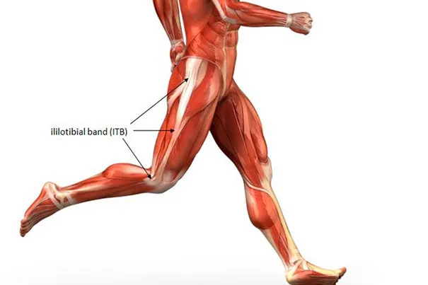
Iliotibial (IT) band
Inserts on Gerdy's tubercle; released during valgus balancing.
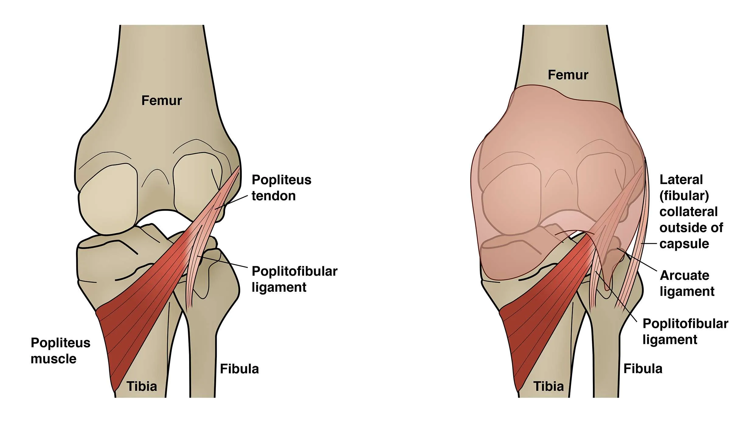
Popliteus tendon
Addressed during lateral soft-tissue balancing.
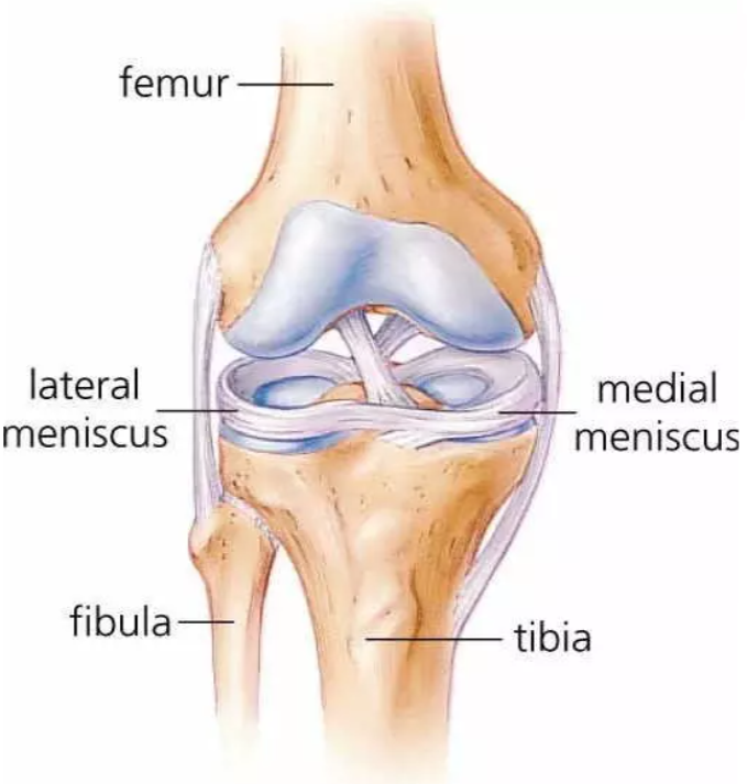
Menisci (medial & lateral)
Fibrocartilage spacers removed during TKA.
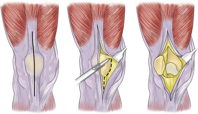
Medial parapatellar arthrotomy
Standard approach: through the quadriceps tendon, around the medial patella, to the tibial tubercle.
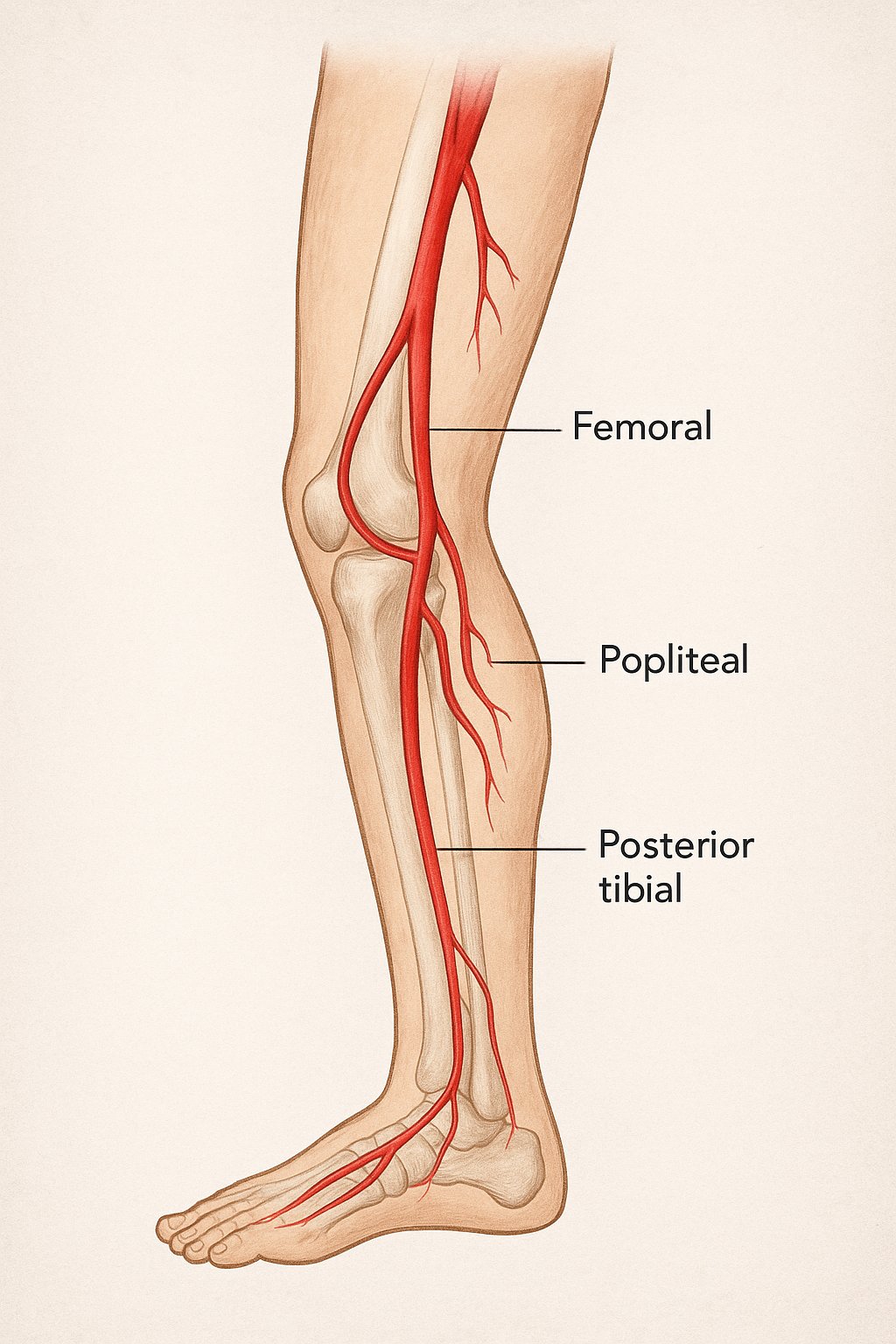
Popliteal artery
Lies just posterior to the knee capsule; at risk during posterior cuts and PCL release.
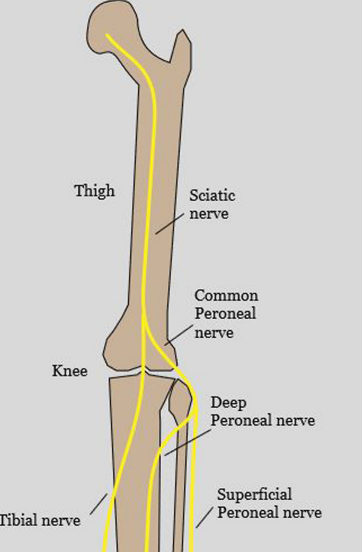
Common peroneal (fibular) nerve
Wraps the fibular neck laterally; at risk when correcting a valgus or flexion deformity.
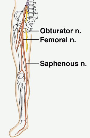
Saphenous nerve (infrapatellar branch)
Crosses the medial incision; commonly causes numbness lateral to the scar.
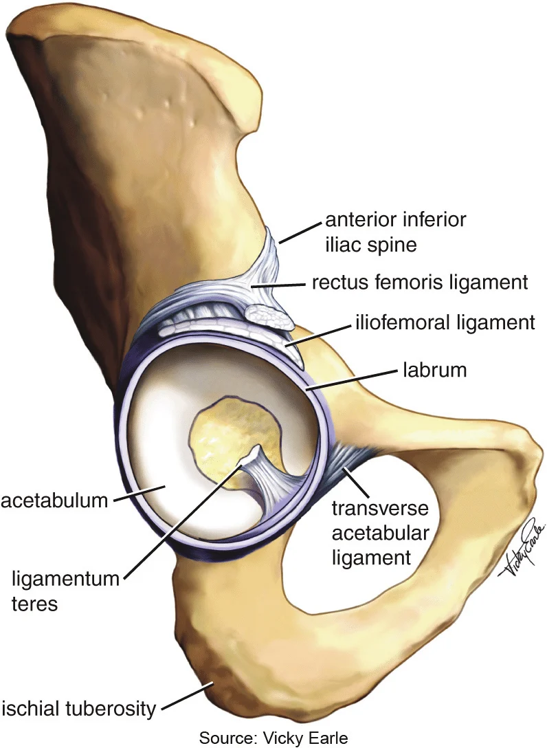
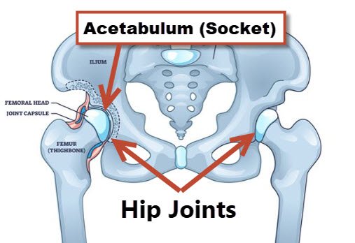
Acetabulum
Reamed to accept the acetabular cup.
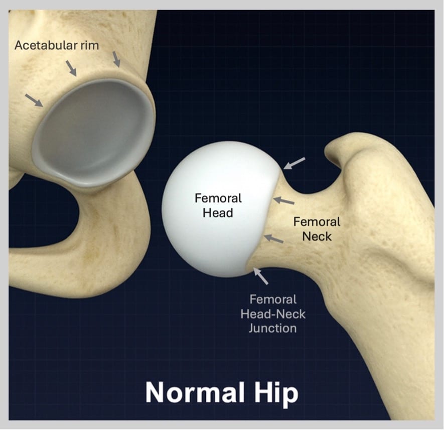
Acetabular rim
Reference for cup seating and osteophyte removal.
Transverse acetabular ligament (TAL)
Inferior acetabular landmark used intra-operatively to set cup anteversion and inclination.