ARRT review - Nuclear Medicine
 Call Kai
 Learn
Learn Practice Test
 Spaced Repetition
 Match
Match Flashcards
 Knowt Play
1/227

There's no tags or description
Looks like no tags are added yet.
Name | Mastery | Learn | Test | Matching | Spaced | Call with Kai | Chat |
|---|
No analytics yet
Send a link to your students to track their progress
228 Terms
Direct interaction
Said to take place when an original ionizing incident happens on that macromolecule (Ex. DNA, RNA, Protein and enzyme) if a macromolecule becomes ionized it is considered abnormal and thus damaged.
Indirect interaction
Occurs if the initial ionizing incident takes place on a distant noncritical molecule, which then transfers that ionization energy to another molecule
Indirect
as the human body is approximately 75% water, it is theorized that most radiation actions with human are
Indirect effects
DNA is the most critical target of radiation, but it is the irradiation of water which cause
Radiolysis of water
If water is irradiated, the water is ionized and separates into molecular products which then produce free radicals
Free radical
Is an uncharged molecule that contains a single unpaired electron in its outermost or valence shell, which it make it chemically unstable and highly reactive

Linear Energy Transfer (LET)
describes a measurable of the rate at which energy is deposited as a charge particle travels through matter.
Low LET radiation
Electromagnetic radiation:
X- and Gamma rays
Produce few and sparse interaction because of their fast moving electrons.
High LET radiation
Particulate radiation:
Alpha particles and neutrons
Highly ionizing and have substantial mass and/or charge, are more likely to interact with tissue
Higher; greater
the ____________ the LET of radiation, the ______________ the chance for a biological interaction
Relative Biological Effectiveness (RBE)
The relative effect of LET.
Measures the biological effectiveness of radiation having different LETs
Factors that influence RBE
-radiation type
-cell or tissue type
-physiologic condition
-biologic result being examined
-radiation dose rate
Stochastic effects
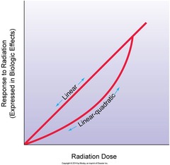
Thought to be nonthreshold, as damage to a few cells or even a single cell could theoretically produce the disease

Stocastic effects
Associated with linear and linear quadratic dose response curves, therefore, even small exposure could carry some increased risk
Ex. Radiation-induced cancer and genetic effect
Deterministic effects
Also known as nonstochastic
Are thought to be threshold, as there are doses below which the effect is not observed
Ex. Cataracts, erythema, fibrosis and hematopoetic damage

Hemopoietic acute effect
100 - 1,000 R will damage this system, also known to be the bone marrow system
Gastrointestinal acute effect
600 - 10,000 R damage this system, especially the small intestine
Central nervous system acute effect
>10,000 R will damage this system, but also starting at a dose of >5,000 R
Somatic
Mutations have consequences for only that person
Genetic
Mutations can affect reproductive organs or the parents gametes, which may affect future generation. It's cumulative.
Law of Bergonie and Tribondeau
immature cells are significantly more radiosensitive than are mature cells
Immature cells
Are known as undifferentiated, precursor or stem cells
Radiosensitive
Mitosis and the passage from G1, into early S-phase, are judged to be the most.
Radioresistant
Mid- to late S-phase is considered to be the most
Pre-implantation
Originates with the joining of the sperm and egg, and continues through day 9 when the zygote becomes deposited in the intrauterine wall. During this stage, the fertilization ovum is repeatedly dividing to form a ball of highly undifferentiated cells.
Radiation during this stage can cause prenatal death.
Major organogenesis
The incidence of congenital abnormalities is more frequent during this period, which is the 2nd - 8th week after conception
Fetal growth stage
This stage starts following the end of major organogenesis (day 45) and continues until term. The incidence of radiation-induced prenatal death and congenital aberrations during this stage is negligible.
Units of Radioactivity
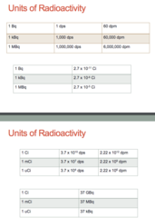
Curie (Ci) and Becquerel (Bq)
Units for Exposure
R and C/kg

Units of Absorbed dose
Rad and Gray
1 Gy = 100 Rad
1 Rad = 0.01 Gy
Units for dose equivalent
Roentgen equivalent (in) man (REM)
Sievert (Sv)
1 Sv = 100 Rem
1 Rem = 0.01 Sv
Film badges
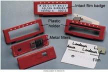
Consist of a small piece of special radiation-dosimetry film, similar to dental film, contained in a light proof packet
changed monthly
Metal filters composed of either copper or aluminum
shallow and deep dose can be calculated according to the amount of the film after processing

Film badge advantages
-simple to use
-inexpensive
-readily processed y commercial labs
-provide a permanent record by labs and in radiology department

film badge disadvantages
-are not reusable
-low limit of sensitivity (approx. 10 mrem)
-accuracy limit +/- 10-20%
-susceptible to heat, humidity and light leaks
Thermoluminescent Dosimeter (TLD)
Contain lithium fluoride or calcium crystals.
The crystals store radiant energy when heated. As they are heated, the crystals release energy as light, which is then measured by a machine that documents the radiation exposure based on how much light is emitted.
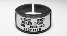
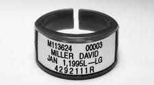
Commonly work as finger rings by nuc med personal to measure occupational exposure to their hands from handling radioisotopes.
Advantages of TLDs
-can be very small
-sealed in teflon, minimizing chance of damage
-low exposure limit, to 5 mrem
-response to X-rays proportional up to approx. 400 R
-Response almost independent of X-ray energy from about 50 kV to 50 mV
-accuracy to approx +/- 5%
-response very similar to tissues
-less sensitive to heat than film badge
-can be worn as a ring on fingers
-can be worn for 3 months
-are reusable
Disadvantages of TLDs
-Cannot be stored as a permanent record
-more expensive than film badges

Optically Stimulated Luminescence Dosimeter (OSL)
Contains filters composed of aluminum, tin, copper. It also houses a thin strip of aluminum oxide. The strip is stimulated by using a laser light and becomes luminescent in relation to the amount of radiation it has received.
Capable of measuring different energy ranges
These various ranges of energy correspond to deep, eye and shallow doses.
Are sensitive to approx 1 mrem. This makes their use especially desirable when monitoring pregnant workers.

Advantages of OSLs
-dose measurement very wide: 1 mrem to 1,000 mrem
-Accuracy +/- 15% shallow and deep exposure
-precision within +/- 1.0 mrem
-energy range 5 keV to over 40 MeV
-complete re-analysis available - can be restimulated many times
-bar coding, color coding, graphic formats and body location icons provide identification
-bimonthly readout offered
-tamper-proof sealed badge
-not affected by heat, moisture, or pressure
-services include badges for whole body, collar, waist, wrist, and tinge exposure to X-rays, gamma rays and beta particles
-reports available in a great variety forms

Disadvantages of OSLs
-more expensive than film badges and TLDs

Pocket Dosimeter
Very sensitive type of personnel monitoring device. The provide instantaneous reading, but must be calibrated daily. they are capable of only predetermine range. If exposure exceeds 200mR, any additional amounts of exposure cannot be documented.
Inside the dosimeter is an ionization chamber. The chamber has a positive and negative electrode. The electrodes are given a positive charge before use.
They are read by viewing a scale through an eyepiece located on the end of the dosimeter.

Annual dose limit for occupational whole body
5 rem (50 mSv)
Annual dose limit for occupational lens of eye
15 rem (150 mSv)
Annual dose limit for occupational skin/extremities
50 rem (500 mSv)
Annual dose limit for occupational whole body lifetime
age x 1 rem (age x 10 mSv)
Annual dose limit for occupational fetus (10 months/280 days)
0.5 rem (5 mSv)
Annual dose limit for occupational fetus (1 month/28 days)
0.05 rem (0.5 mSv)
Annual dose limit for public infrequent exposure
0.5 rem (5 mSv)
Annual dose limit for public frequent exposure
0.1 rem (1 mSv)
Medical event
A radiopharmecutical misadministration
Defines this term using two factors: error and exposure.
Errors of a misadministration/medical event
- The wrong radiopharmaceutical is administered, the dosage is administered to the wrong person, the dosage is administered by the wrong route or the dosage differs from the prescribed dosage by more than 20% or falls outside the prescribed range.
Excess exposure
Defined as exceeding the following limits:
5 rem (50mSv) effective dose equivalent
50 rem (500mSv) SDE to skin
Next calendar day
The NRC must be notified of a medical event by telephone no later than the
15 days
A written report for a medical event should be done within
5 years
Records of a medical event should be kept for
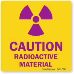
Caution: Radioactive materials
-indicates any area in which certain quantities of radioactive materials are used or stored
-found on entrances to work areas and nuclear medicine labs
-indicate the potential presence of radiation sources and/or contamination
-ingestion of food or drink strictly prohibited in these areas
-no smoking or application of cosmetics allowed in these areas
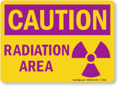
Caution: radiation area
-denote area in which an individual could receive more than 5 mrem (0.05 mSv) in 1 hour at 30 cm from the radiation source
-commonly seen at the entrances to nuclear medicine labs
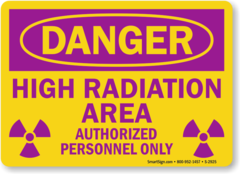
Caution: high radiation area
-designates areas in which an individual could receive more than 100 mRem (1 mSv) in 1 hr at 30 cm from the radiation source
-Commonly seen in areas where radiation therapy is performed
Grave danger: Very high radiation area
-designates an area in which an individual could receive an absorbed dose of more than 500 rad (5 Gy) in 1 hr at 1 m from the radiation source
-not commonly seen in the hospital settings

Survey trigger level for a restricted area
5 mrem/hr (0.05 mSv/hr)
Survey trigger level for unrestricted area
0.2 mrem/hr (2 uSv/hr)
Wipe test
This test must be analyzed using a well counter or an uncollimated gamma camera
The trigger level for a wipe test
22,000 dpm/100 cm^2
NRC regulation for survey and wipe records
Must be retained for 3 years. The record must include the date the survey was performed, the results, the instrument used, and the name of the individual who performed the survery
Minor spills procedure
- Notify all persons in the immediate vicinity that a spill has occurred
- Contain the spill by placing absorbent material over liquids. Limit movement in the area until the extent of the spill is known and the are is isolated
-If clothing is contaminated, remove it before leaving the spill site. If skin is contaminated, was as soon as possible with warm water and soap. Do not use abrasive cleansers, brushes, and hot water to clean the skin because this can increase absorption
- wear appropriate protective clothing, such as disposable gloves and shoe covers
-place all materials used to clean the spill into plastic bags and dispose of them as radioactive waste
-use a survey meter and wipe test to determine whether any removable contamination remains
-Use absorbent paper and/or shielding to cover the area of spill if activity remains after decontamination. Label the paper or shield with "Caution: radioactive material"
-Report the spill the RSO
Major spill procedure
-Clean the immediate area. Notify all personnel not involved with the spill to leave the area
-use absorbent paper to prevent the spread of contamination, but do not attempt to clean up the spill
-limit movement of contaminated personnel to prevent spread of contamination. Have personnel remove contaminated clothing before leaving the area
-Evacuate and close all doors to the area. Lock doors or secure the area to prevent entry
-Notify the RSO immediately. Cleanup and decontamination of major spills should be conducted under the supervision of the RSO
-decontaminate personnel as described previously. Treating serious injuries always takes precedence over decontamination
DPM (Calculation)
net cpm
___________
efficiency
Radioactive package trigger level when surveying
If the exposure rate exceeds 200 mR/hr at surface or 10 mR/hr at 1 m, the NRC and the carrier delivering the shipment must be notified immediately through the RSO
Radioactive package trigger level when wipe testing
If contamination exceeds 24 dpm/cm^2 or 7,200 dpm for a wiped surface of 300cm^2 for beta, gamma and low toxicity alpha emitters and 2.4 dpm/cm^2 or 720 dpm for a wiped surface of 300cm^2 for all other alpha emitters, notify the NRC and the carrier through the RSO
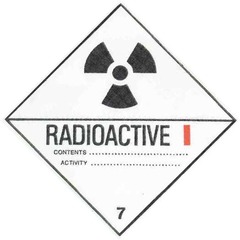
White I
Exposure rate at surface:
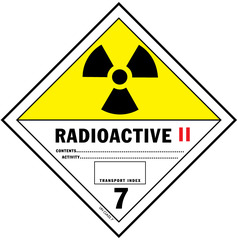
Yellow II
Exposure rate at surface:
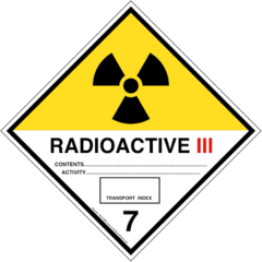
Yellow III
Exposure rate at surface:
120 days
Radioactive materials with a half-life of less than ______________ can be retained in a shielded storage are until they have decayed to a level of activity that equals background
10 half-lives
a common rule of thumb is to decay the radioactive product for approximately _____________________ before discarding it in regular or biological hazard trash
Atomic Structure
Atoms of which matter is composed contain electrons of negative charge and negatively charged electrons orbiting a central group of particles, have the positively charged nucleus
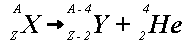
Alpha decay
(excessive nuclear mass)
- Very large unstable atoms, atoms with high atomic mass, may split into nuclear fragments. The smallest stable nuclear fragment that is emitted is a particle consisting of two neutrons and two protons, equivalent to the nucleus of a helium atom
- Has too many protons and too many neutrons
- Carries a highly ionizing electric charge (+2)
- In this decay process the Z- number is reduced by 2 and the A- number reduced by 4
beta negative decay
(Unstable neutron-proton ratio)
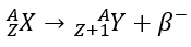
- Nuclei with excess neutrons can achieve stability by a process that amounts to the conversion of a neutron into a proton and an electron. The proton remains in the nucleus, but the electron is emitted.
- Unstable nuclei having an excess number of neutrons (Neutron-rich) will tend to decay by this
- In this decay process the Z- number of the parent product is increased by 1, but the A- number does not change
- The excess neutron is turned into a proton, ___ - particle, and an anti-neutrino.
beta postive decay (positron decay)
- Unstable nuclei having an excess number of protons (proton-rich) will tend to decay by this and/or electron capture.
- In this decay one of the excess protons is converted into a neutron, a positive electron or positron, and a neutrino.
- For the positron to be formed, the parent nucleus must have at least 1.02 MeV of energy available for the decay process to occur.
- In this decay the Z- number of the parent product is decreased by 1, but the A- number remains unchanged

Gamma decay
- The decay mode for those unstable nuclei that just need to rid themselves of excess energy
- In this decay both the Z and A number remain unchanged
- Electromagnetic photons carry neither mass nor charge
- For electromagnetic to interact with matter they must have a direct hit
(There is no difference between an electromagnetic photon, a y-photon and x-ray photon except for their origin)

Electron capture
- An alternative mode of decay if proton-rich nucleus cannot meet the 1.02 MeV of energy required for a positron emission
- Combines with an excess proton to produce a neutron and a neutrino
- Z- number is decreased by 1, but A- number remains unchanged.
- Note that positron decay and this decay will produce the exact same daughter product in the end
Decay schematics
Decay of a nuclide from unstable state to stable state can occur in a series of steps. Standard notation is used to describe these steps.

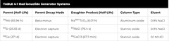
Generator (parent-daughter)
Parent isotopes are the isotopes of a particular chemical element that can undergo radioactive decay to form a different isotope from a different chemical element. Daughter isotopes, on the other hand, are the products of radioactive decay of parent isotopes.
Ex.
99Mo - 99Tc
Used in most RPs for Nuc med.
T1/2 parent - 66 hr
T1/2 daughter - 6 hr
T1/2p / T1/2d - 11
82Sr - 82Rb
Used in cardiac perfusion imaging (PET)
T1/2 parent - 25.5 days
T1/2 daughter - 75 secs
T1/2p / T1/2d - 29,000
68Ge - 68Ga
Used in neuroendocrine imaging (PET)
T1/2 parent - 271 days
T1/2 daughter - 68 mins
T1/2p / T1/2d - 5,800
photoelectric absorption
This process occurs when the incident photon has energy that is just a little greater than the binding energy of an inner shell electron.
When the incident photon interacts directly with the inner shell electron, it gives up its entire energy to the inner shell electron and ejects it with a certain amount of kinetic energy.
is an ionizing type of interaction that represents complete absorption of the incident photon.
The ejection of a shell electron results in the emission of characteristic x-rays.

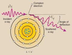
Compton scattering
A medium-energy photon interaction in which the incident photon ejects an outer shell electron that has a lower binding energy.
In addition to the ejected electron, this produces a lower-energy scattered photon. This lower-energy scattered photon will most likely be photoelectrically absorbed in its next interaction.
Is also an ionizing type of interaction and it tends to be the predominant mode of interaction of diagnostic energy photons in soft tissue
Pair production
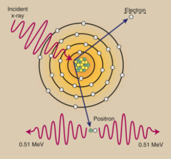
High-energy photon interaction in which the incident photon interacts with the intense electromagnetic field of the nucleus. The photon disappears and in its place, an electron-positron pair appears.
( not relevant in nuc med )
Compton scattering
The dominant type of interaction in materials with lower atomic numbers, such as human tissue (Z = 7.5)
Photoelectric effect
Is the dominant type of interaction in materials with higher atomic number, such as lead (Z = 82)
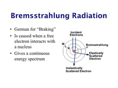
Bremsstrahlung
Most of the x-rays produced the at target are ____.
Small charged particles such as electrons or positrons may be deflected by nuclei as they pass through matter. Which may be attributed to the positive charge of the atomic nuclei.
Annihilation
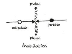
This interaction in matter involves a positron (positive electron) and an electron (negatron). After a positron has transferred most of its kinetic energy by ionization and excitation, it combines with a free or loosely bound negative electron.
This interaction is explosive, as the combined mass of the two particles is instantly converted to energy in the form of two oppositely directed photons, each energy 511 keV
Major spills by dose and RPs
>1 mCi of I-131, Sr-89
>10 mCi of Ga-67, In-111, I-123
>100 mCi of Tc-99m, Tl-201
Reactor produced radionuclides
Tc99m
Mo99
I-131
Xe-133
Sm-153
Sr-89
Accelerator produced radionuclides
I-123
In-111
Tl-201
Ga-67
Radionuclide purity
Mo-99
Effect: Radiation dose, poor image quality. When administered to patient is taken up by the parenchymal cells of the liver and delivers unnecessary radiation dose to the organ.
Limits:
Chemical purity
Al3+
Effect: Poor image quality. Biodistribution of RP.
Limits:
Radiochemical impurities
Is defined as the fraction of total radioactivity present in its desired chemical form.
Ex. Unbound (free) Tc99 tech and hydrolyzed reduced Tc99
Unbound (free) Tc99 tech will appear as increased tracer in stomach, thyroid and salivary gladn
Hydrolyzed reduced Tc99 Will have unexpected uptake in the reticuloendothelial system, especially the liver.
This can be determined by using radiochromatography
Geiger counter
High voltage, each ionization causes an avalanche of electrons and hence a large electrical pulse; measure as counts per min; best for detecting low level radiation; conversion to exposure rate is not necessarily accurate due to variability of response depending on radiation energy.
Used to survey hands, feet, contamination and lost thyroid marker source
Capable of detecting exposure rates from 0.1 mRem/gr (1 uSv/hr) to 100 mRem/hr (1 mSv/hr)
Calibration is required annually - once a yr
proper operation is checked each day of use with a dedicated check source

Ionization chamber - cutie pie
It is superior to the G-M counter in measuring high dose rates. It is most useful for accurately determining the exposure rate being produced by a patient who has received a radionuclide, particularly a therapeutic dosage.
Calibration is required annually - once a yr
proper operation is checked each day of use with a dedicated check source

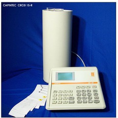
Dose calibrator
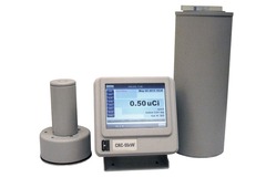
An ionization chamber that measures radioactivity in curies or becquerels rather than in counts per min. It can accurately measure dosage as low as 0.01 uCi, as well as activities in the curie range.
Can readily detect gamma photons but. are less sensitive for beta radiation, because only high-energy beat radiation can pass through glass chamber walls.
Correction factors are usually required for measuring beta radiation
Dose calibrator accuracy QC
Measure of the readings of the dose calibrator in comparison with well-accepted standards.
Two long-lived nuclide sources, such as 137Cs (T1/2 = 30 yrs) and 57Co (T1/2 = 270 days) are measured repeatedly in the calibrator and the average readings are compared with values issued by the NIST for that particulate source.
If the reading differ from the standards by more than 10%, the dose calibrator should not be used.
This QC should be checked at installation, annually and after repairs.
Record kept for 3 yrs
Dose calibrator constancy QC
To ensure that the calibrator readings are constant from day to day, and fall within an acceptable range compared to prior readings
done daily
sources used:
Co57 - 122 keV
Ba133 - 356 keV
Cs137 - 662 keV
Acceptable results +/- 10%
Records kept for 3 yrs