F26 NEU C61 Midterm #1 Flashcards
 Call Kai
 Learn
Learn Practice Test
 Spaced Repetition
 Match
Match Flashcards
 Knowt Play
1/136

There's no tags or description
Looks like no tags are added yet.
Name | Mastery | Learn | Test | Matching | Spaced | Call with Kai | Chat |
|---|
No analytics yet
Send a link to your students to track their progress
137 Terms
Earliest Origins of the Mind
Cave Paintings (~35,000 years ago). Showed early humans could imagine things not physically in front of them and create symbolic images. Showed they had complex mental capabilities, creativity, culture.
Trepanation
Definition: Drilling Holes in Skull for Intentional Medical Purposes. Practice in Ancient Egypt (5000-7000 years ago). Ancient Egypts believed that heart was the location of memory and consciousness.
Hippocrates
~400 B.C.; from Ancient Greece. Didn’t believe disease was caused by gods. Believed that brain is involved in sensation and seat of intelligence. Proposed that humors (blood, phlegm, yellow bile, black bile) are body’s vital fluids.
Aristotle
~300 B.C.; from Ancient Greece. Didn’t believe that brain was involved in emotion, sensation, and movement, instead those responsibilities were the responsibility of the heart. Brain only necessary to cool the blood from the heart.
Galen
~130-200 A.D.; from Roman Empire. Most important Roman physician. Believed Brain’s fluid-filled cavities (ventricles) is where the humors mixed. Believed Cerebrum is soft → can have sensations “imprinted” on it → cerebrum receives sensation. Cerebellum is hard → commands muscles. Found ventricles in dissection, thought fluid moved from nerves to ventricles to cause sensation/perception
What was required for the rise of modern neuroscience?
Microscopes (1655)
Cell Theory (1830s)
Modern Chemistry (1800s)
What was Santiago Ramón y Cajal’s contribution to neuroscience?
Proved brain was made of individual, distinct cells rather than a continuous, fused network.
Descartes
Early 1600s; Believed center for intellect was outside of brain, in the mind (Brain and mind are different). Body is controlled by mind, received commands from the pineal grand (in the middle of brain). Humans are different from other animals because of intellect and god-given soul. His theory is called substance dualism.
Mind-Body Problem
What is the relationship between the mind/mental (thoughts, feelings, consciousness) and the physical (body/brain)?
Dualism
The brain and mind are two separate kinds of things, fundamentally different.
Physicalism
Brain and mind are the same thing; the mind is a physical property.
Property Dualism
Mind and brain are one substance, but has 2 properties: physical and mental. Mental properties happen as a result of the physical brain.
Key Problem with Dualism
Interaction Problem: How does a non-physical mind cause physical neurons to fire, and vice versa?
Physicalism
Mental states are physical states of the brain. There is nothing extra required.
Identity Theory
Theory under physicalism. Mental states are completely identical to physical brain states
(firing of neurons). Changing brain chemistry can change thoughts, emotions, moods since they are one and the same.
Functionalism
Theory under physicalism. The physical doesn’t matter- mental states are defined by their functional roles. The same mental state can be created by multiple different physical arrangements of neurons. Expands the definition of minds
Why do we care about the Mind-Body Problem?
Neuroscience assumes methodological physicalism because that’s what makes questions scientifically testable.
Nervous System
Includes brain, spinal cord, and nerves throughout the body. Many animals have a nervous system without any true brain. Consists of central nervous system and peripheral nervous system.
Brain
One organ in the nervous system. Serves as control center. Allows intelligent thought (e.g. memory), specialized behaviors (e.g. hunting, mating), specialized senses (e.g. smell, taste, touch), coordinates sensor inputs and motor outputs (bigger body → bigger brain).
Central Nervous System (CNS)
Brain and Spinal Cord
Peripheral Nervous System (PNS)
Nerves and cells that connect the CNS to the arms, legs, and organs.
Somatic Nervous System
Under the PNS. Voluntary. Consists of motor neurons and sensory neurons (e.g. skin, muscles, eyes)
Autonomic Nervous System
Under the PNS. Involuntary. Split into Sympathetic (Fight or flight response), Parasympathetic (Relaxed non-threatening conditions), and Enteric (In the gut, regulates digestion)
Brain Weight + Oxygen and Glucose Consumption
<3% of the body’s weight, consumes 25% of body’s oxygen and 70% of it’s glucose
What is a gyrus?
Ridge in the Brain

What is a fissure?
Deep groove in the brain

What is a sulcus?
Groove in the brain

Grey Matter in the Brain
Processes Information and Thoughts, sits on outer surface of the brain
White Matter in the Brain
Transmits signals from gray matter to other parts of the brain and body, white matter lies beneath gray matter
What does the occipital lobe do in the brain?
Vision, visual processing.

What does the frontal lobe do in the brain?
Decision making, language
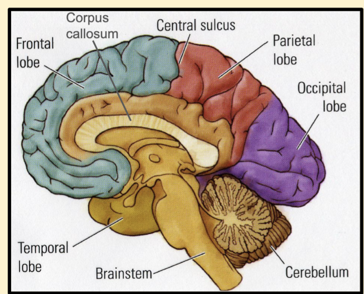
What does the temporal lobe do in the brain?
Memory, emotion, language

What does the parietal lobe do in the brain?
Sensory processing, spatial reasoning
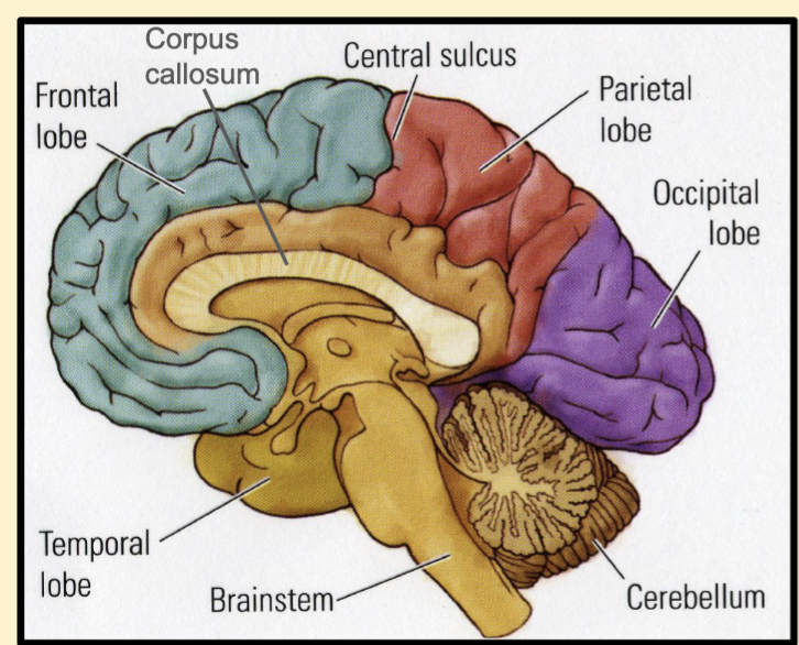
What does the cerebellum do in the brain?
Voluntary movement, balance

What does the corpus callosum do in the brain?
Connects both hemispheres

What does the brain stem do?
Connects brain to spinal cord, consists of midbrain, pons, medulla oblongata, and spinal cord. Sensory and motor info have to pass through brainstem to get to and from spinal cord and body (PNS). In spinal cord, motor info travels down the spinal cords to get to PNS and sensory info travels up to get to the brain. Spinal cord also center for coordinating certain reflexes.

Phrenology
Measured bumps on skull and claimed their location impacted cognitive function. Developed my Franz Josef Gall, pseudoscience today.
Paul Broca
1858, His Patient was able to understand but not speak → found out that different brain regions have specific functions
Carl Wernicke
1874, Patient spoke gibberish but did not comprehend language
Phineas Gage
Discovered that brain damage to the frontal lobe would lead to anger/behavior changes
Wilder Penfield
1930s, Developed “cortical homunculus” map that mapped motor control. Simulated different parts of motor cortext and observed movements in different parts of the body.
Nissl Stain
First effective dark-blue stain of brain tissue that let us view cell bodies (but couldn’t see inside, no structure). Created by Camilo Golgi in the 19th century, he believed that brain cells were physically continuous with each other in a single network
Golgi Stain
Stains only a subset of neurons → allowed for clear visualization of the network of brain cells
Neuron Doctrine
Evidence discovered through the electron microscope. Brain is composed of cells (neurons) which are primary info processing units. Independent from one another. Information is transmitted through synapses.
What is a synapse?
Gap between two neurons. Transmission site from the synaptic to the post-synaptic neuron. Communication at synapses both electrical and chemical. Has three components = presynaptic membrane, synaptic cleft, postynaptic membrane
What are dendrites and where they are located in the neuron?
Branched, tree like extensions of a neuron that act as the main receiving antenna for incoming signals from other nerve cells.

What is the cell body and where is it located in the neuron?
Houses the nucleus and produces energy and proteins. Circle-ish part of the neuron.

What is the axon hillock and where is it located in the neuron?
Connects the cell body of the neuron with the axon of the neuron. Also gathers and integrates information from all the synapses on a neuron’s dendrites and cell body and coverts that information into a “action potential”. Looks like a cone, right below the cell body.
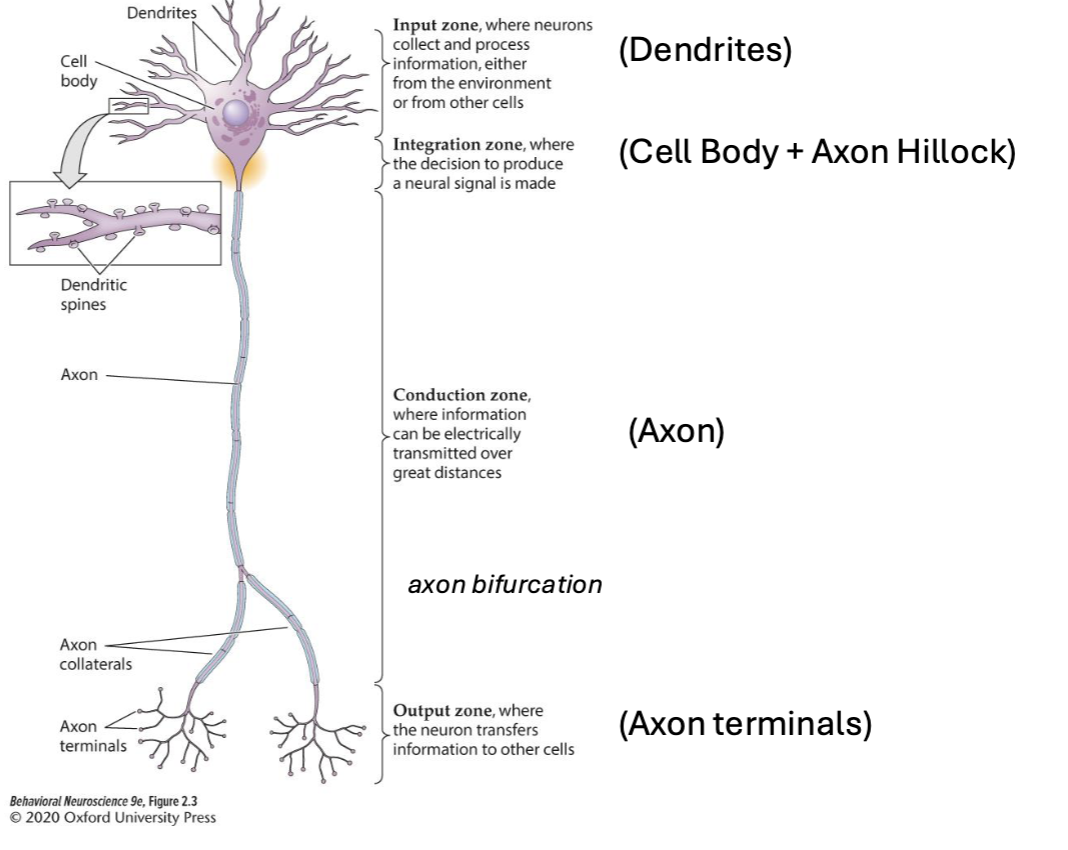
What is the axon and where is located in the neuron?
Where information is electrically transmitted down from the cell body. Longest part of a neuron.
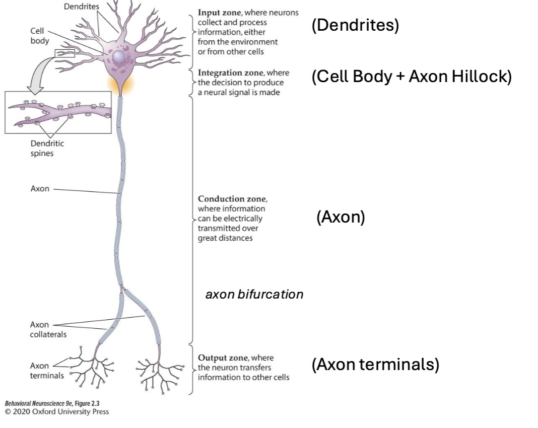
What are axon terminals and where are they located in the neuron?
Output zone where neuron transfers information to other cells. Looks kind of like dendrites but like on the bottom.
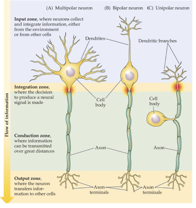
Polarities of a Neuron
Multipolar: Many dendrites, single axon
Bipolar: One dendrite, one axon
Unipolar: A single extension that branches in two directions
Glia
Non-neuronal cells that are support cells in the brain. Four types:
Microglia: “Clean-up crew” that destroy debris via phagocytosis. From immune system.
Macroglia:
Astrocytes: Star-shaped support cells. Supports neuron by providing energy and prevent spillover. Also present at neuron to neuron and blood vessel to neuron connections. Don’t overlap. Also envelop synapses.
Oligodendrocytes: In CNS. Form the myelin sheaths that insulate the axons of some neurons to speed up neural communication. 1 oligodendrocyte can wrap myelin around ~50 axons.
Schwann Cells: in PNS. Also forms the same myelin sheaths as oligodendrocytes. But can only wrap myelin around one axon.
Ependymal Cells
Line the ventricles, create the circulate cerebrospinal fluid (CSF)
Why is the brain heavily folded?
More folds increase the surface area of the cerebral cortext. Bigger surface area → more neurons → more processing power without needing a bigger skull.
Electrons communicate using electrical and chemical signals
Electrical Signals: Flow within a neuron via an action potential
Chemical Signals: Between neurons through neurotransmitters at the synaptic cleft
Neuron has a very _ resting potential
Negative
Key Players in Neuron Communication
Water: In both intracellular and extracellular fluid, polar solvent which means charged substances can dissolve in water
Ions: Electrically charged atoms or molecules
Cations: Positively charged ions
Anions: Negatively charged ions
Cell Membrane
Channels
Current
Build up of ions on one side of a membrane. Aka “potential”. Measured in volts/mV
Voltage
Flow of ions across that membrane. Measured in Amps
Cell Membrane
Made of phospholipid bilayer: hydrophilic heads out, hydrophobic tails inside. Ions can only cross through embedded proteins (channels & pumps). What allows the cell to hold a voltage across it. Semi-permeable (some ions can pass through, but others cannot)
Ion Channels
Proteins in the membrane that allow ions to pass through. Highly specific, their pores allow only 1 or few types of a ions to flow. Examples: Potassium leak channels + sodium-potassium pump.
How to measure membrane potential of a neuron?
At rest, membrane potential = -65 mV. Maintained by cell membrane’s lipid bilayer and semi-permeable ion channels
Electrostatic Forces
Ions flow toward opposite charges, away from similar charges
Diffusion
Ions move from areas of high concentration to areas of low concentration
Concentration Gradient
Difference in ion concentration
Equilibrium
Rate of which ions are leaving = same as rate as they are entering
Ions concentration inside and outside a neuron
Na+, CL-, CA2+, concentrated outside the cell, K+ and proteins concentrated inside the cell
Membrane potential is primarily determine by _ ions
K+, Determined this way because potassium pump pushes Na+ ions out and K+ ions in, creating a build up of K+ ions in the cell. Leaky ion channels allow K+ ions to diffuse back out of the cell across their gradient. This creates a negative membrane potential. The number of K+ ions diffusing out is balanced by K+ ions pulled back in by electrical force, and the cell is at equilibrium in this state.
Potassium Channels
Potassium leak channels: Allow K+ ions to freely diffuse across the membrane according to their ion gradient
Sodium-Potassium pump: Pumps 3 Na+ ions outside the cell for every 2 K+ ions pumped into the cell
Uses 1 molecule of ATP for each exchange
What is K+ movement affected by?
Diffusion (Concentration gradient): The difference in concentration of K+ inside & outside the cell
Electrical gradient: The difference in charge between the inside & outside of the cell

Identify each area and its function

Identify each area and it’s function
What is an action potential?
Very brief but large change in membrane potential
All-or-nothing event (size and duration are always the same)
Originates in the axon initial segment
Propagates at high speed along the axon
Carries info to post-synaptic cells, even across long distances
How info is carried in networks of neurons, allows for long distance communication
What does it mean to be polarized?
Very negative compared to outside
Hyperpolarization Meaning
Decrease in membrane potential (inside of the cell is more negative compared to outside)
Depolarization Meaning
Increase in membrane potential (inside of the cell becomes more positive relative to outside)
What ions’ movements define the phases of an action potential?
K+ and Na+
Stage 1 of Action Potential
Resting State
Leak channels are open (both Na+ and K+ but
way more K+ leak channels)
Resting membrane potential is maintained
Net negative charge inside cell
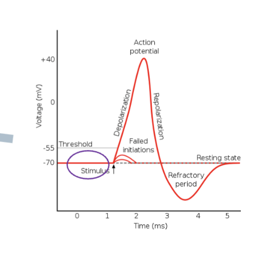
Stage 2 of Action Potential
Depolarizing Phase
Neurotransmitter from pre-synaptic neuron opens ligand-gated Na+ channels
These channels only open when cell depolarizes past threshold
Na enters the cell and depolarizes the membrane
Fires action potential at -55 mV

Stage 3 of Action Potential
Rising Phase
Voltage-gated sodium channels (cytoplasmic inactivation gate physically blocks inner pore once equilbrium is reached) open once threshold is reached → what makes action potential happen because Na+ flows into the cell in huge amounts due to electrical and concentration gradients
Rapidly depolarizes neuron to +40 mV

Summary of Synaptic Inputs
Dendrite → Soma → Hillock → AP! → Axon
What is EPSP?
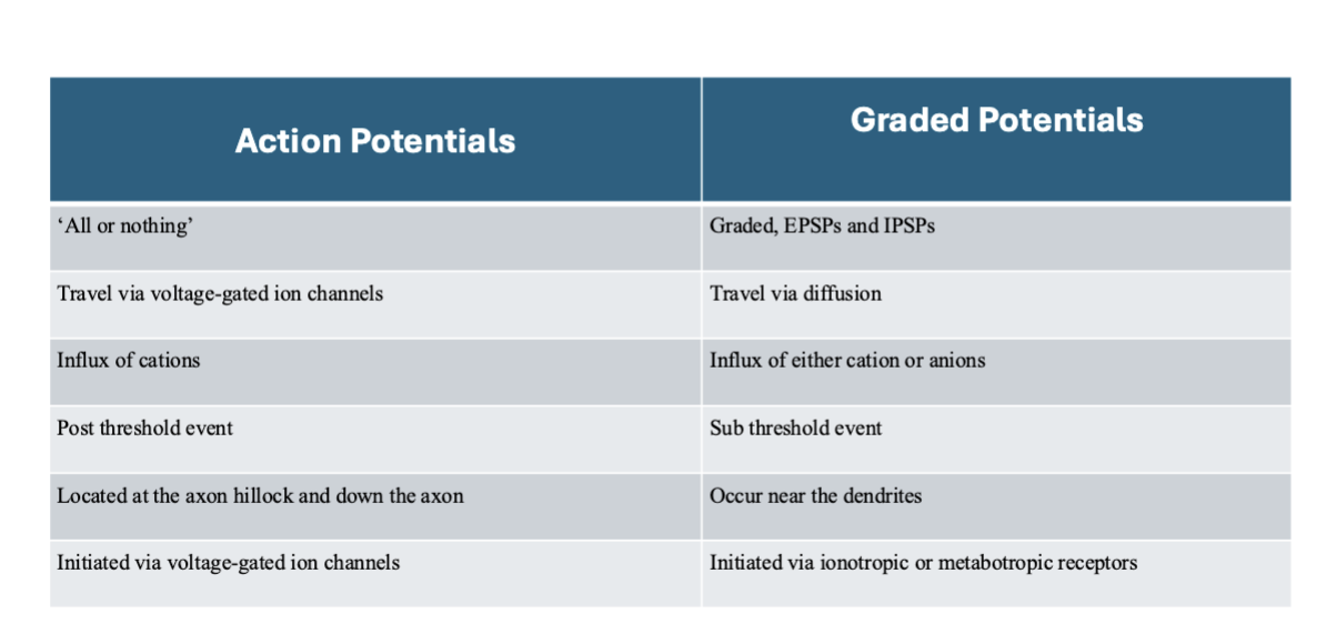
Excitatory post-synaptic potential → let cations in → depolarize → closer to reaching threshold. EPSPs and ISPSs are summed at axon hillock
What is IPSP?
Inhibitory post-synaptic potential → let in anions → hyperpolarize → farther from reaching threshold. EPSPs and ISPSs are summed at axon hillock
What is Stage 4 of an Action Potential?
Falling Phase
Voltage-gated Na+ channels inactivated (NOT closed)
Voltage-gated K+ channels finally open
K+ rapidly leaves the cell
Membrane potential begins to fall back down toward -65 mV

What is Stage 5 of an Action Potential?
Undershoot/ Refractory Period
Voltage-gated Na+ channels inactivated
Voltage-gated K+ channels remain open (enabling undershoot aka becomes even more negative)
Refractory period: So negative (below resting potential) that no APs can be fired until it’s over! Prevents AP from propagting backwards
In unemyelinated axons…
Slow Conduction
Action potentials (AP) propagate continuously
Influx of sodium in one AP triggers another AP beside the first
The refractory period & the undershoot keep APs traveling in only 1 direction
In myelinated axons…
Insulates the axon which allows AP to move much faster by reducing current leaks
Kept going by small gaps in myelin called nodes of ranvier where ions can move in/out
AP “jumps” between nodes via saltatory conduction
What channels are open during the rising phase?
Voltage gated Na+ channels
Na+ is moving into the cell
What channels are open during the falling phase?
Voltage-gated K+ channels
K+ is moving out of the cell
What channels are open during the refractory/undershoot phase?
Voltage-gated K+ channels
K+ is moving out of the cell
What channels are open during resting potential?
None
Who was Otto Loewi?
Won an Novel Prize proving that transmission of nerve impulses was chemical, not electrical
What is dendritic arborization?
Dendritic branching that reflects the complexity of a neuron’s input capacity
What is a neurotransmitter?
Chemical messages sent from the presynaptic neuron to the postsynaptic neuron. Released from synaptic vesicles
What causes neurotransmitters to release across the synapse?
Action potentials, specifically the depolarization that opens voltage-gated calcium channels which causes influx of calcium (Ca2+ ions)
What direction do calcium ions flow?
Flow into the cell, down its concentration gradient. Ca2+ binds and activates a protein called synaptotagmin which triggers neurotransmitter release
Synpatic Transmission Overview - What are the steps?
Action potential reaches the synaptic terminals → triggers neurotransmitter (ex. dopamine, serotonin, GABA) release into the synapse → triggers a graded potential (EPSP/IPSP) for the following neuron which undergoes the same process. More specifically, the calcium ions binds and activate the protein synaptotagmin which triggers neurotransmitter release via SNARE complexes
How can neurotransmitters be cleared from synaptic cleft?
Can be re-uptaken into the axon terminal or uptaken by glia
Can be degraded by enzymes
Can just diffuse out of the synaptic cleft
Electrical vs Chemical Communication in Neurons
Action potentials are electrical communication within a neuron, chemical communication involving neurotransmitters is between neurons
What are graded potentials and what is the differences between them and action potentials?
They determine if an action potential fires or not. They can depolarize (EPSP) or hyperpolarize (IPSP) a neuron.