Encoding Pt 2 - Hippocampus and Long Term Memory
1/20
There's no tags or description
Looks like no tags are added yet.
Name | Mastery | Learn | Test | Matching | Spaced |
|---|
No study sessions yet.
21 Terms
Memory retrieval
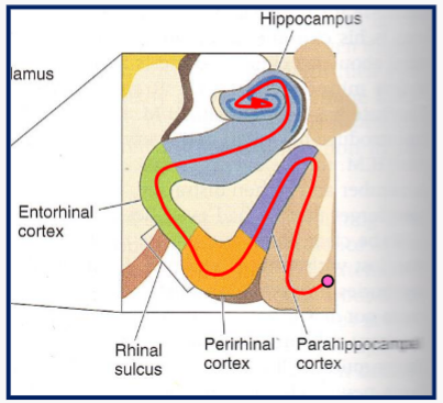
Parahippocampal and rhinal cortices → Hippocampus
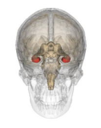
What structure is shown in the image?
Hippocampus
Medial Temporal Lobe - Anatomy and constituents
Parahippocampal and perirhinal are connections between Association cortices (all 3) and hippocampus
Removal of the medial temporal lobe
Results in severe anterograde amnesia (Clive Wearing)
What does the hippocampus do?
Form declarative/explicit memories
Ultimately ‘stored’ in the cortex
Tells you where you are in space
Spatial memory/learning
Often lost early in Alzheimer’s
Active site of neurogenesis
Multiple sub-regions
Encoding patterns
Pattern completion
Recognising something from a partial representation
Pattern Separation
Learning to distinguish between similar
The hippocampus is very important for both
Not just for visual memories
Neuronal Signals
Electrical signal
Travels along neurons as an ‘action potential’
Chemical Signal
Travels between neurons as neurotransmitters
Excitatory neurotransmitters
Increase probability of target neuron firing action firing action potential
Inhibitory neurotransmitters
Reduce probability of target neuron firing action potential
What could a ‘code’ look like?
Which neurons fire
What causes them to fire
How long they fire for
Where they project to
How many other neurons they are connected to
Numbers/types of neurotransmitter receptors
What is Hippocampal indexing?
The basis for ‘retrieval’
Pattern completion
Pattern separation
Hippocampal indexing
Pattern of neuronal activity from cortex activates a specific subpopulation of neurons in CA3
These are densely, reciprocally, connected
Partial input (part of the pattern) - activates the whole group of CA3 neurons
The connections between this group can be modified, for example when new information is learned
What is synaptic plasticity?
The cellular basis for learning and memory
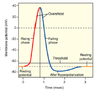
Action potentials
Presynaptic neuron releases neurotransmitter
Enough ligand-gated channels open
Membrane reaches the threshold
Action potential will fire
Long-term potentiation definition
Frequent firing strengthens synapses
Long-term potentiation
Brief, intense firing by presynaptic neuron
Abundant glutamate release
This causes changes in post-synaptic neuron
Opens NMDA-type glutamate receptors
Increases expression and insertion of AMPA-type glutamate receptors
(other changes as well)
Strengthens the synapse
Increases the likelihood that neurons fire together
Changes are long-lasting
Calcium influx prompts gene expression
Happens across the brain
Best understood in the hippocampus
Synapse between CA3 (Schaffer collaterals) and CA1 (Pyramidal cells)
Long-term Depression
Connections between neurons become weaker
Prolonged, low-intensity firing of presynaptic neuron
Pre and postsynaptic neuron do not then ‘fire together’
Multiple cellular mechanisms
Decreased expression/insertion of postsynaptic AMPA receptors
Decreased presynaptic glutamate release
Also occurs across the brain
Not as well understood as LTP
From Synapse to code to memory
One theory is that the ‘code’ is a pattern of firing of a specific group of neurons in a specific hippocampal region (CA3?)
Firing pattern in association cortices activates that specific group - partial firing still activates full group
The members of the group, and their firing pattern, can be modified by LTP and LTD - Allows for two distinct groups to be formed as part of pattern separation
Directly linking LTP/LTD to memories is hard to do
Boosting Memory
Eat glutamate?
Stimulate NMDA and AMPA glutamate receptors?
NMDA and AMPA receptors are everywhere
Systemic stimulation can cause seizures
Too much glutamate is excitotoxic and a major mechanism of cell death
Types of Spatial Representation
Allocentric (non-egocentric)
A map of the environment
object-to-object
hippocampus
Egocentric
Where am I in the environment
Me-to-object
left/right, up/down etc.
posterior parietal cortex and prefrontal cortex
Place Cells
Pyramidal neurons in hippocampus (CA1 + CA3)
Activated by allocentric environmental cues
Visual, olfactory, other senses
Also activated by ‘replay’ of cues (thinking about the map)
Encode a ‘place field’
Field is plastic - can change
Some are spatially oriented
(front, back, etc)
Other navigational neurons
Head position cells
Subiculum
Fire when oriented towards a specific direction
Border cells
Place cells that are specifically activated by barriers
Reward-place neurons
learn about a reward in a particular place. Not value of rewards (happens in Orbitofrontal cortex), or place, but connection between the two
Bringing together where it is and what it is, in the same network
Grid cells
Entorhinal cortex
Hexagonal map
Only requires one co-ordinate to change location, compared to Cartesian (X-Y) mapping
Adjacent grid cells map adjacent grid
Dead reckoning or path integration
Calculating current position relative to a previous position
Distance travelled, speed, direction