Plasma Cell Dyscrasias
 Call Kai
 Learn
Learn Practice Test
 Spaced Repetition
 Match
Match Flashcards
 Knowt Play
1/24

There's no tags or description
Looks like no tags are added yet.
Name | Mastery | Learn | Test | Matching | Spaced | Call with Kai | Chat |
|---|
No analytics yet
Send a link to your students to track their progress
25 Terms
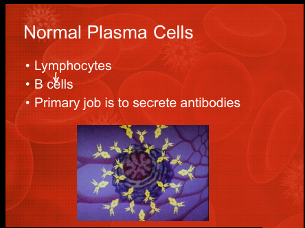
Normal plasma cell function
B‑cells that differentiate into plasma cells whose primary job is antibody secretion.
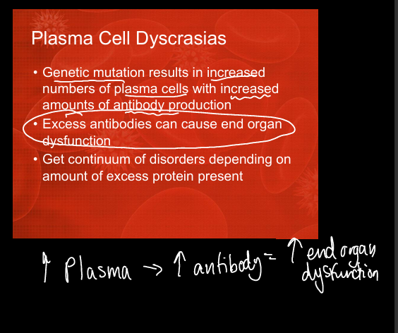
Definition of plasma cell dyscrasia
Clonal plasma cell expansion causing excess monoclonal immunoglobulin(inc in antibodies) and potential end‑organ damage.
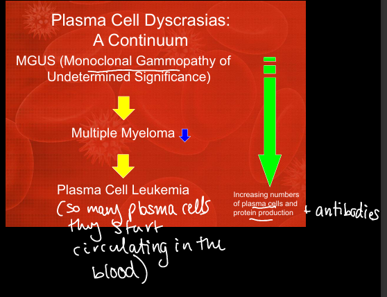
Continuum of plasma cell disorders
Monoclonal Gammopathy of Undertmined Significance (MGUS) → Multiple Myeloma → Plasma Cell Leukemia (increasing plasma cells and protein burden).
Diagnostic test to quantify plasma cells
Bone marrow biopsy and aspirate.
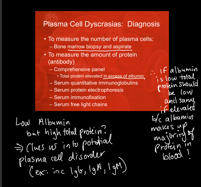
Diagnostic tests to quantify monoclonal protein
SPEP, immunofixation, quantitative immunoglobulins, serum free light chains.
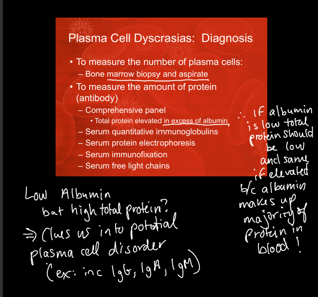
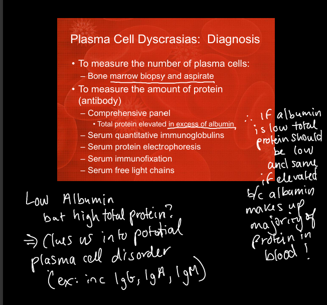
Clue suggesting plasma cell disorder on CMP
High total protein with normal/low albumin (albumin should dominate total protein).
Polyclonal vs monoclonal immunoglobulin pattern
Polyclonal = broad gamma region= normal
Monoclonal is very narrow(all the same protein so they all blot in same spot)
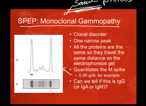
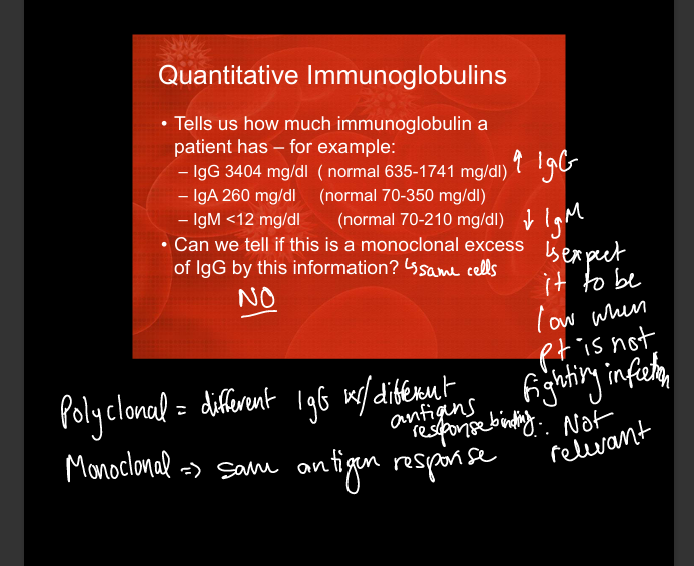
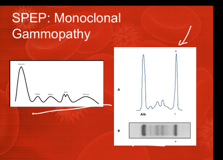
SPEP monoclonal gammopathy pattern
Single sharp gamma‑region spike representing identical immunoglobulins.
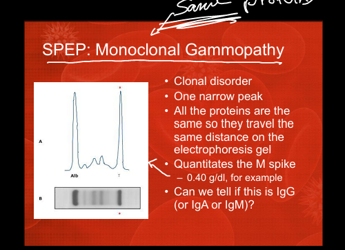
Purpose of immunofixation/Immunoelectrophoresis
Confirms clonality and identifies heavy chain (IgG/IgA/IgM) and light chain (kappa/lambda).
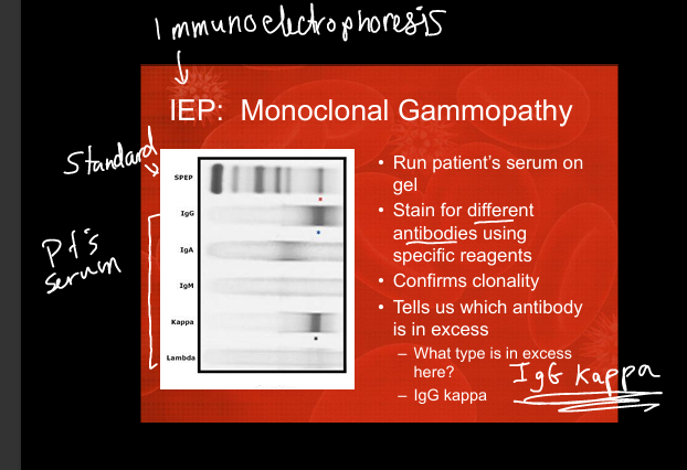
Serum free light chain abnormality in myeloma
Markedly elevated kappa or lambda with abnormal ratio (>1.65 or <0.26).
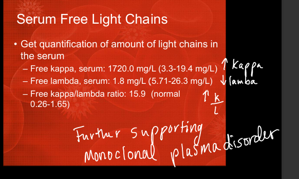
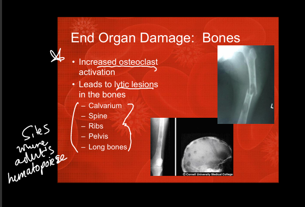
Bone involvement in myeloma
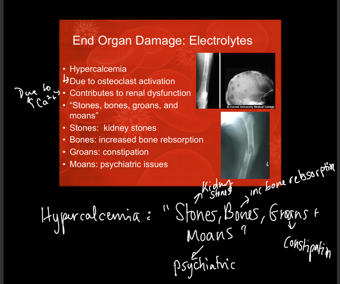
Lytic lesions due to increased osteoclast activation (skull, spine, ribs, pelvis, long bones).
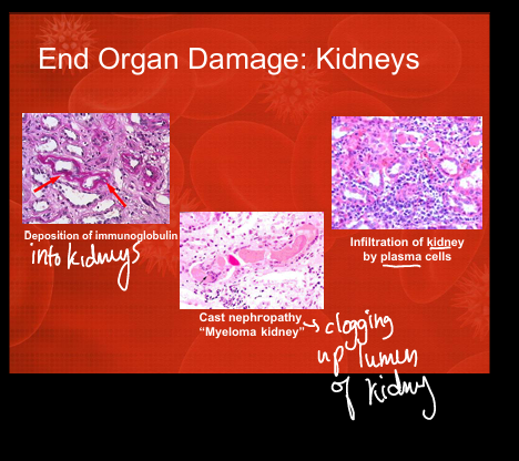
Kidney involvement in myeloma
Cast nephropathy (“myeloma kidney”), immunoglobulin deposition, plasma cell infiltration.
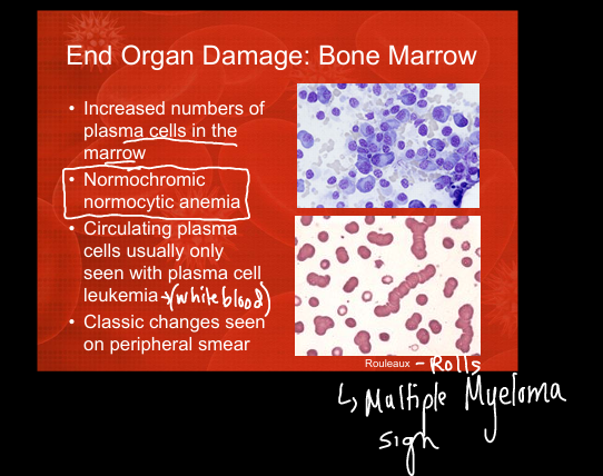
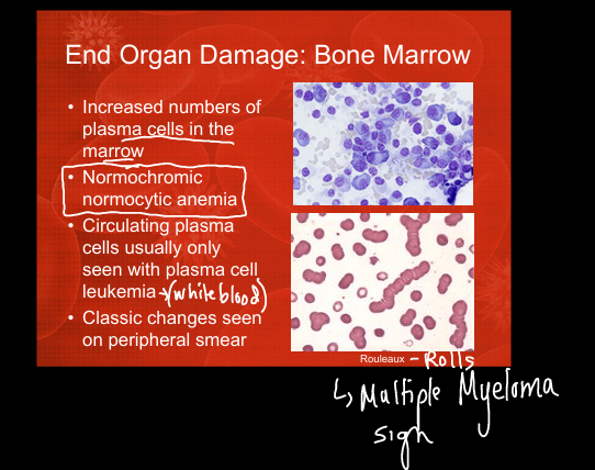
Peripheral smear finding in myeloma
Rouleaux formation due to increased serum proteins.
Electrolyte abnormality in myeloma
Hypercalcemia from increased osteoclast activation (“stones, bones, groans, moans”)=kidney stones, increased bone resorption, constipation, psych issues
Anemia type in myeloma
Normocytic, normochromic anemia due to marrow infiltration.
Treatment approach for plasma cell dyscrasias
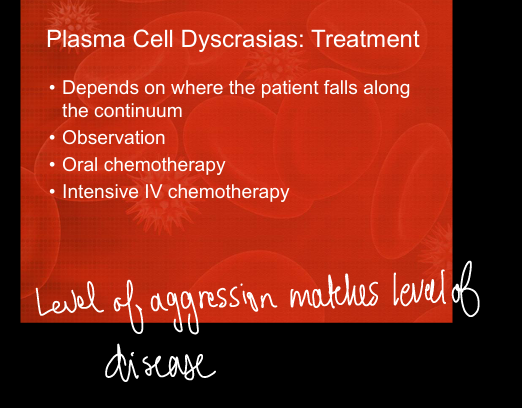
Depends on disease severity: observation → oral chemo → intensive IV chemo.
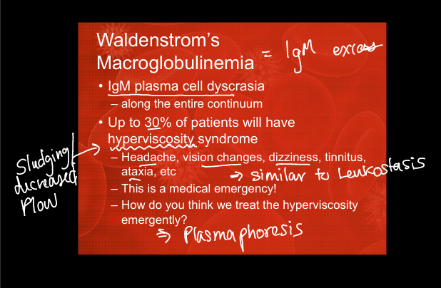
Waldenstrom’s macroglobulinemia definition
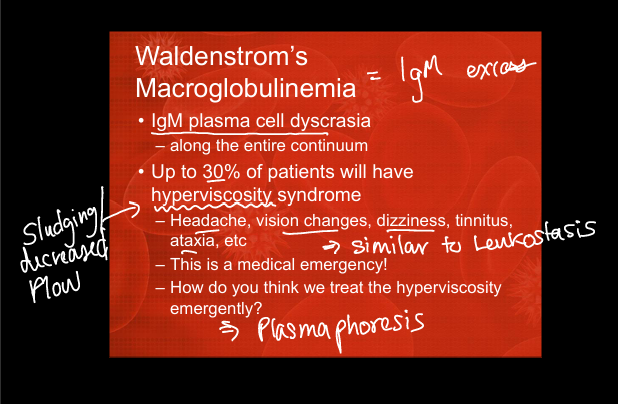
IgM‑producing plasma cell dyscrasia(IgM excess) along the same continuum as myeloma.
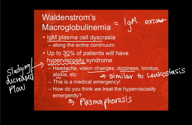
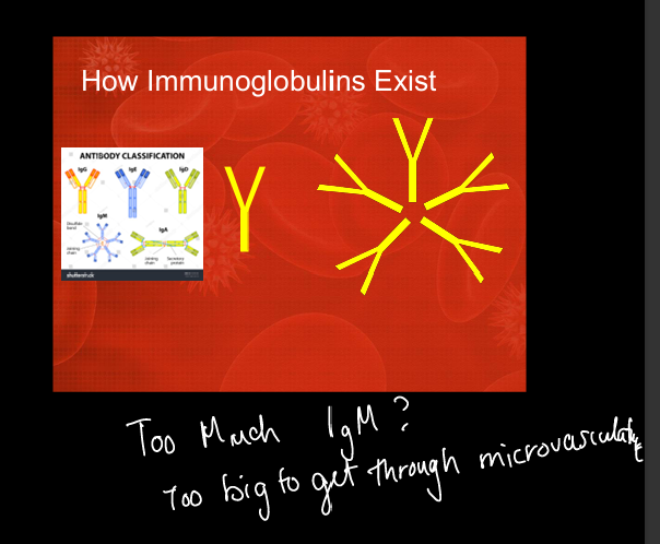
Why IgM causes hyperviscosity
IgM is a large pentamer that increases serum viscosity and impairs microvascular flow in really high volumes
Symptoms of hyperviscosity syndrome
Headache, vision changes, dizziness, tinnitus, ataxia — medical emergency. (similar concept to leukostasis)
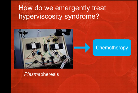
Emergent treatment for hyperviscosity
Plasmapheresis (not chemotherapy).
SPEP pattern in infection
Broad gamma region increase from polyclonal immunoglobulin response.

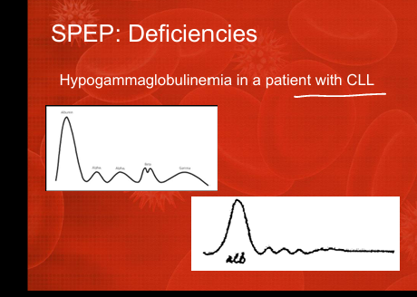
SPEP pattern in hypogammaglobulinemia
Low/flat gamma region (e.g., CLL).
SPEP pattern in multiple myeloma(monoclonal gammopathy)
Sharp monoclonal spike in gamma region (M‑spike).
CRAB mnemonic in Plasma Cell Dyscrasias
HyperCalcemia, Renal dysfunction, Anemia, Bone lesions.
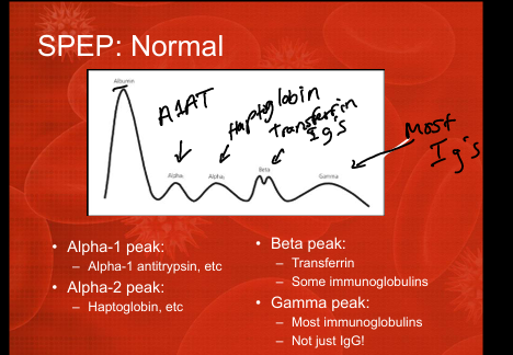
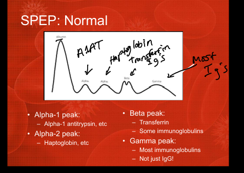
SPEP Peaks and their indications
1: Albumin peak
2: Alpha-1 peak= alpha antitrypsin
3: Alpha-2 peak: Haptoglobin
4: Beta peak: Transferrin and some immunoglobulins
5: Gamma peak: most immunoglobulins