skull, vertebral column, and thoracic cage
 Call Kai
 Learn
Learn Practice Test
 Spaced Repetition
 Match
Match Flashcards
 Knowt Play
1/131

There's no tags or description
Looks like no tags are added yet.
Name | Mastery | Learn | Test | Matching | Spaced | Call with Kai | Chat |
|---|
No analytics yet
Send a link to your students to track their progress
132 Terms
What part of the skeleton is the skull apart of?
How many individual vertebra in the vertebral column?
How many pairs of ribs are in the thoracic cage, what else is there?
The skull is apart of the axial skeleton
There are 24 individual vertebra in the vertebral column
There are 12 pairs of ribs and the sternum within the thoracic cage
Give the definition of each of these terms
Foramen
Fissure
Canal
Process
Fossa
Condyle
Facet
CN
Foramen - an opening: in the skull there are several foramina that allow for the passage of nerves and blood vessels between the inside and outside of the skull
Fissure - an opening: in the skull fissures allow for the passage of nerves and blood vessels between the inside and outside of the skull. The difference between a Foramen and a fissure - a Foramen tends to be round or oval in shape whereas fissures are more slit-like.
Canal - A tunnel or enclosed pathway: in the skull, these tunnels allow for nerves and blood vessels between the inside and outside of the skull. The difference between a Foramen and a canal os that a Foramen is simply an opening whereas a canal is a tubular tunnel-like passageway
Process - A projection or outgrowth of bone
Fossa - a depression in the bone
Condyle - a round-ish ending of a bone where it articulates with another bone at a joint
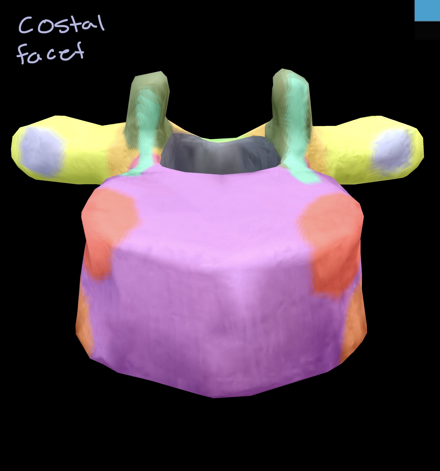
Facet - A facet is a flattened ending of a bone it articulates with another bone at a joint. Although there are severe all “facets” usually refers to the facet joints in the vertebral column
CN ‘ when you read CN this is referring to a cranial nerve when writing cranial nerves the proper way to write it is using Roman numerals such as the trigeminal nerve CN V (cranial nerves 5)
Note : where you find outgrowths of bone on the skeleton, these are often formed by the stress of a muscle or ligament pulling on the bone in this region this applies to not only the skull but the whole body. Examples include “process”, “tubercle”, “tuberosity”, “trochanter”, “epicondyle”, “spine”, and “line”
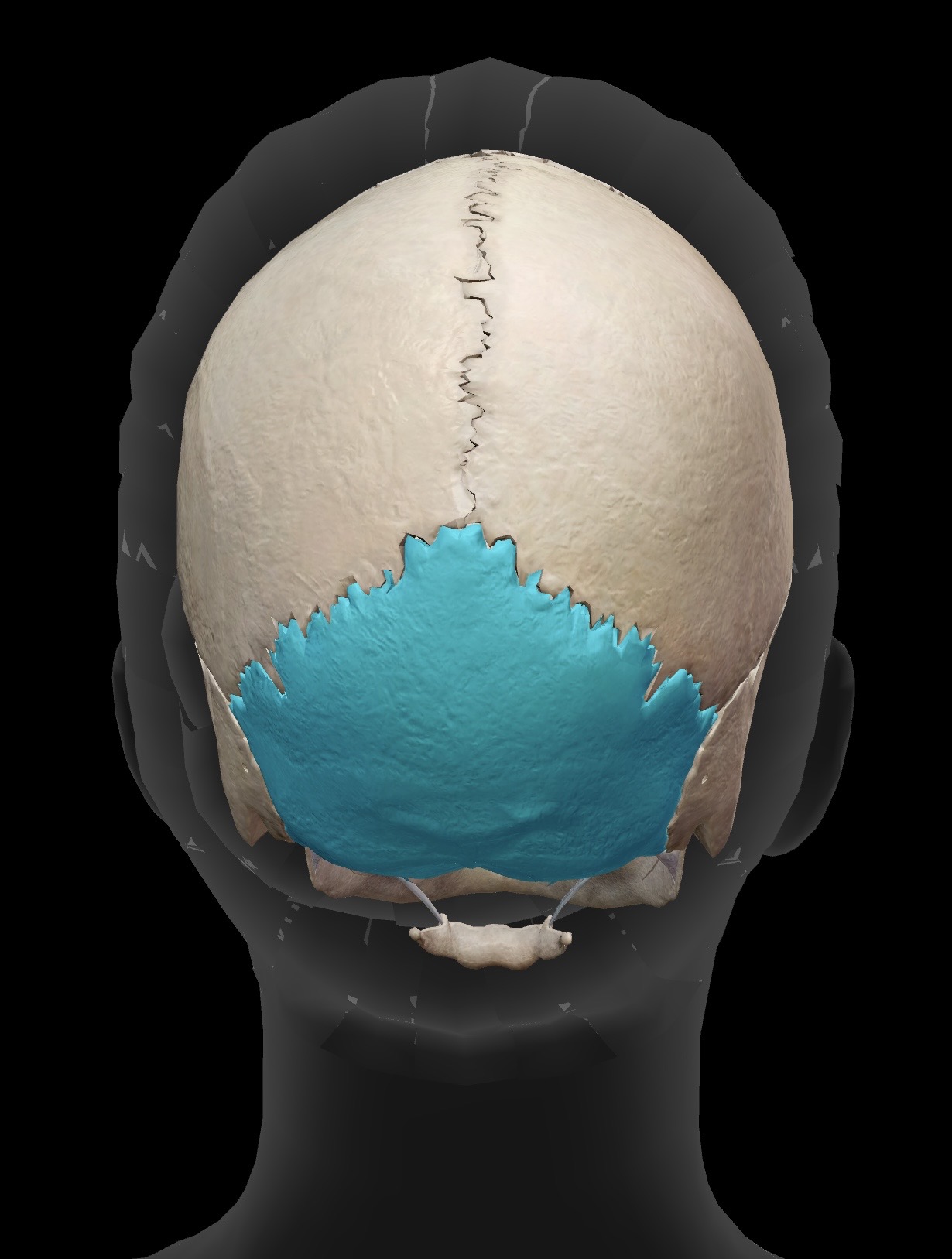
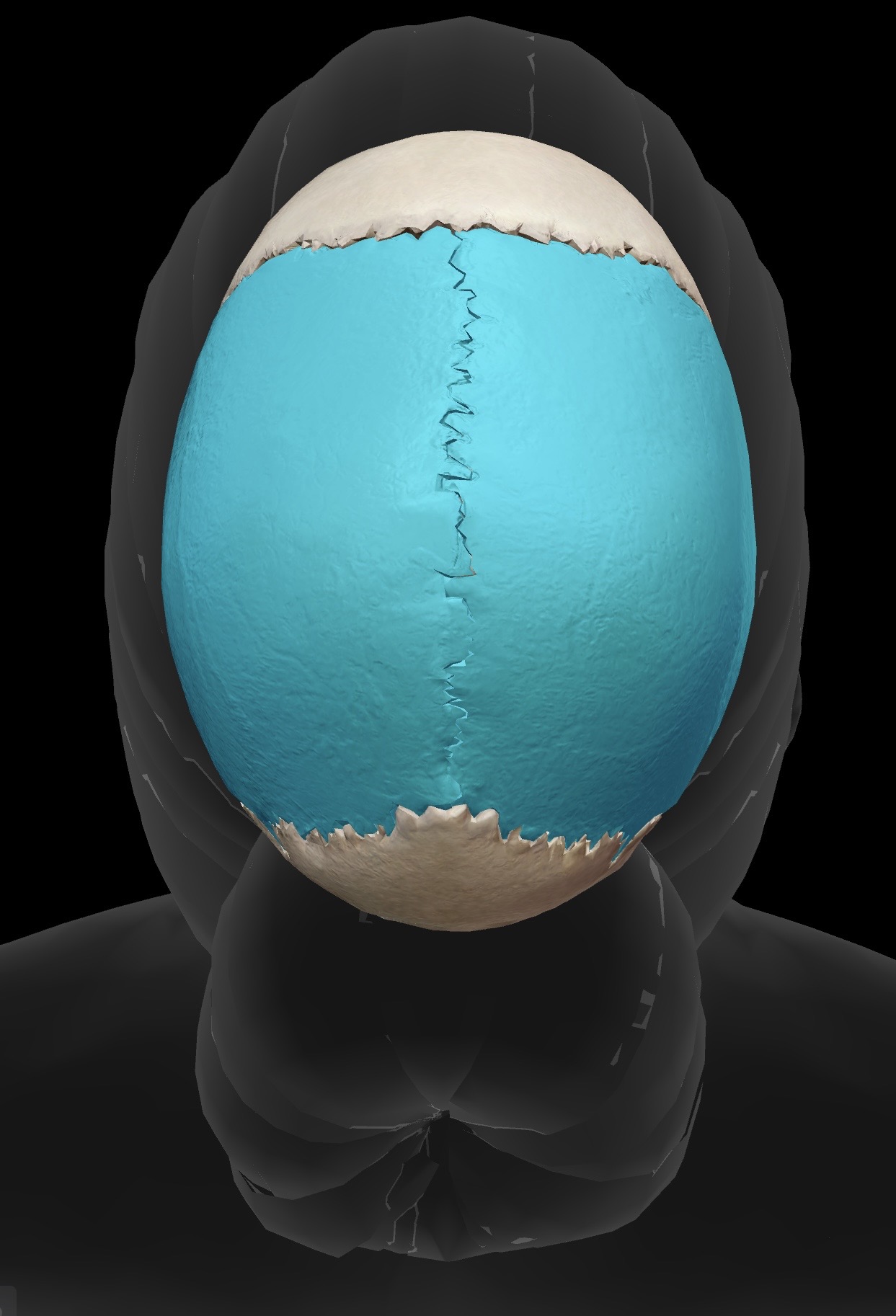
Parietal bones (2)
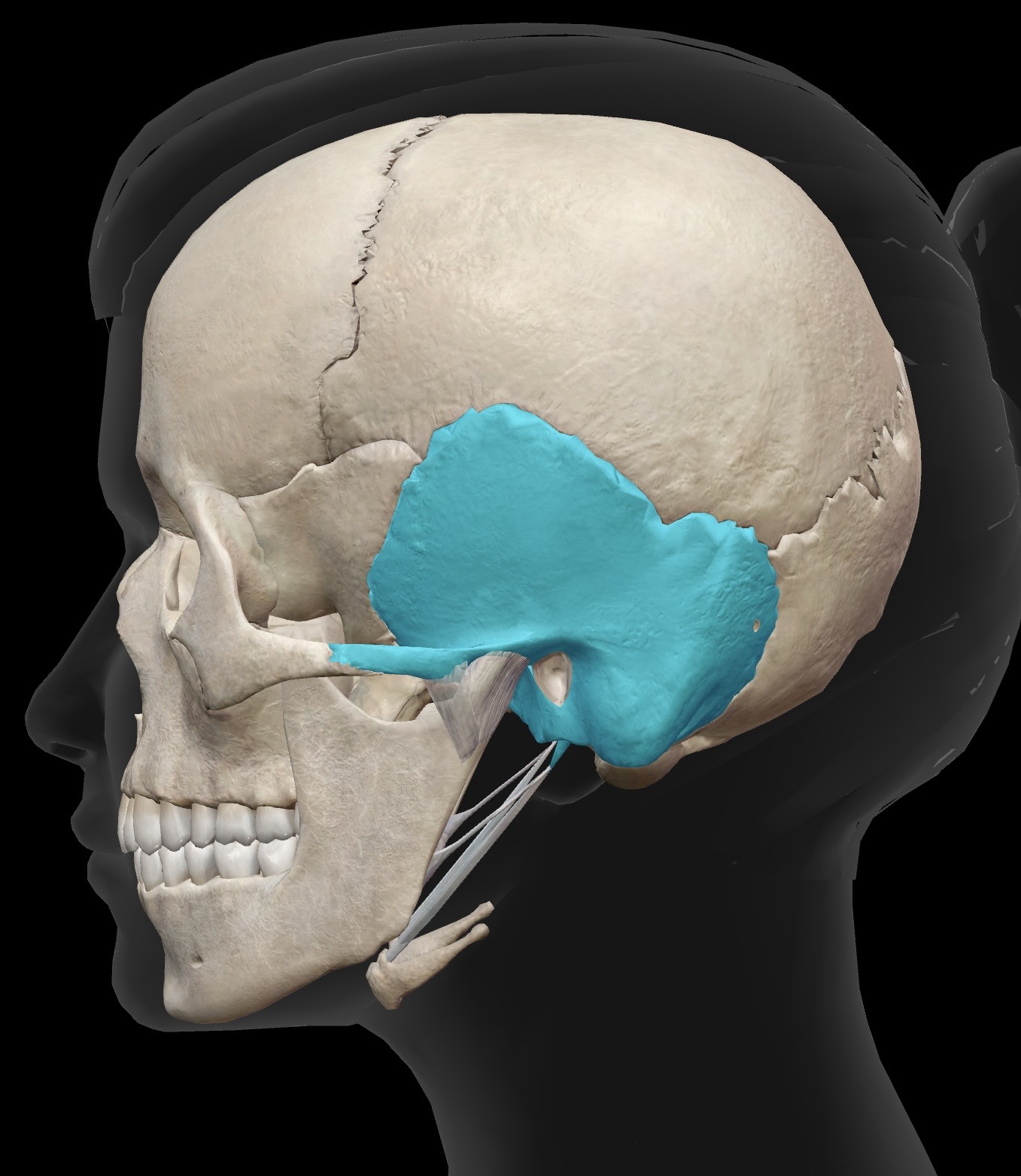
Temporal bones (2)
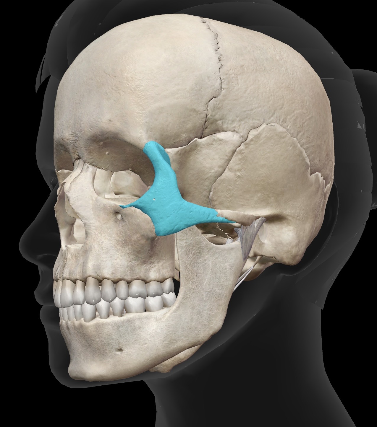
Zygomatic bone (2)
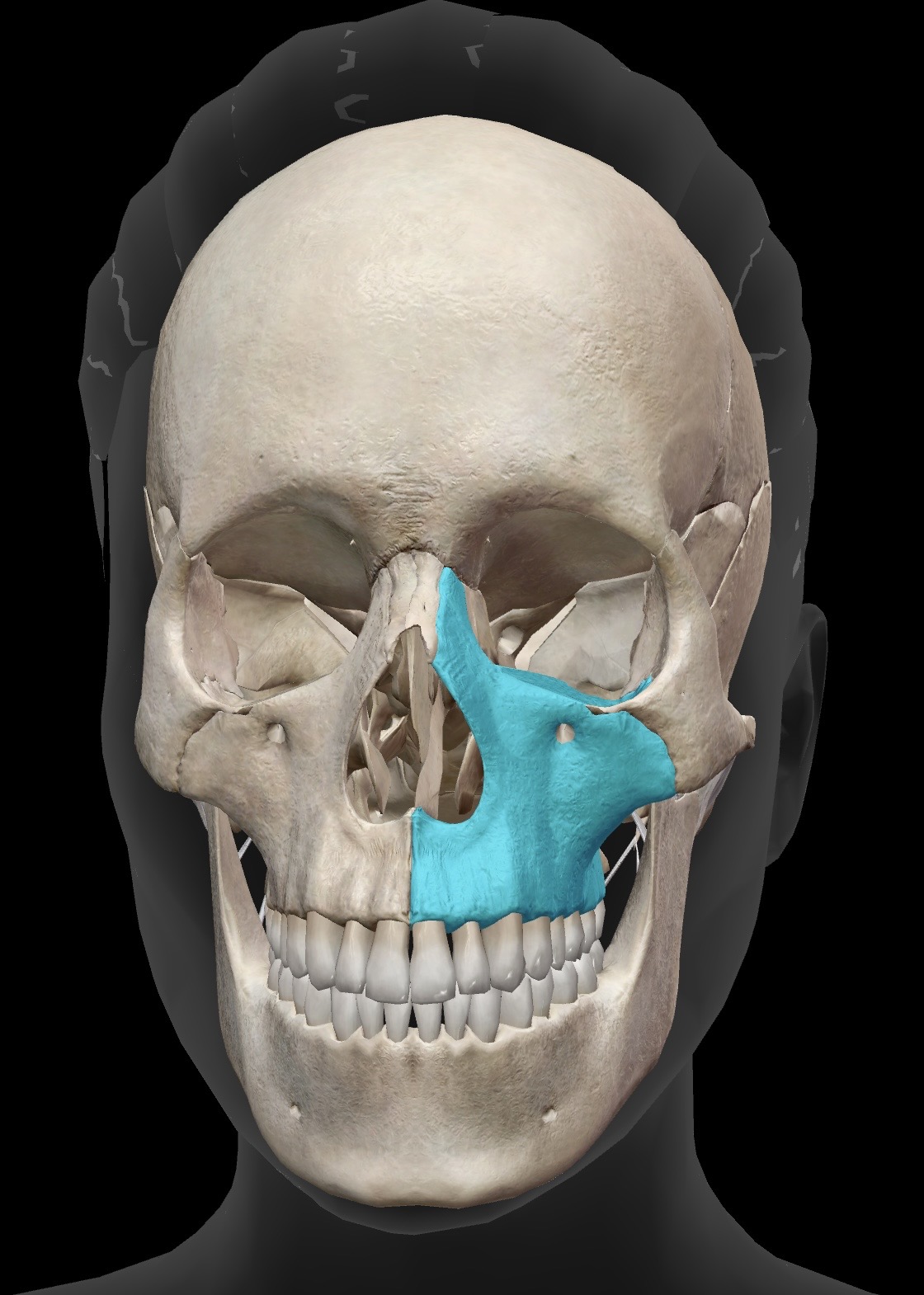
Maxilla (2)
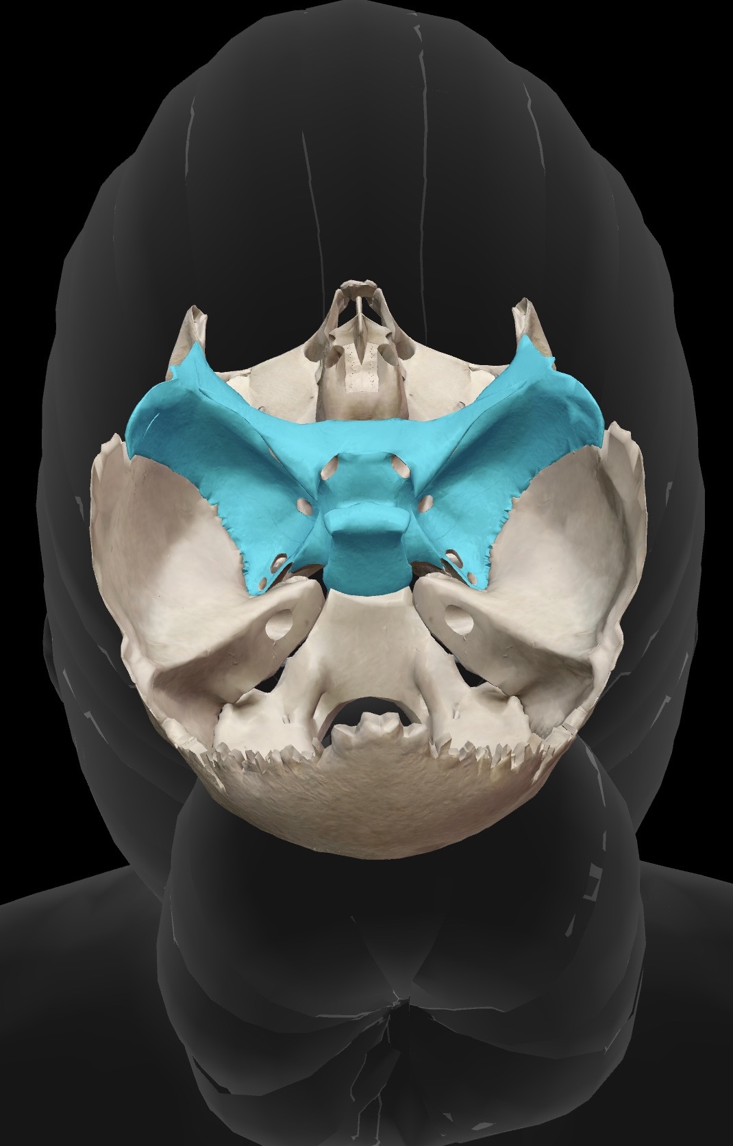
Sphenoid bone
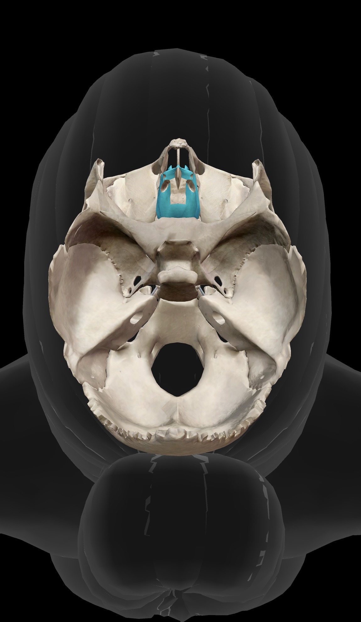
Ethmoid bone
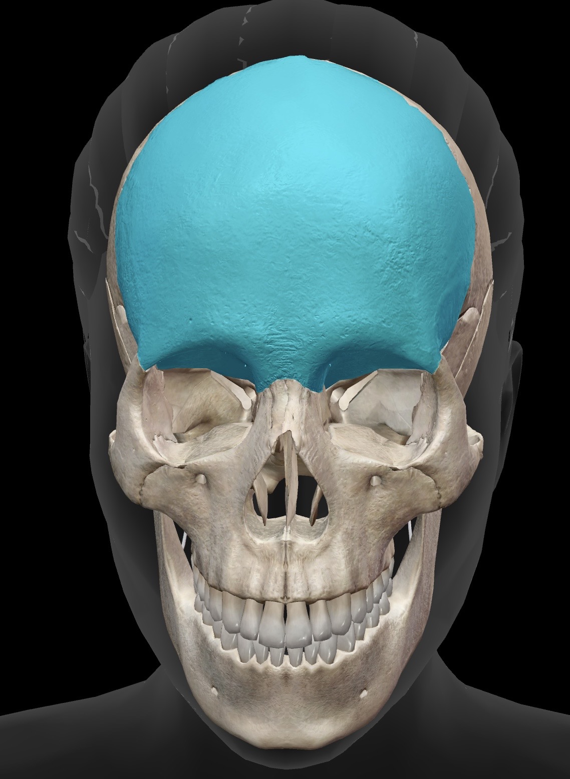
Frontal bone
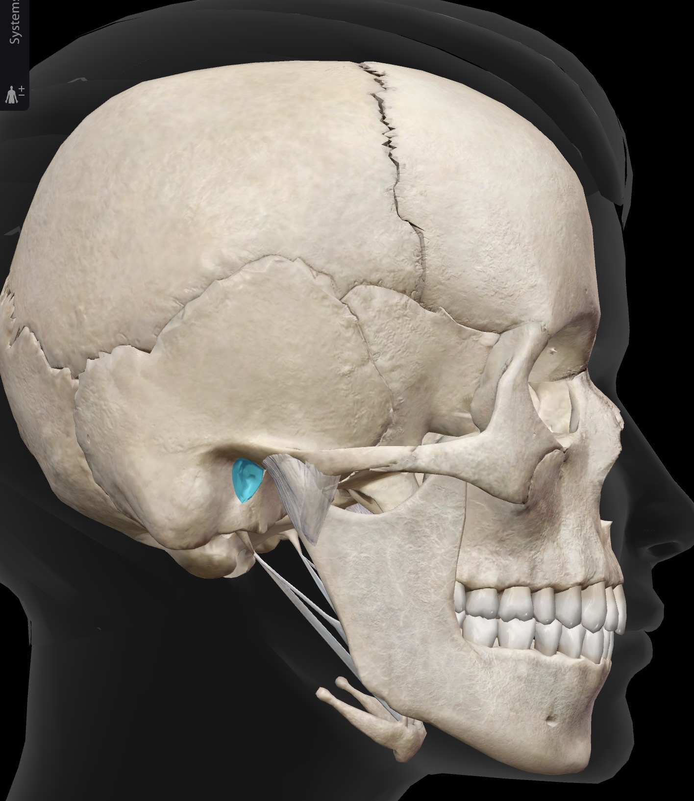
What is this and what is its function?
External auditory (acoustic) meatus - tube-like passage away from the outer ear to the ear drum
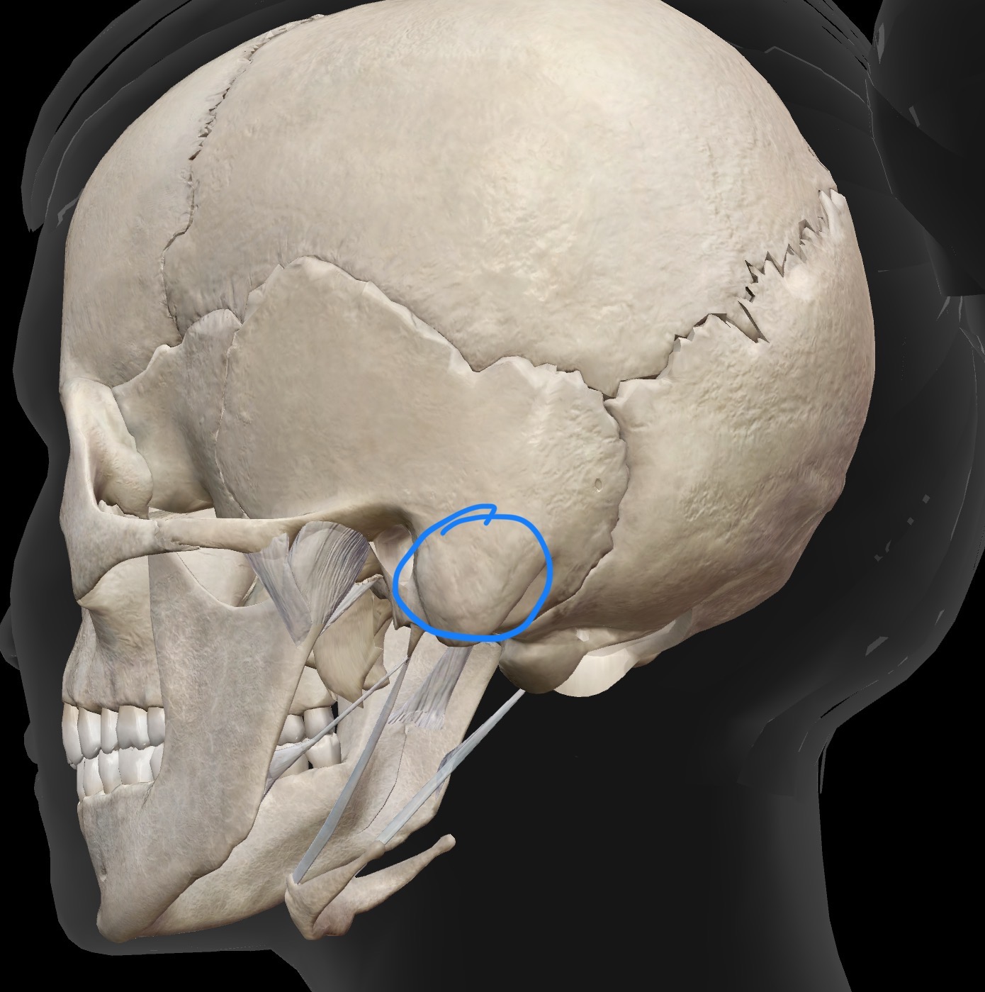
What is this and what is its function?
Mastoid process - one significance for the structure: attachment site for sternocleidomastoid
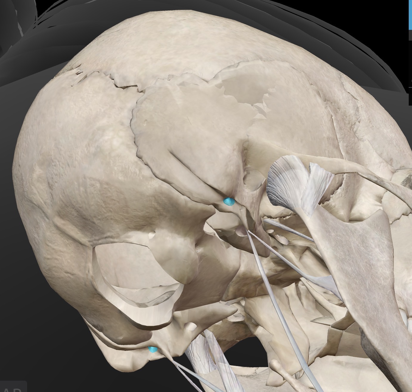
What is this and what is its function?
Stylomastoid Foramen - passageway for the facial nerve (CN VII)
Note: inflammation of the facial nerve as it passes through the stylomastoid process may result in Bell’s palsy

What is this and what is its function?
Mandibular fossa - Articular surface on the temporal bone (appears as a depression), that forms a joint with the condylar process of the mandible to form the temporomandibular joint (TMJ)
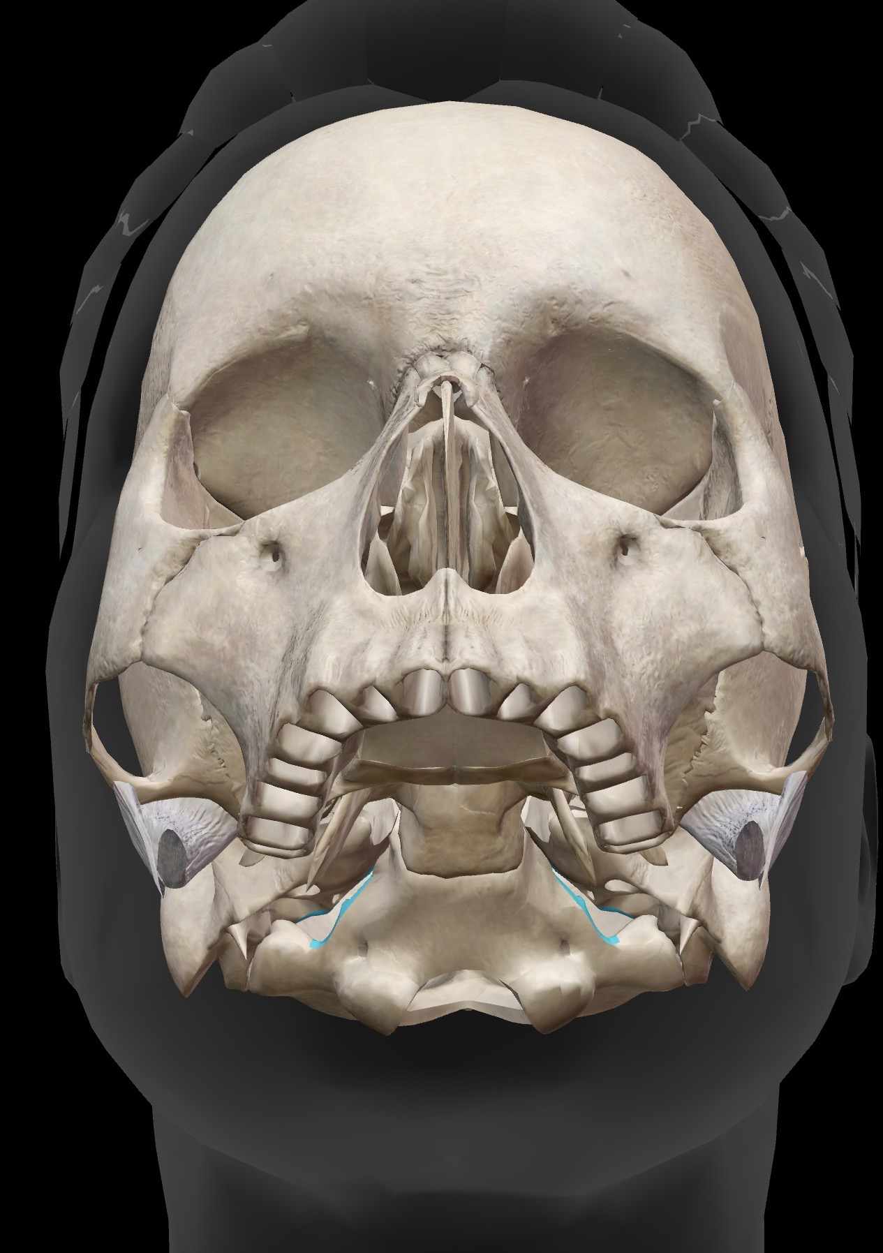
What is this and what is its function?
Jugular foreman - located posterior to the carotid canal, the jugular foreman appear as a large irregularly-shaped openings
One significance of the structure: the internal jugular vein (carrying deoxygenated blood from the brain to the heart)
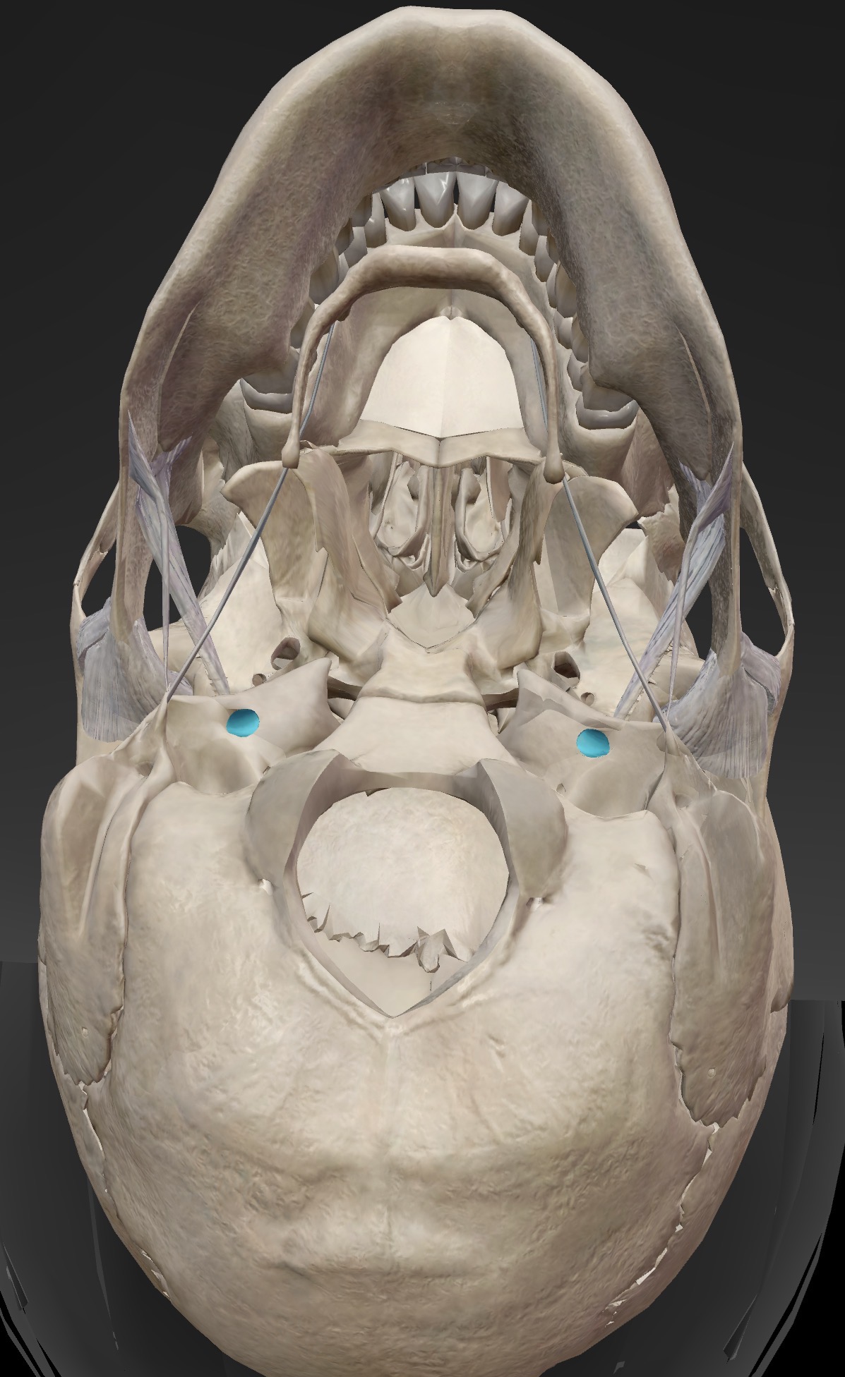
What is this and what is its function?
Carotid canal - located on the base of the skull, anterior to the jugular Foramen. The carotid canal runs vertically at first, then runs forward and medially to enter the skull.
Significance: the carotid artery travels through here

What is this and what is its function?
Glabella - region between the eyebrows.

What is this and what is its function?
External occipital protuberance - bony protuberance on the posterior aspect of the occipital bone.
Significance of the structure - serves as an attachment site of trapezius
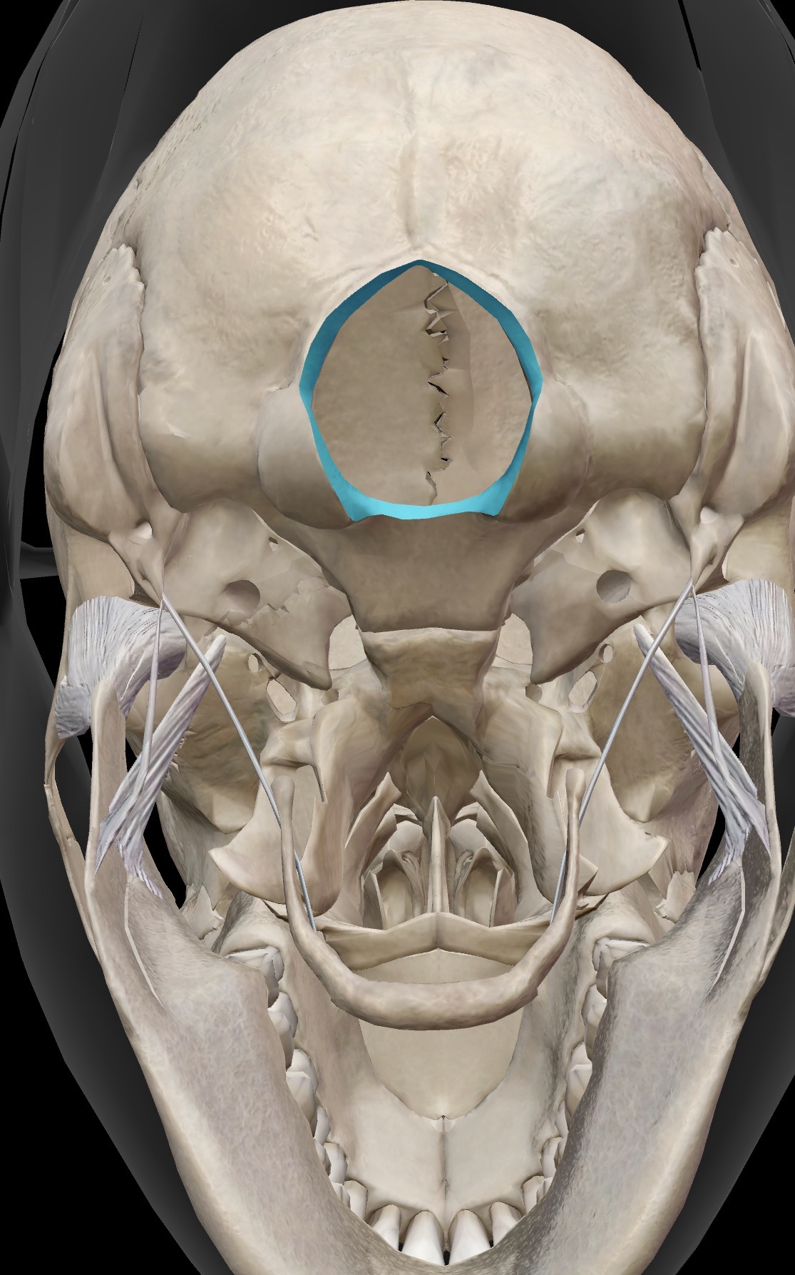
What is this and what is its function?
Foramen magnum - opening at the base of the skull
Significance- the spinal cord passes through here

What is this and what is its function?
Occipital condyles (2) - oval-shaped joint surfaces on the inferior aspect of the occipital bone, on either side of the Foramen magnum
Significance - the occipital condyles articulate with the superior articular facets of C1 (the atlas) to for, the Atlanto-occipital joint (a condyle/ellipsoid joint)

What is this and what is its function?
Greater wings of the sphenoid (2) - posterior portion of the sphenoid when looking into the skull from the top down; appear as butterfly wings

What is this and what is its function?
Lesser wings of the sphenoid - anterior portion of the sphenoid when looking into the skull from the top down.

What is this and what is its function? What is the deepest part called?
Sella turcica - a depression in the central part of the body of the sphenoid bone (sella turcica = Turkish saddle)
The deepest part of the sella turcica is called the hypophyseal fossa, where the pituitary gland sits (the pituitary gland is also called the hypophyseal gland)
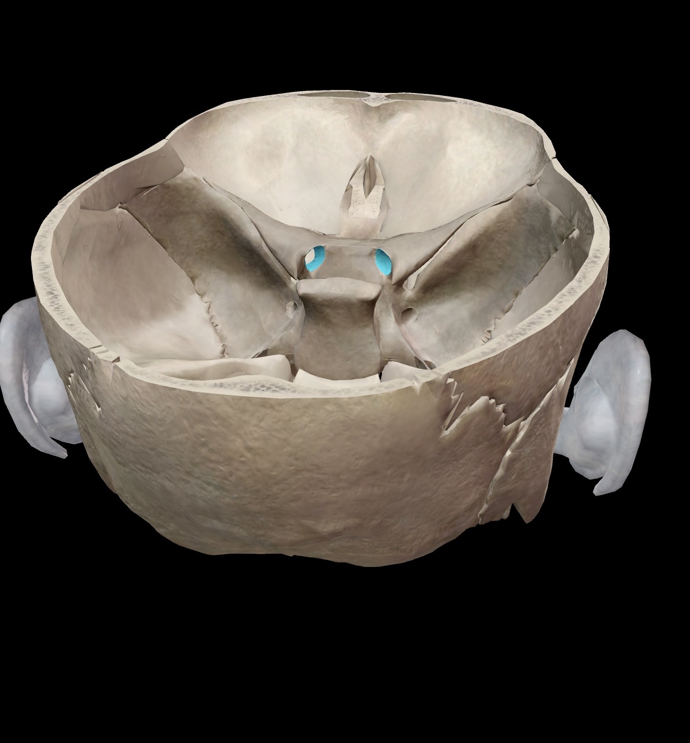
What is this and what is its function? What nerve passes through here?
Optic foremen- openings in the sphenoid bone.
Significance - optic nerves passes through here (cranial nerve 2 AKA CN ll)
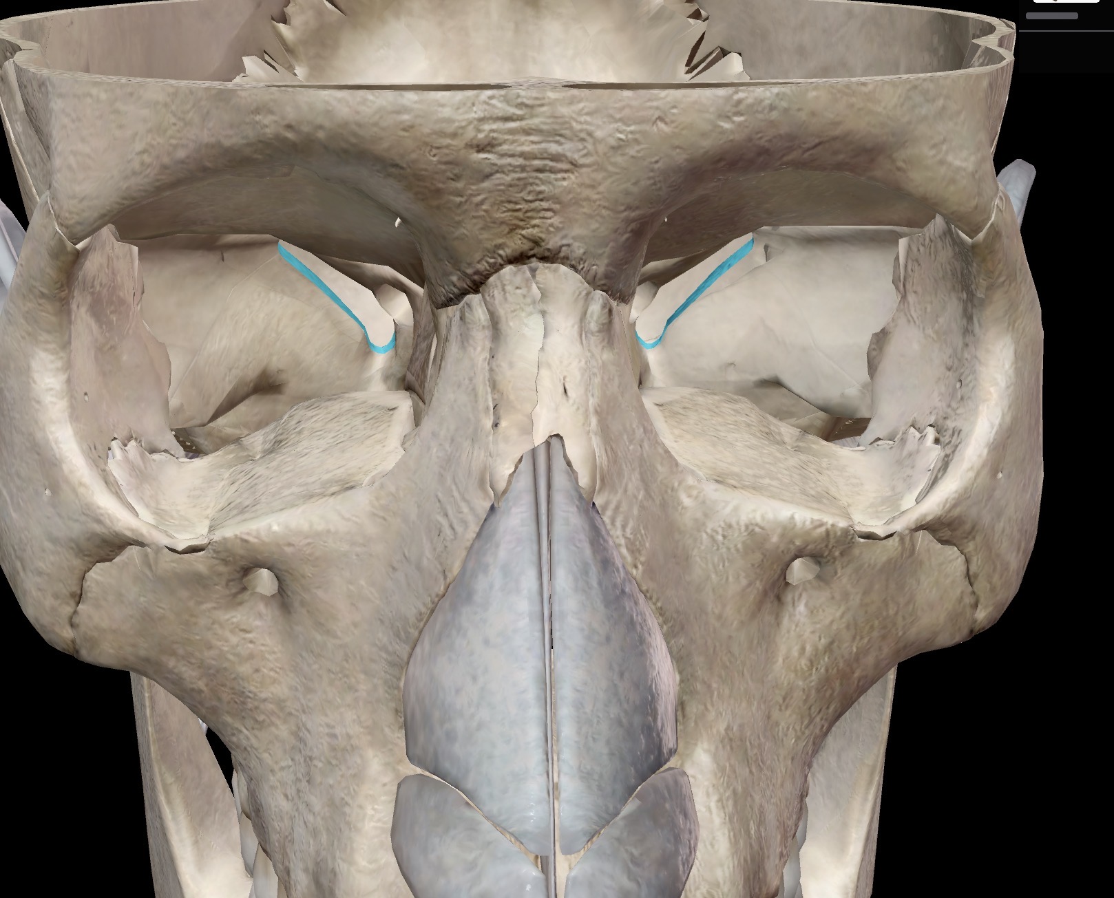
What is this and what is its function? What nerves pass through here and what is their function?
Superior orbital fissure (2) - best viewed through the orbit of the eye
Importance nerves that passes through here
Branch of CN V (trigeminal nerve) carrying sensory information about touch and temperature from the skin of the face and scalp towards the somatosensory cortex of the brain
CN lll (oculomotor nerve) carrying motor neurons as part of the pathway from the brain to 4 of 6 eye muscles for moving the eyeball (medial rectus, superior rectus, inferior and inferior oblique muscles)
CN lV (trochlear nerve), carrying motor neurons as part of the pathway from the brain 1 of 6 eye muscles for moving the eyeball
CN Vl (abducens), carrying motor neurons as part of the pathway from the brain to 1 of 6 eye muscles for moving the eyeball (superior oblique muscle)

What is this and what is its function? What nerves pass through here and what is their function?
Foramen ovale (2) - viewed as two oval shaped openings
Sensory branches of CN V (trigeminal nerve) travel through here, as part of the sensory pathway carrying sensory information about touching temperature from the skin of the face and scalp to the somatosensory (sensory) cortex of the brain
Motor branches from CN V (trigeminal nerve) travel through here, part of the motor pathway carrying motor information from the motor cortex of the brain to the muscles of mastication (chewing muscles), including the masseter and temporalis muscles
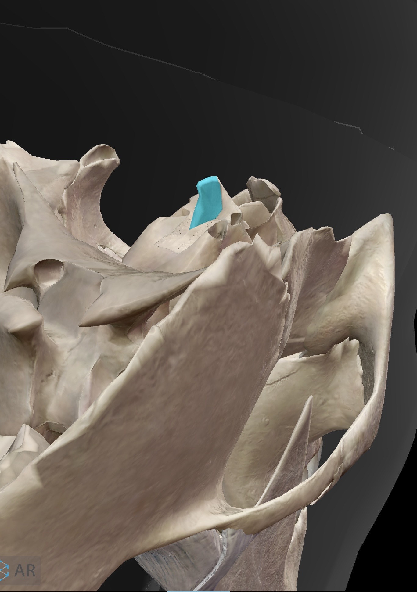
What is this and what is its function?
Crista galli - the Crista galli is a triangular projection at the superior aspect of the ethmoid bone. (it rises from the cribiform plate of the ethmoid bone)
It is the site of the flax cerebri (the falx cerebri is part of the dura mater)
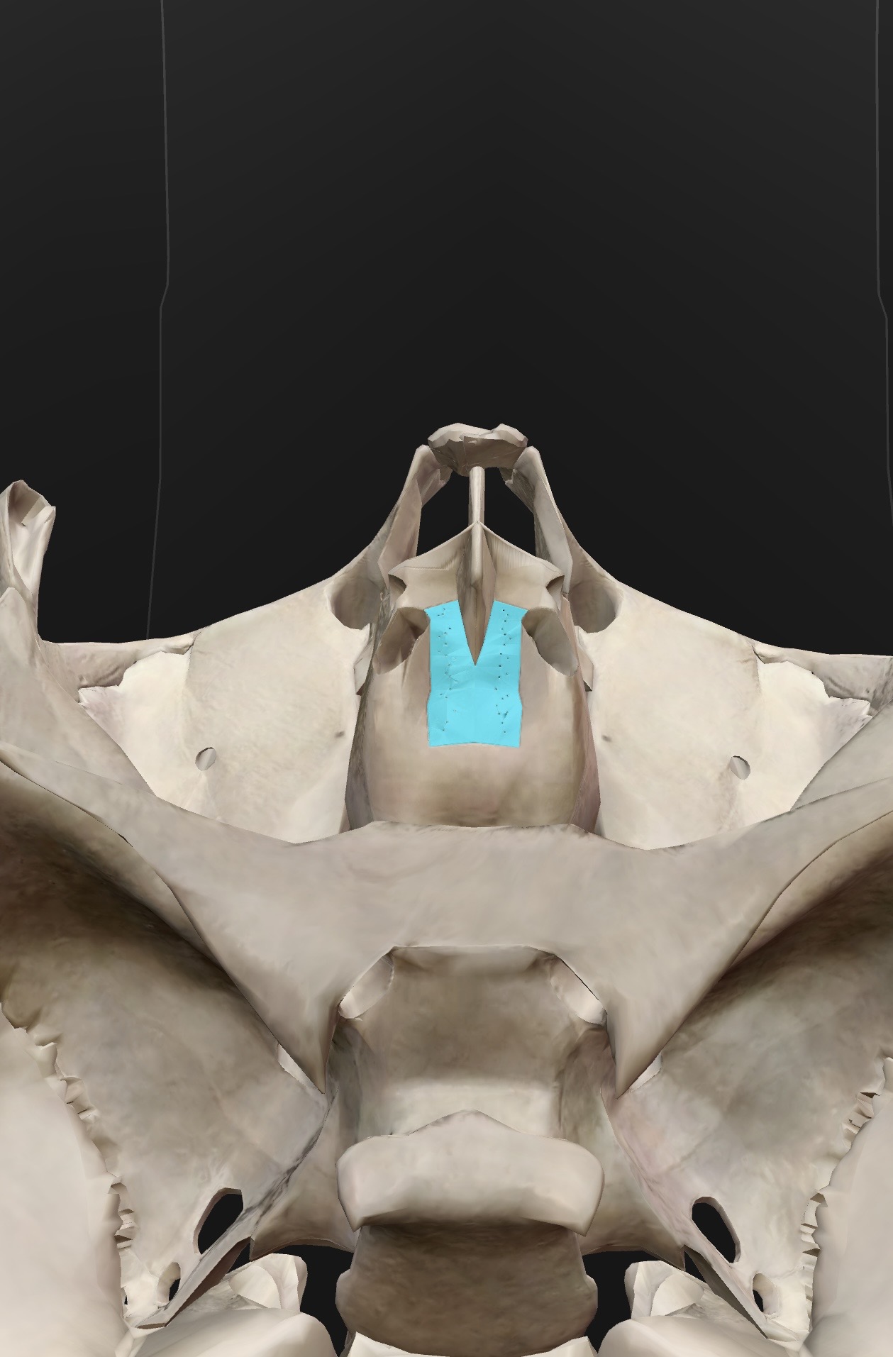
What is this and what is its function?
The cribiform plate is a perforated portion of the ethmoid bone that sits on either side of crista galli (at the superior aspect of the ethmoid bone).
The olfactory bulbs sit on top of the cribiform plates. Travelling through the perforations are neurons of the olfactory nerve (CN l) carrying sensory information about the smell from the nasal cavity toward the brain.
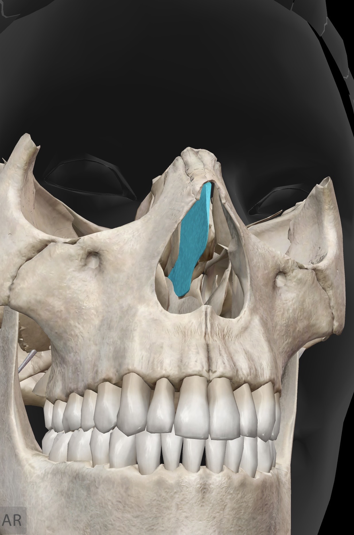
What is this and what is its function?
Perpendicular plate - the perpendicular plate is the perpendicular aspect of the ethmoid bone that can be viewed by looking into the nasal cavity. It can be seen superior to the vomer.
The vomer, the perpendicular plate and a plate of cartilage make up the nasal septum (which divides the nasal cavity into right and left portions)
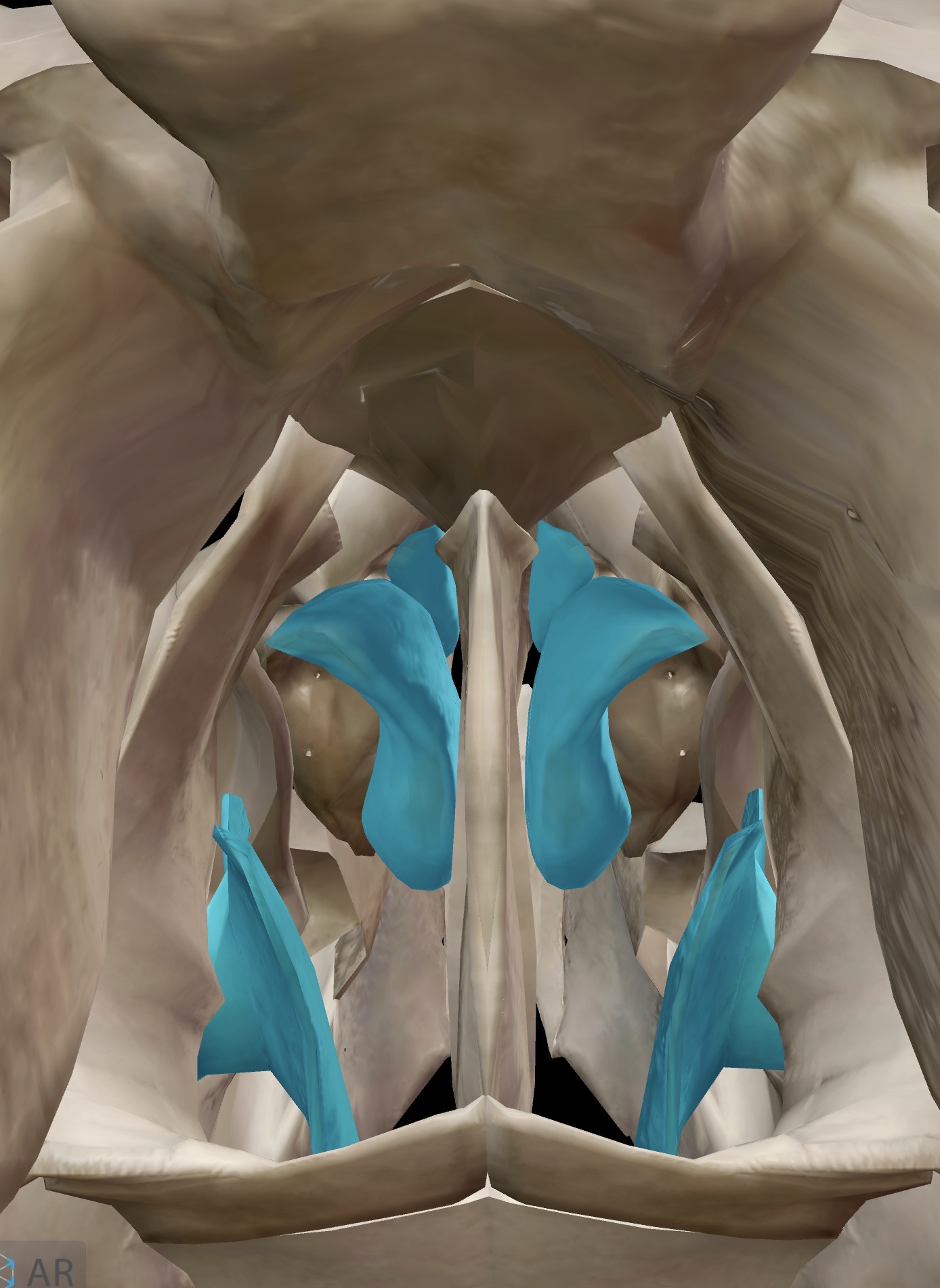
What is this and what is its function?
Superior and middle nasal conchae (turbinates, 2 of each) - appear as little shelves on the sides of the nasal cavities
Their purpose is collective: to slow down the flow of air as we breathe in through our nose. The benefit of slowing down the air is so that the nasal epithelium can do its job better: to warm, filter and humidify the air before it passes to the lungs for gas exchange.
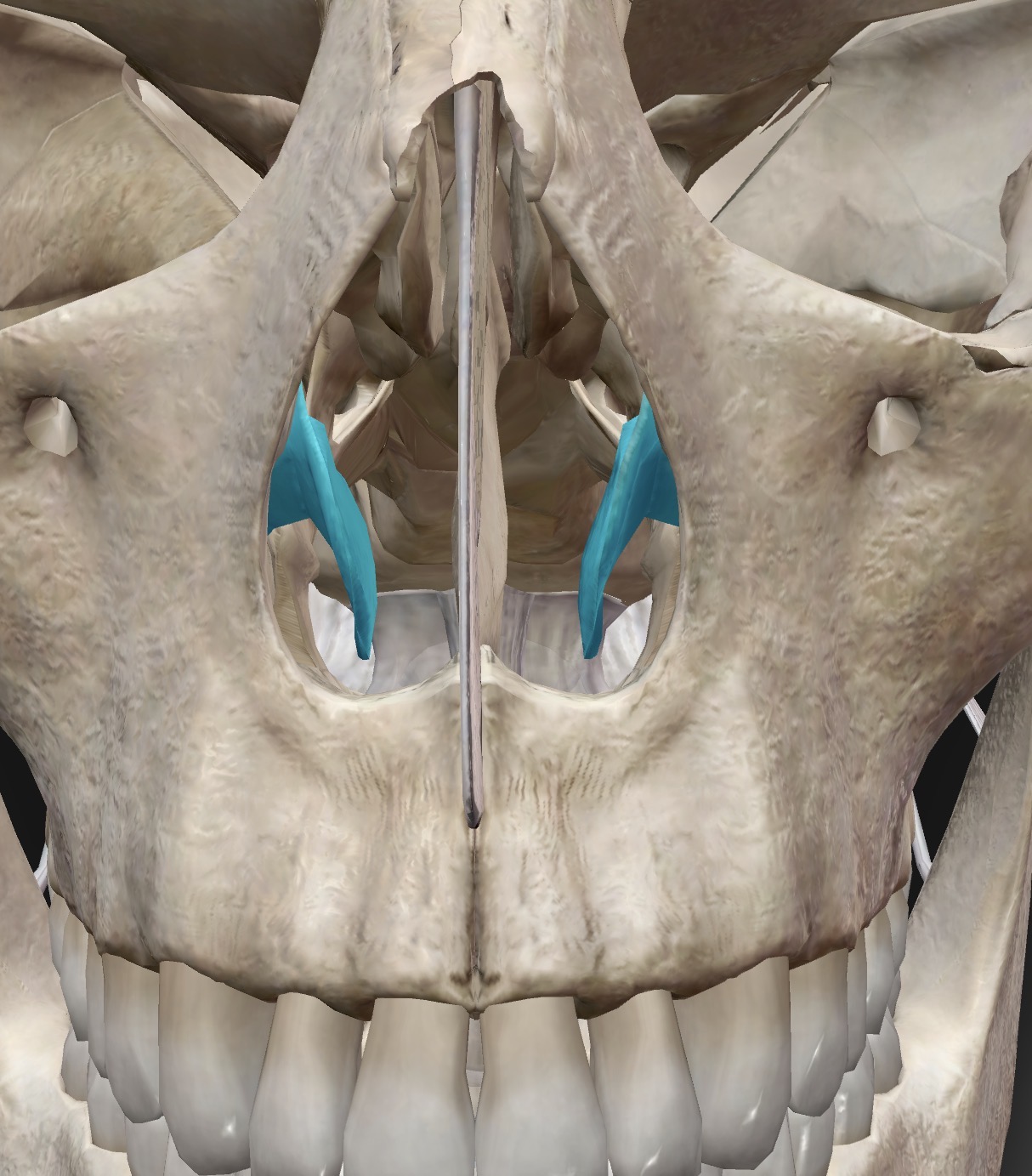
Name this bone
Inferior nasal conchae
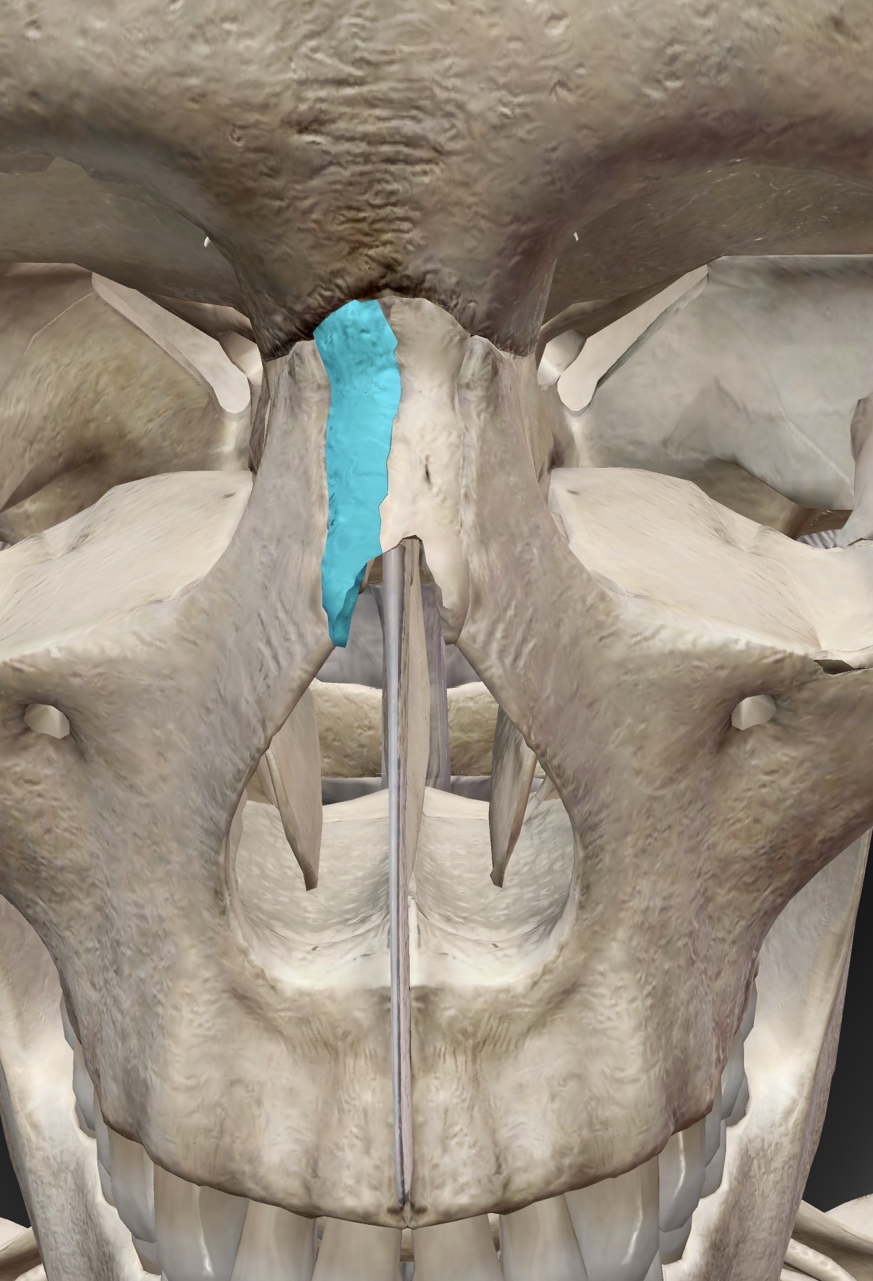
Name this bone
Nasal bones
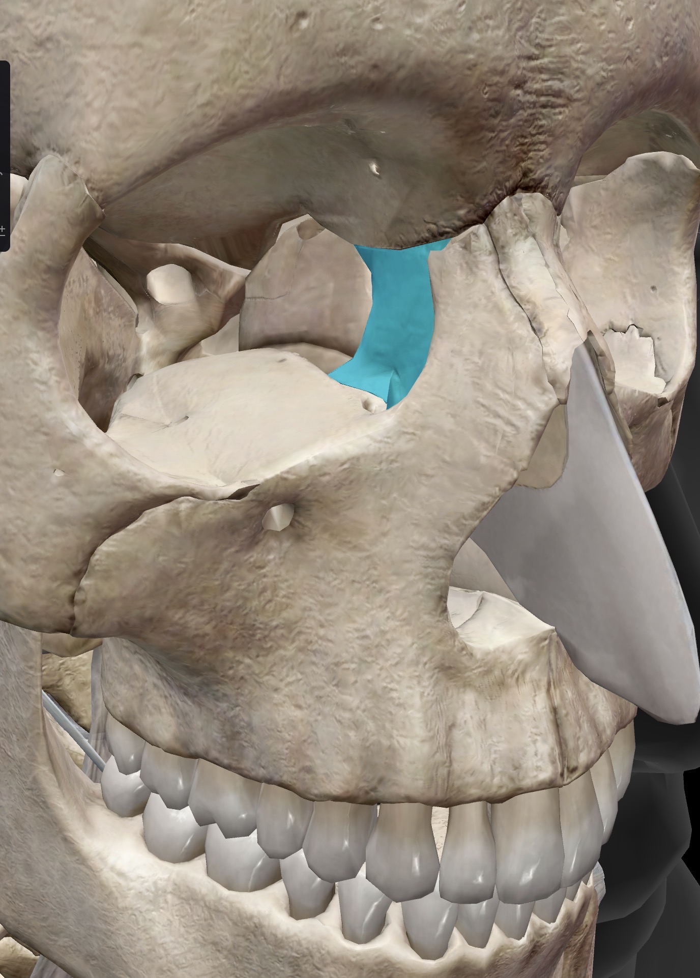
Name this bone
Lacrimal bone
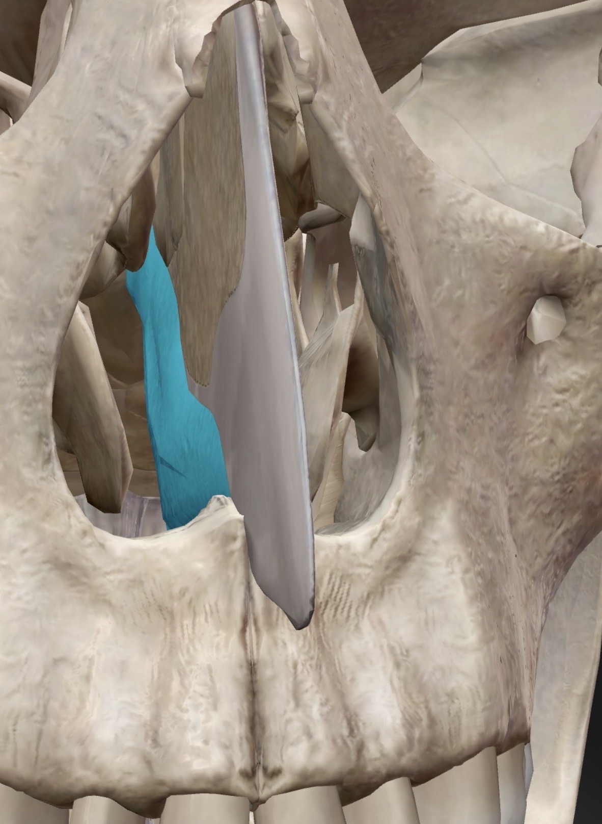
Name this bone
Vomer
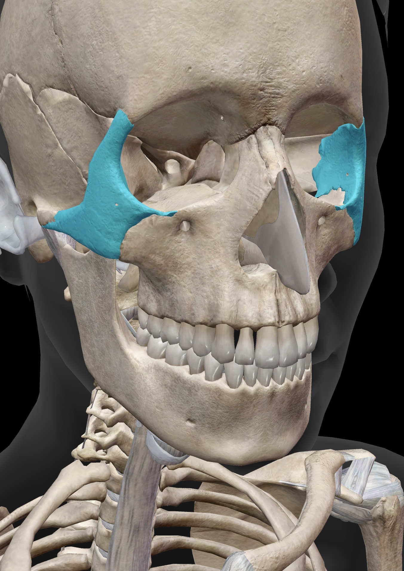
Name this bone
Zygomatic bone
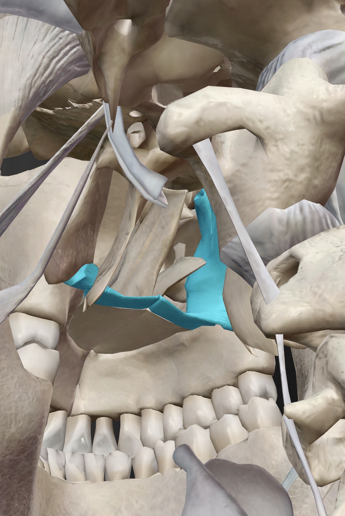
Name this bone
Palatine bones
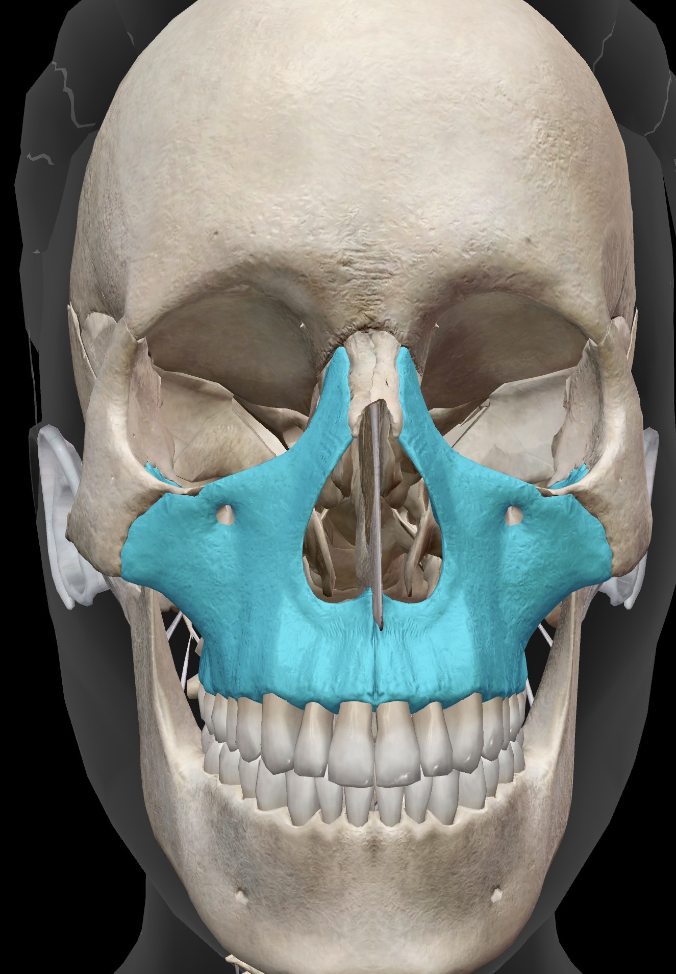
Name this bone
Maxilla
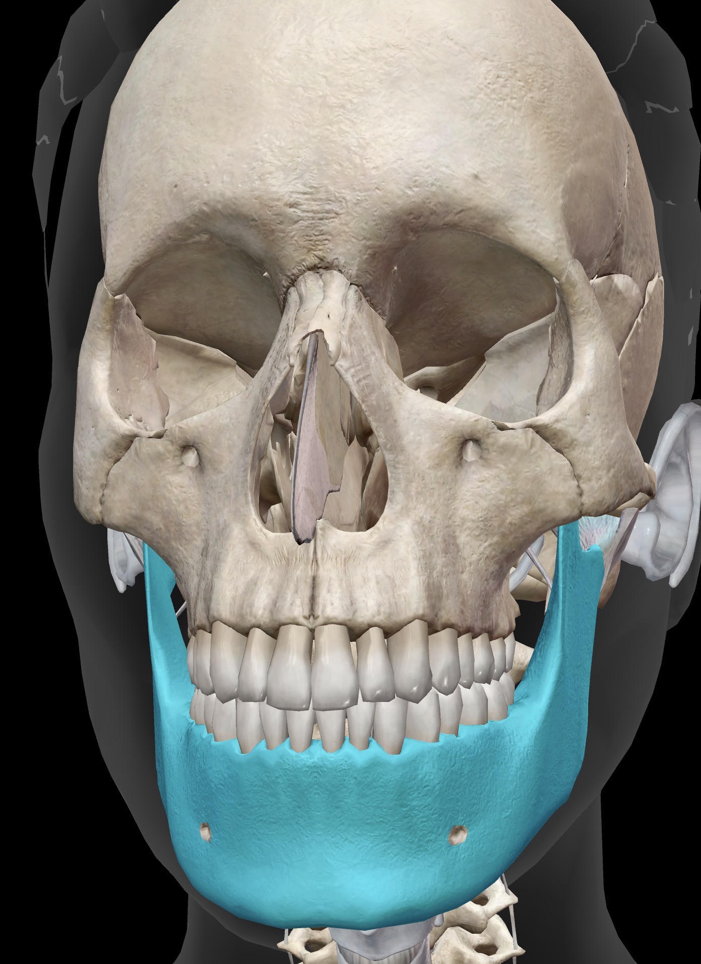
Name this bone
Mandible
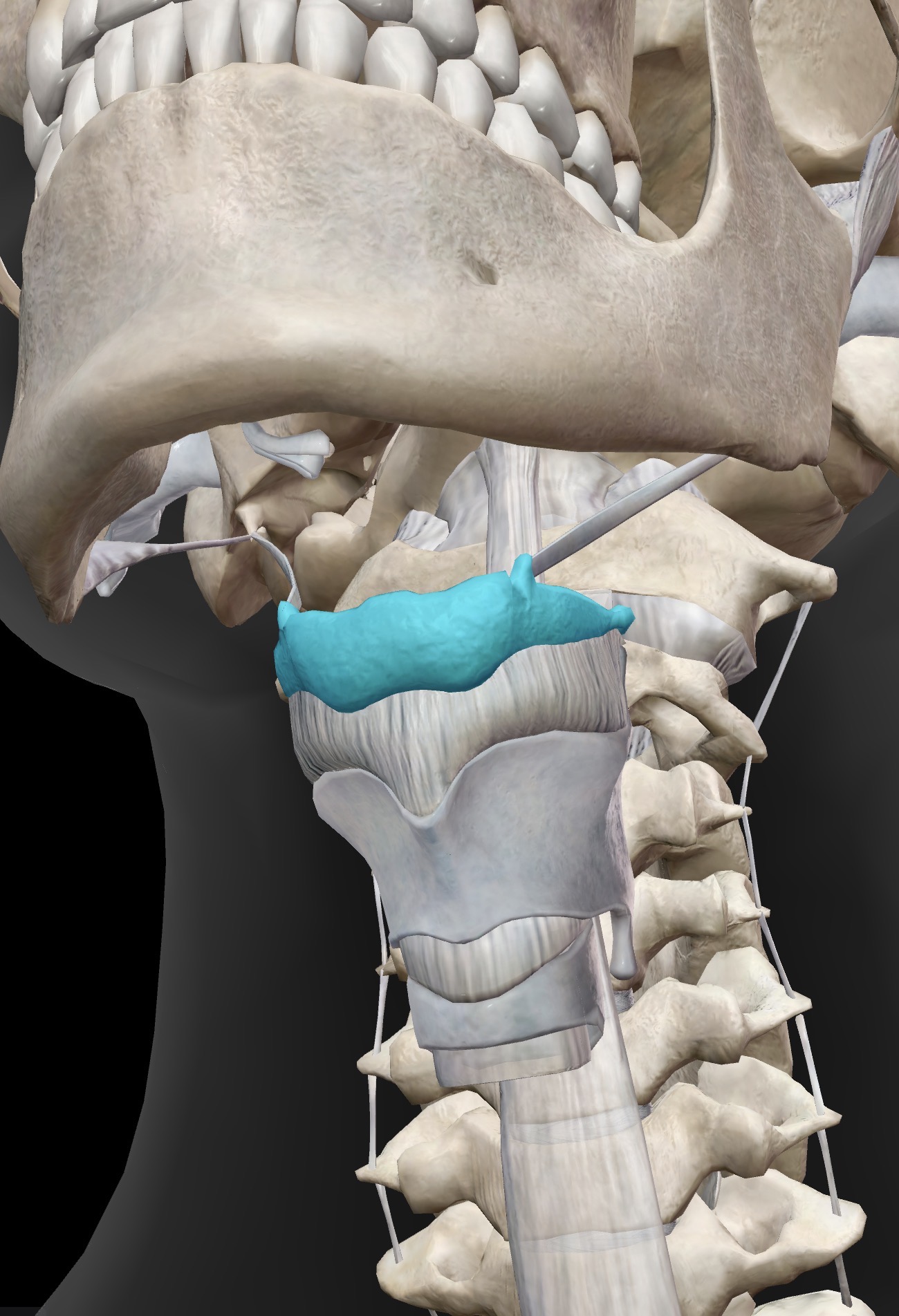
Name this bone
Hyoid

What is the name for this bone and bone landmark, what does the landmark do?
Lacrimal bone and lacrimal fossa - The lacrimal fossa is a depression in the lacrimal bone that contains the lacrimal sac which drains nasolacrimal duct or tear duct)
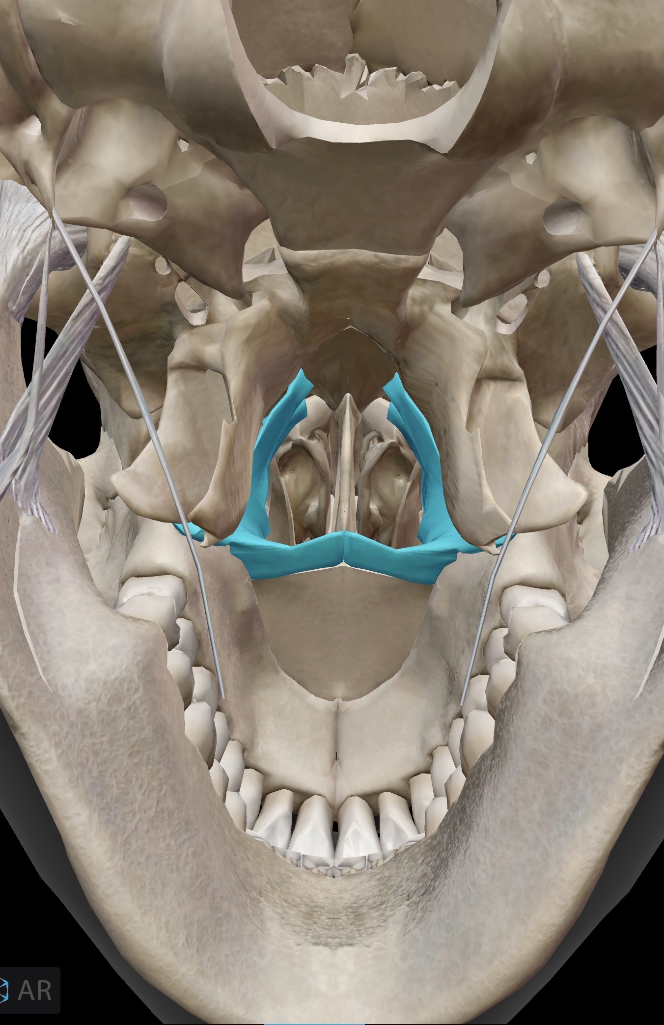
What is the name for this bone and bone landmark, what does the landmark do?
Maxilla, palatine bone - The palatine processes of the maxilla bone are the best viewed by rotating the skull and looking up at the “hard palate”
The palatine processes form the anterior portion of the hard palate, and the palatine bones form the posterior portion
Cleft palate is a congenital condition whereby the palatine processes of the maxilla bone don’t fuse properly during development in the womb

What is the name for this bone and bone landmark, what does the landmark do?
Mandible bone - Ramus (2) vertical “arms” of the mandible
Significance: where the chewing muscle masseter attaches
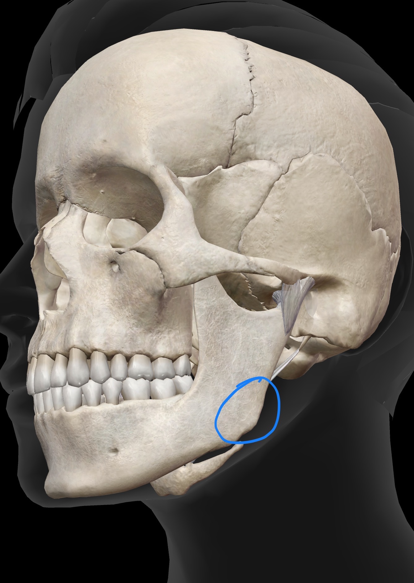
What is the name for this bone and bone landmark, what does the landmark do?
Mandible bone, Angle (2) - where the body meets the ramus, one on each side
Significance - Where the chewing muscle masseter attaches

What is the name for this bone and bone landmark, what does the landmark do?
Mandible bone Mental protuberance - Central part of the body (chin)
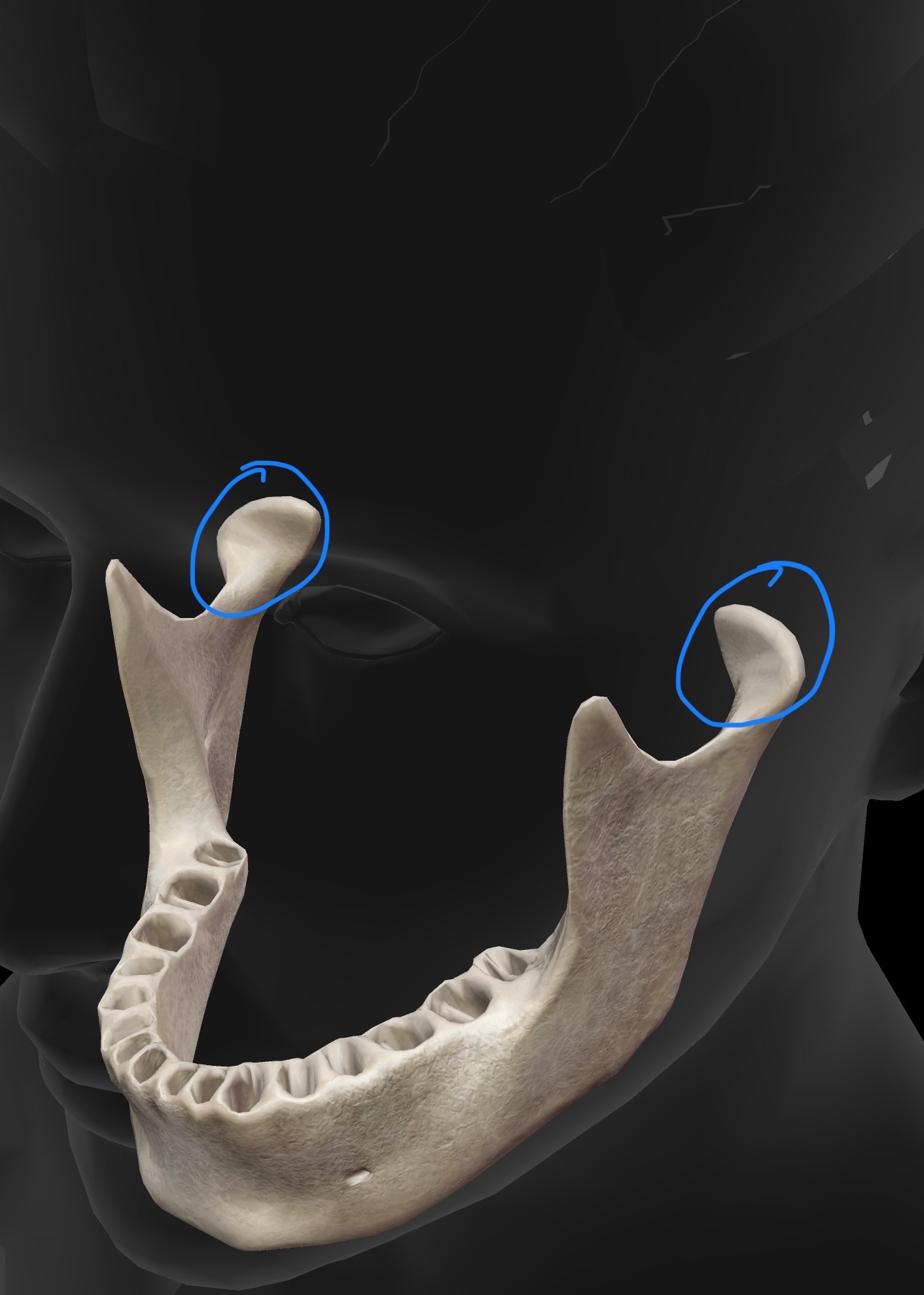
What is the name for this bone and bone landmark, what does the landmark do?
Mandible bone Condylar process - projection from the ramus where the mandible articulates with the mandibular fossa of the temporal bone to form the temporomandibular joint (TMJ)
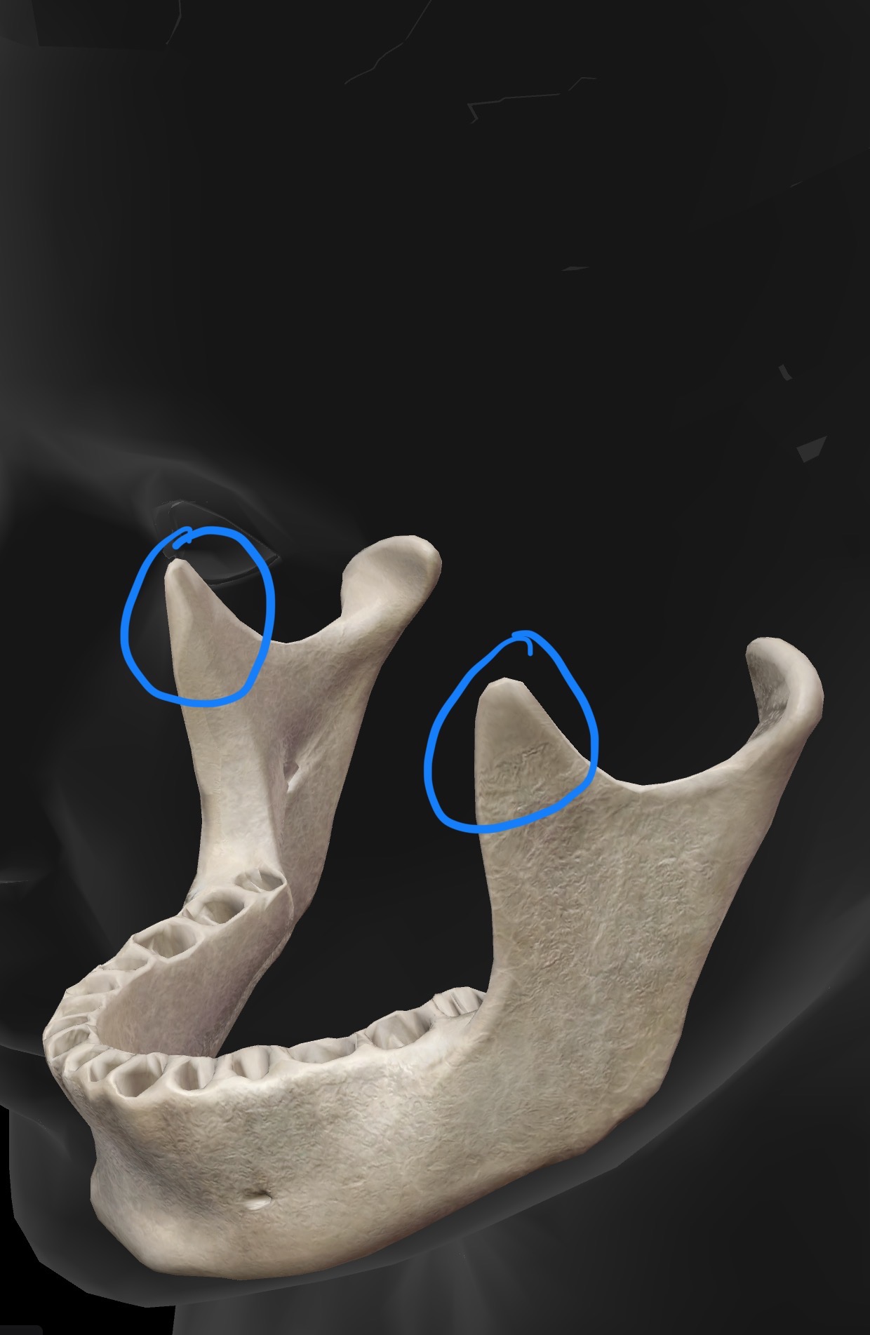
What is the name for this bone and bone landmark, what does the landmark do?
Mandible bone, coronoid processes (2) - projection from the ramus, anterior to where the mandible forms a joint with the temporal bone, that serves as a muscle attachment site (for temporalis on its medial surface and masseter on its lateral surface)
Note: There are also coronoid processes on the ulna
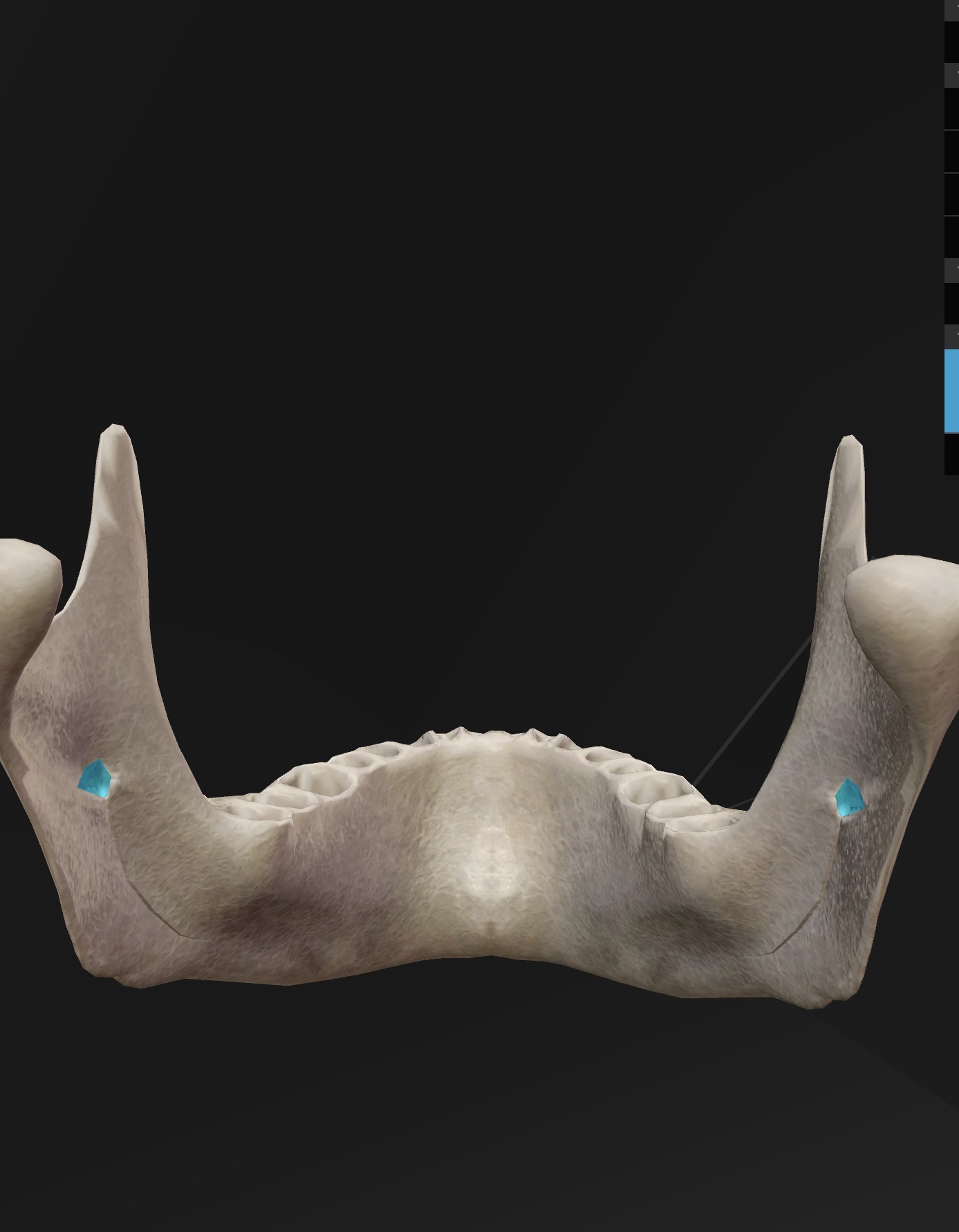
What is the name for this bone and bone landmark, what does the landmark do?
Mandibular Foramen (2) - Openings on the medial surface of the rami of the mandible that allow for passage of a branch of CN V (the trigeminal nerve) carrying sensory information about touch and temperature form the skin of the face and scalp toward the sensory (somatosensory) cortex of the brain.
In some dental procedures, anaesthetic may be administered near this opening to block the sensory branch of trigeminal nerve passing through this region
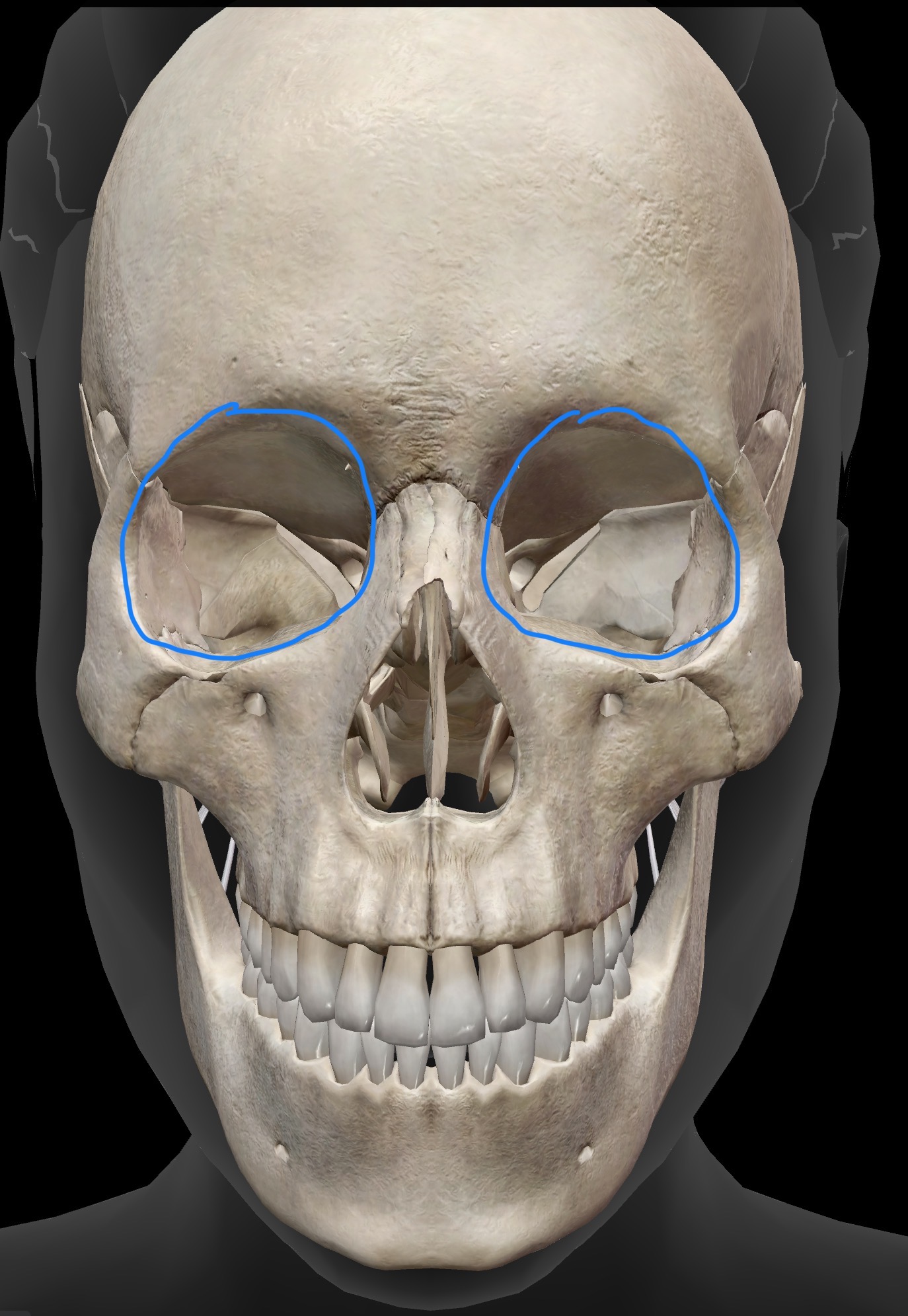
What is the name of this region what is its purpose?
The orbit of the eye (eye socket) that houses the eyeball
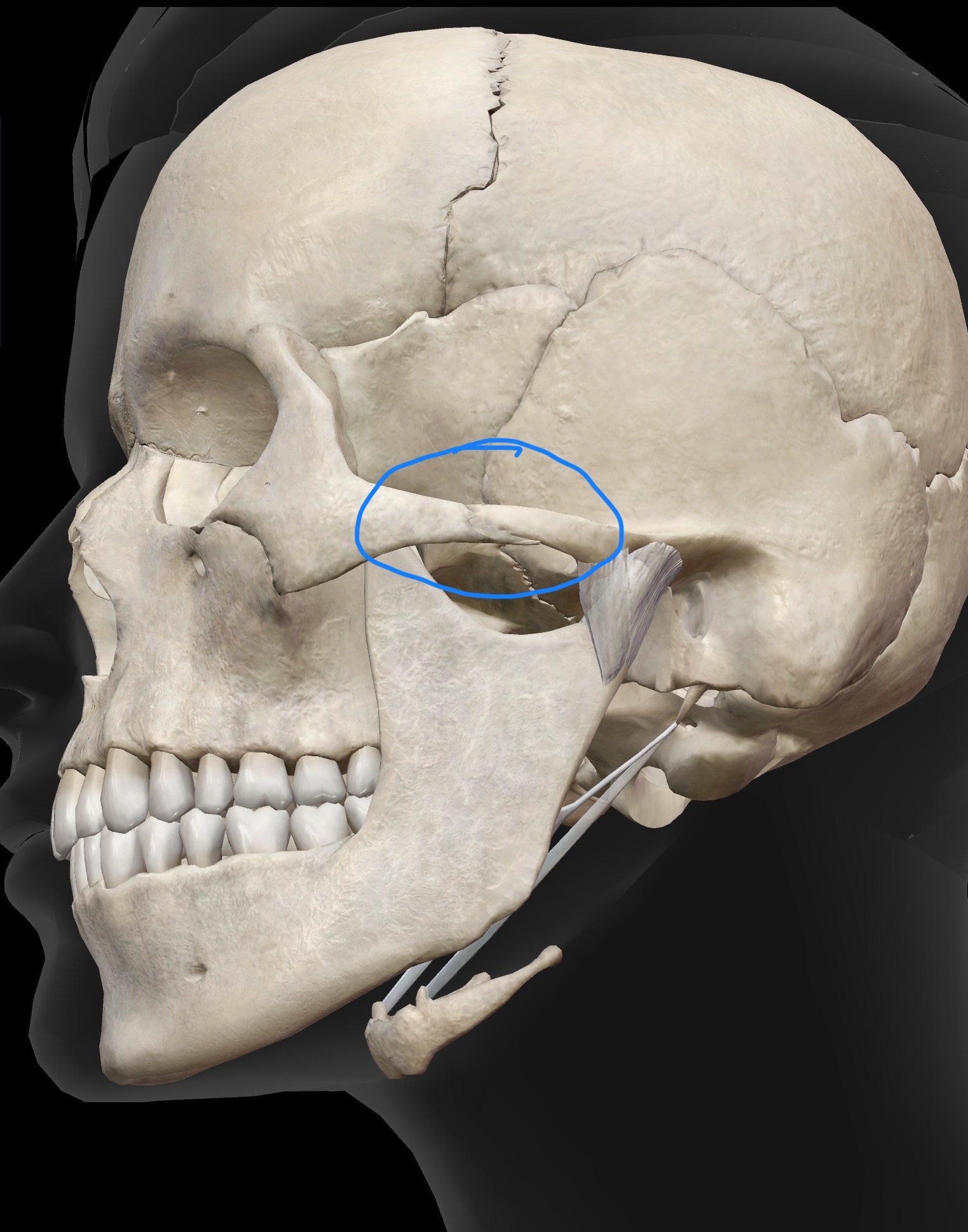
What is the name of this region what is its purpose?
Zygomatic arch - The zygomatic arch is formed partly by the temporal bone and partly by the zygomatic bone
Significance - Masseter has an attachment site along the zygomatic arch
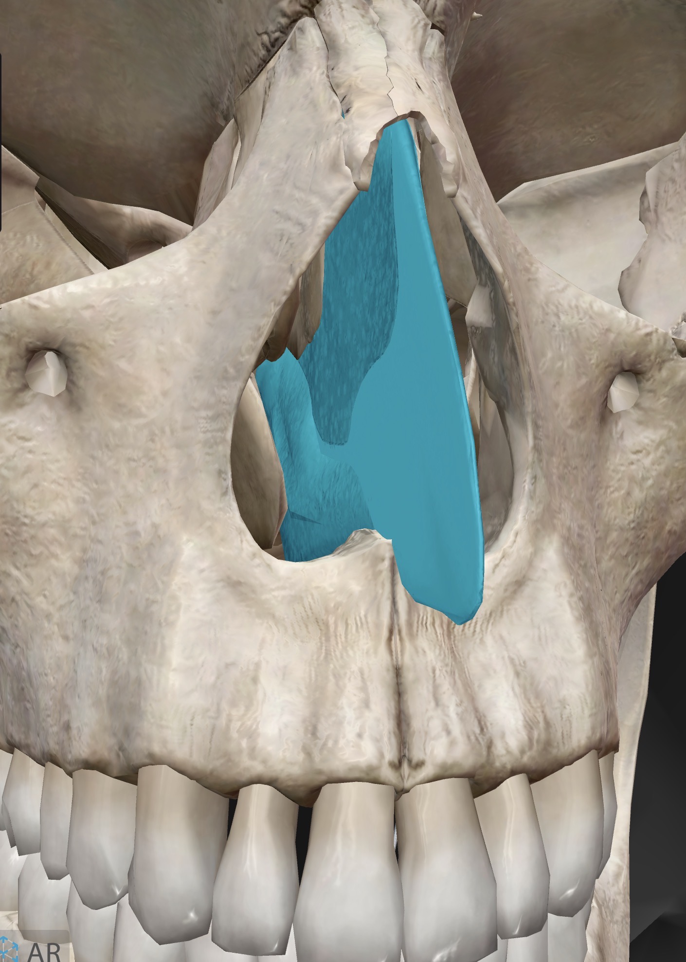
What is the name of this region what is its purpose? What is it composed of?
Nasal septum - The nasal septum is the partition within the nasal cavity that separates the nasal cavity into left and right aspects
It is composed of: The perpendicular plate (part of the ethmoid), The vomer (a facial bone), and cartilage

Name the joint, the structural type (fibrous, cartilaginous, or synovial), and the functional type (synarthotic, amphiarthrotic, or diarthrotic) . Name the bones it articulates with as well
Coronal suture
Articulates with the frontal bone, and left and right parietal bones
Structural type - Fibrous suture
Functional type - synarthrotic
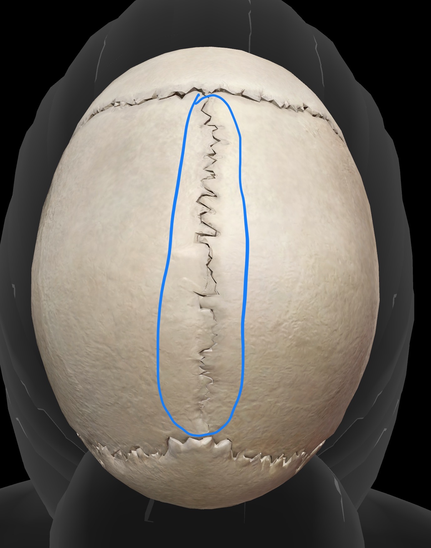
Name the joint, the structural type (fibrous, cartilaginous, or synovial), and the functional type (synarthotic, amphiarthrotic, or diarthrotic)
Sagittal suture
Articulates with left and right parietal bones
Structural type - fibrous suture
Functional type - synarthrotic

Name the joint, the structural type (fibrous, cartilaginous, or synovial), and the functional type (synarthotic, amphiarthrotic, or diarthrotic)
Lambdoidal suture
Occipital bone articulates with left and right parietal bones
Fibrous structure - fibrous suture
Functional type - Synarthrotic

Name the joint, the structural type (fibrous, cartilaginous, or synovial), and the functional type (synarthotic, amphiarthrotic, or diarthrotic)
Squamous sutures (2)
Temporal bone articulates with parietal bones Fibrous structure bilaterally
Structural type - Fibrous suture
Functional type - synarthrotic

Name the joint, the structural type (fibrous, cartilaginous, or synovial), and the functional type (synarthotic, amphiarthrotic, or diarthrotic)
Temporomandibular joint (TMJ)
Temporal bones articulates with the mandible bilaterally
Structural type - synovial condyloid (ellipsoid)
Functional type - Diarthrotic; elevation/depression, protrusion/retrusion (protraction/retraction), lateral and medial excursion

Name the joint, the structural type (fibrous, cartilaginous, or synovial), and the functional type (synarthotic, amphiarthrotic, or diarthrotic)
Gomphosis (several)
Mandible with teeth and maxilla with teeth
Structural type - fibrous
Functional type - synarthrotic

Name the joint, the structural type (fibrous, cartilaginous, or synovial), and the functional type (synarthotic, amphiarthrotic, or diarthrotic)
Atlanto-occipital joint
Occipital condyles of skull with with superior articular facets of C1 (the atlas)
Sturctural type - synovial condyloid (ellipsoid)
Functional type - diarthrotic: flexion/extension, right and left side flexion
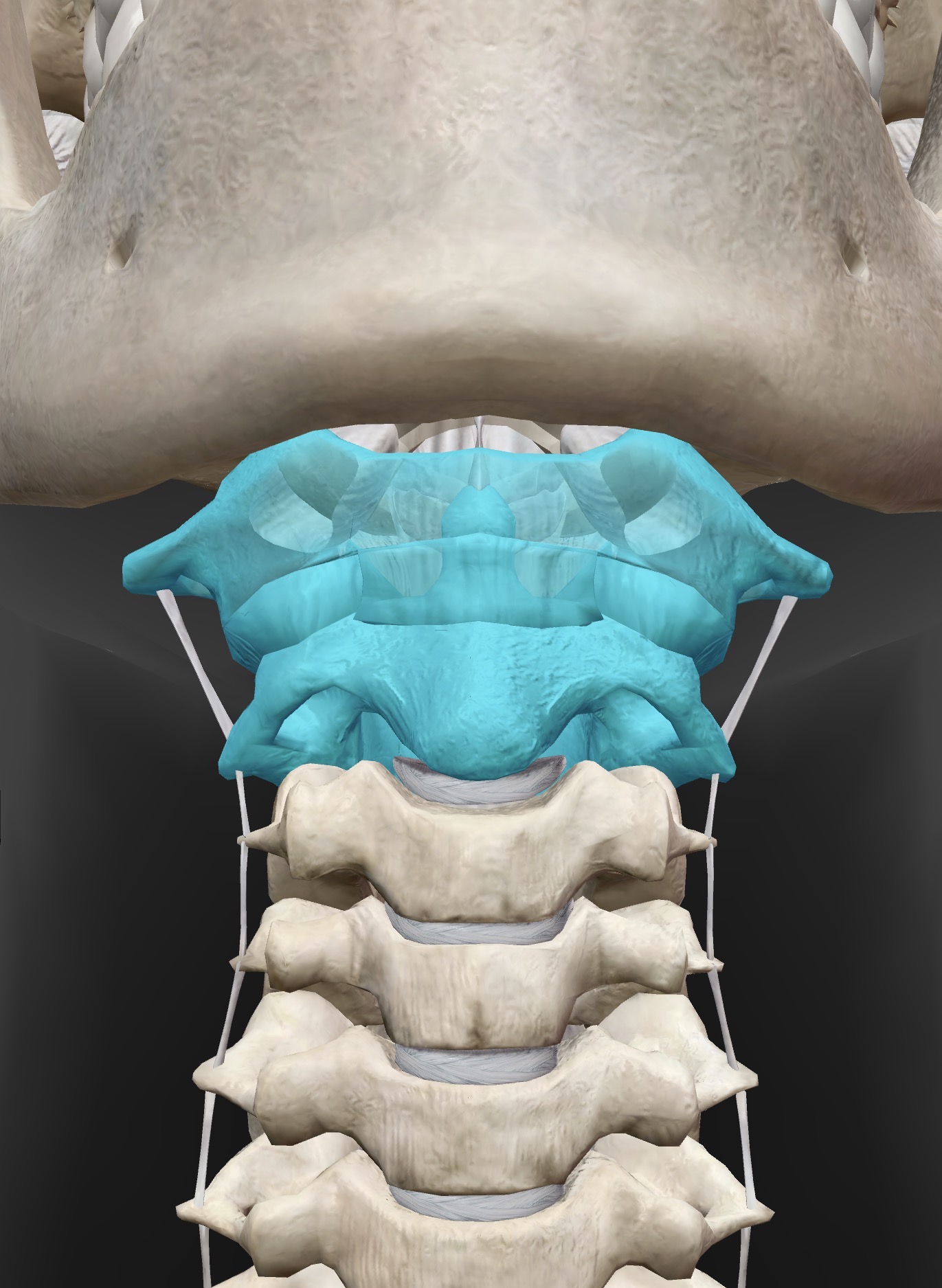
Name the joint, the structural type (fibrous, cartilaginous, or synovial), and the functional type (synarthotic, amphiarthrotic, or diarthrotic)
Atlantoaxial
C1 (atlas) & dens (odontoid process) of C2 (axis)
Structural type - synovial pivot
Functional type - diarthrotic: allows left and right rotation

Name the joint, the structural type (fibrous, cartilaginous, or synovial), and the functional type (synarthotic, amphiarthrotic, or diarthrotic)
Intervertebral facet joints of the cervical spine (several)
Articulations between inferior and superior articular facets of adjacent vertebrae
Structural type - synovial plane
Functional type - diarthrotic: gliding movements
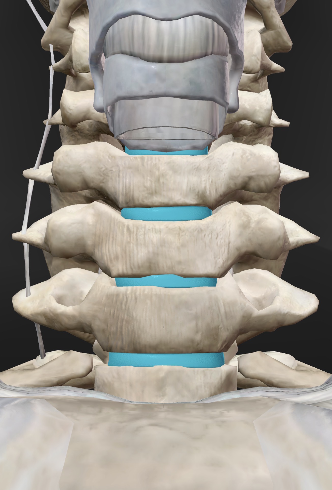
Name the joint, the structural type (fibrous, cartilaginous, or synovial), and the functional type (synarthotic, amphiarthrotic, or diarthrotic)
Intervertebral discs of the cervical spine
articulations between adjacent vertebral bodies
Structural type - Cartilaginous symphysis
Functional type - amphiarthrotic
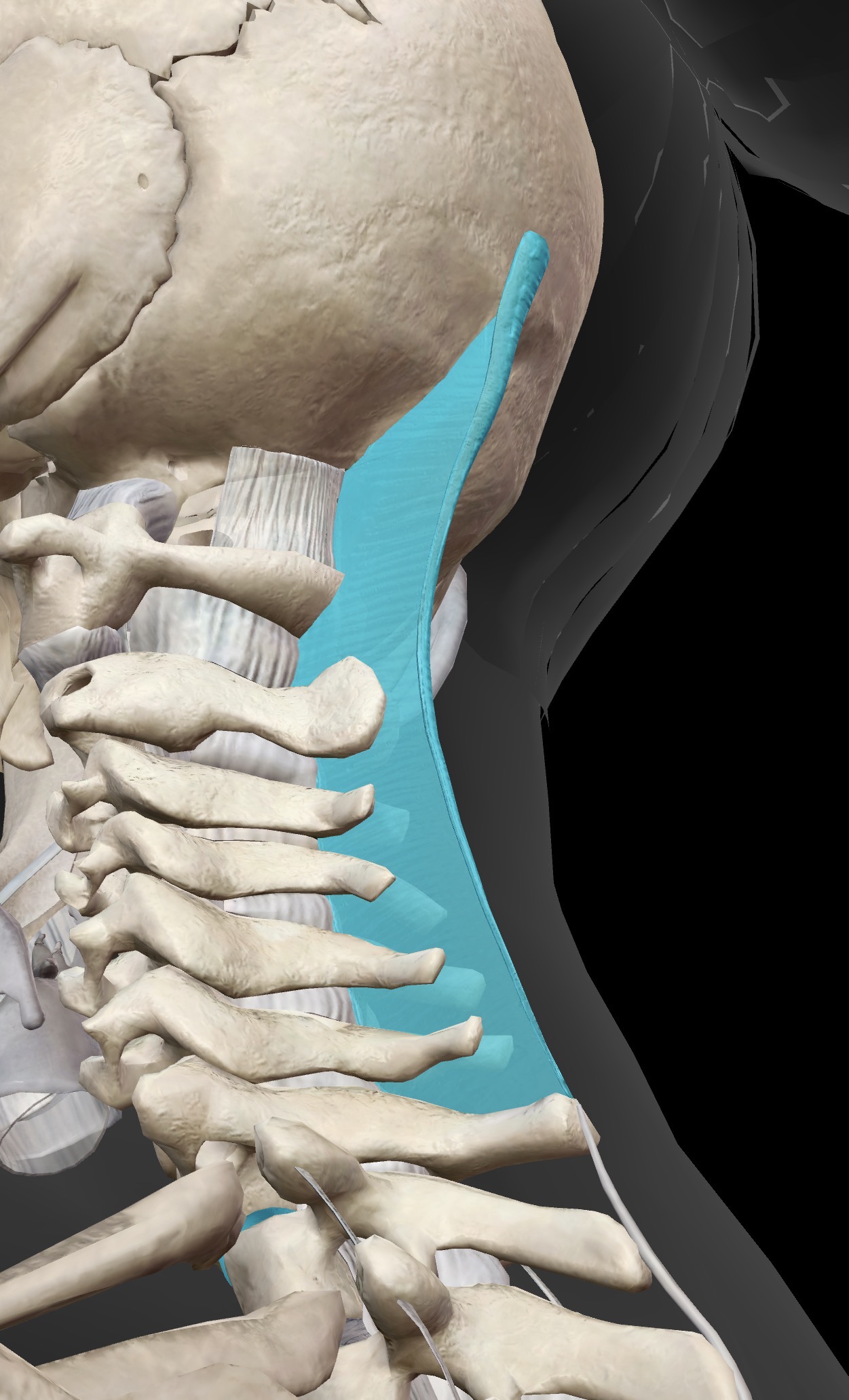
Name this ligament, the area it resides in and its point of attachment and purpose
Ligamentum nuchae
Ligament between the external occipital protuberance and the seventh cervical vertebrae is known as the ligamentum nuchae. Resists flexion of the vertebral column/trunk
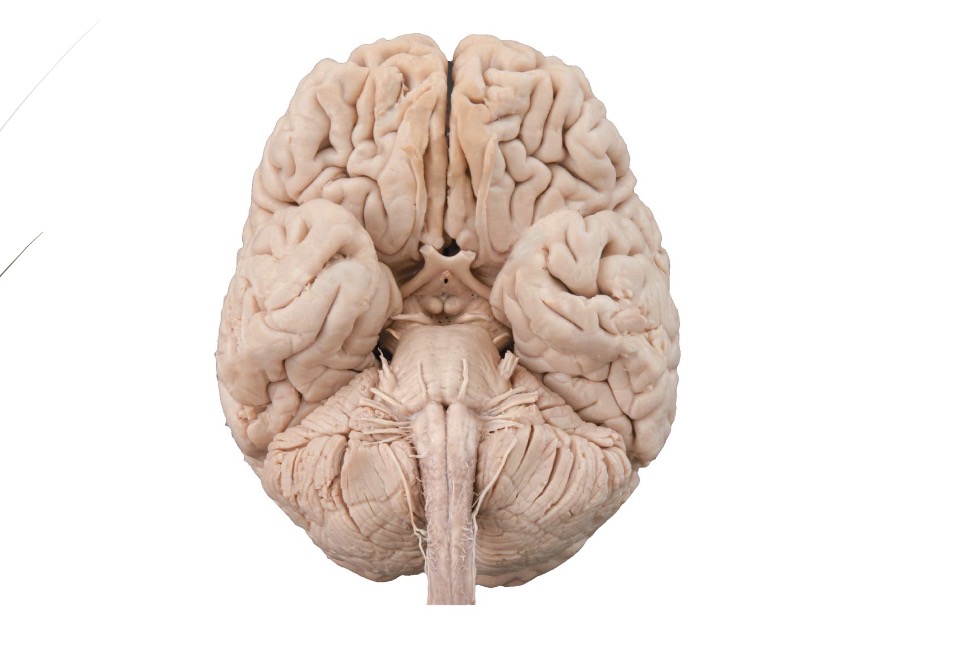
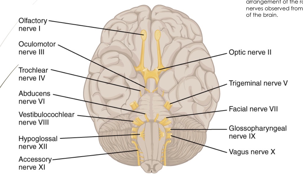
Name each cranial nerve and list the type of nerve as well as its basic function (with stars)
Olfactory (CN l)
Type - sensory
Basic function - smell
Optic (CN ll)
Type - sensory
Basic function - vision
Oculomotor (CN lll)
Type - motor
Basic function - Motor (somatic): eye muscles (levator palpebrae superioris, medial rectus, superior rectus, inferior rectus, inferior oblique)
Trochlear (CN lV)
Type - motor
Basic function - eye muscles (superior oblique)
Trigeminal (CN V)
Type - mixed (sensory and motor)
Basic function - Motor: muscles of mastication (chewing) - eg. temporalis, masseter. Sensory: skin of face/scalp
Abducens (CN Vl)
Type - motor
Basic function - eye muscles (lateral rectus)
Facial (CN Vll)
Type - mixed (sensory and motor)
Basic function: motor (somatic): muscles of facial expression - eg: platysma, orbicularis oris, orbicularis oculi, frontalis
Accessory (Xl)
Type - motor
Basic function - innervates sternocleidomastoid and trapezius
Hypoglossal (Xll)
Type - motor
Basic function - muscles of tongue
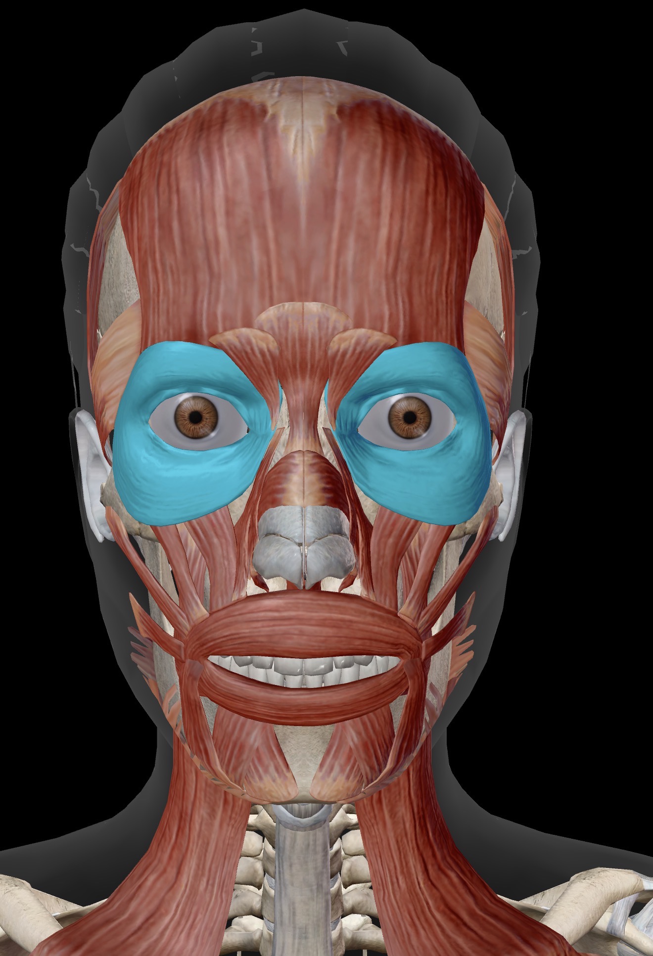
Name the muscle and corresponding letter ( O = origin, I = insertion, A = actions, IN = innervation)
A
IN
Orbicularis oculi:
A. Closes eyelid (as in a wink)
IN. Branch of facial nerve (CN Vll)
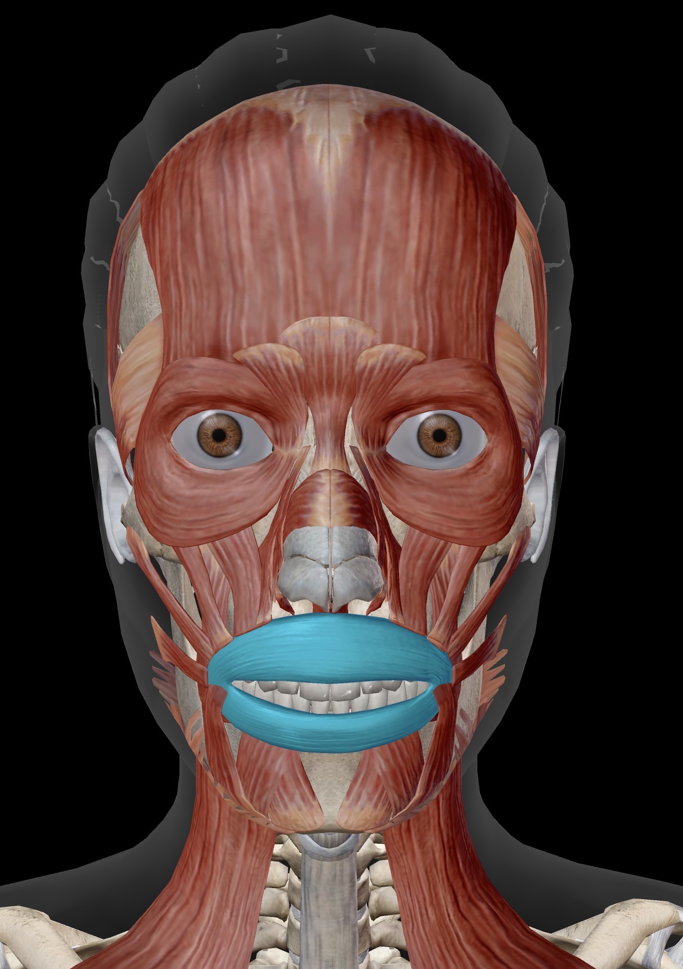
Name the muscle and corresponding letter ( O = origin, I = insertion, A = actions, IN = innervation)
A
IN
Orbicularis oris:
A: Closes lips, protrudes lips (as in a kiss)
IN. Branch of facial nerve (CN Vll)
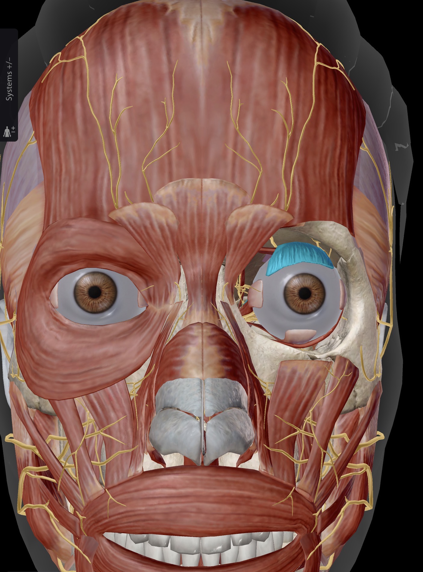
Name the muscle and corresponding letter ( O = origin, I = insertion, A = actions, IN = innervation)
A
IN
Levator palperbrae superioris:
A. Elevates the upper eyelid
IN. Oculomotor nerve (CN lll)

Name the muscle and corresponding letter ( O = origin, I = insertion, A = actions, IN = innervation)
A
IN
Frontalis:
A. Raises the eyebrows
IN. Branch of facial (CN Vll)
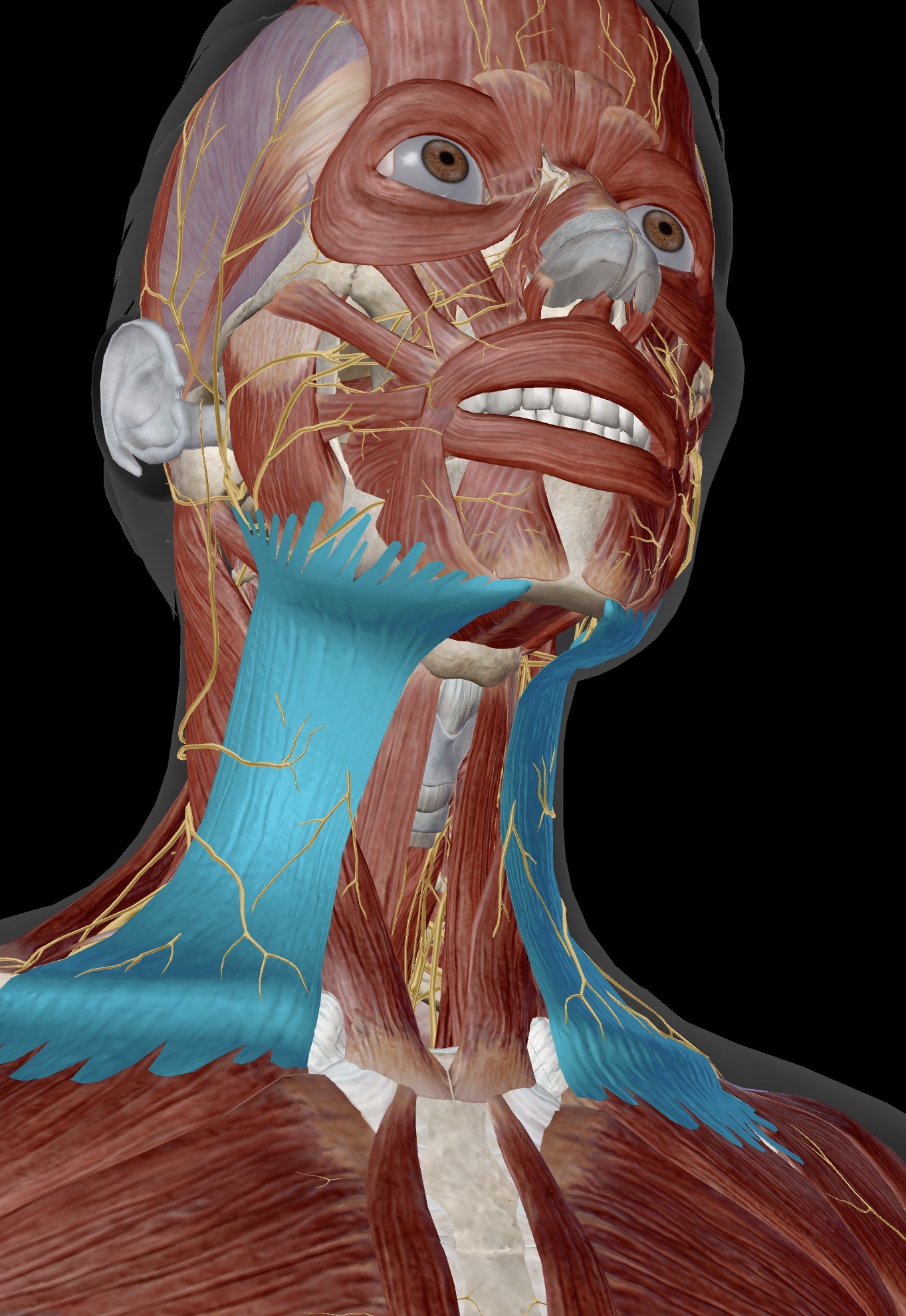
Name the muscle and corresponding letter ( O = origin, I = insertion, A = actions, IN = innervation)
A
IN
Platysma:
A: Depresses the lower jaw
IN. Branch of facial nerve (CN Vll)
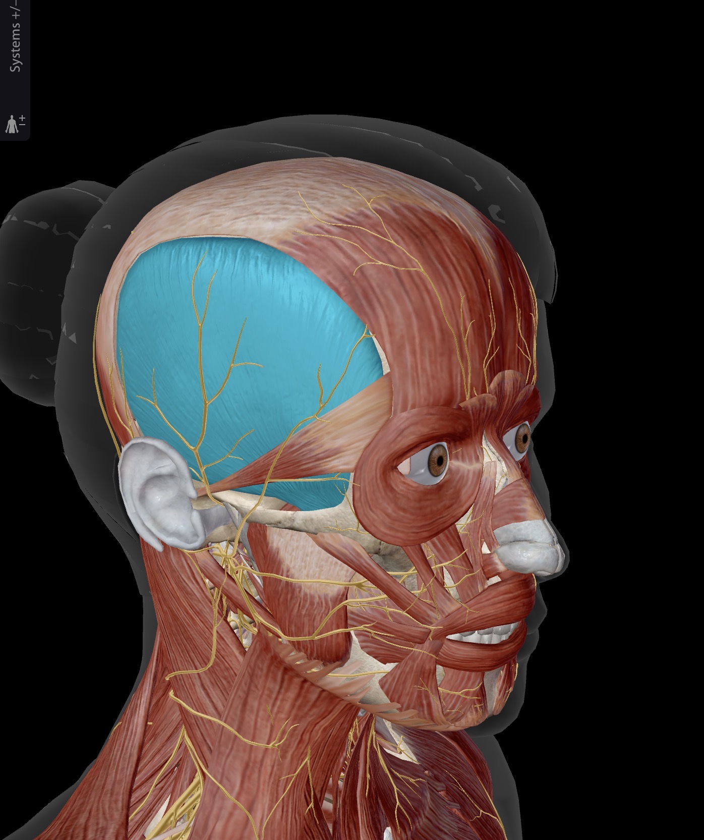
Name the muscle and corresponding letter ( O = origin, I = insertion, A = actions, IN = innervation)
O
I
A
IN
Temporalis:
O. Temporal fossa
I. Under zygomatic arch to coronoid process and anterior ramus of the mandible
A. Vertical fibres elevate and horizontal fibres retract the mandible against the maxilla with great force; closes jaw
IN. Branch of trigeminal nerve (CN V)
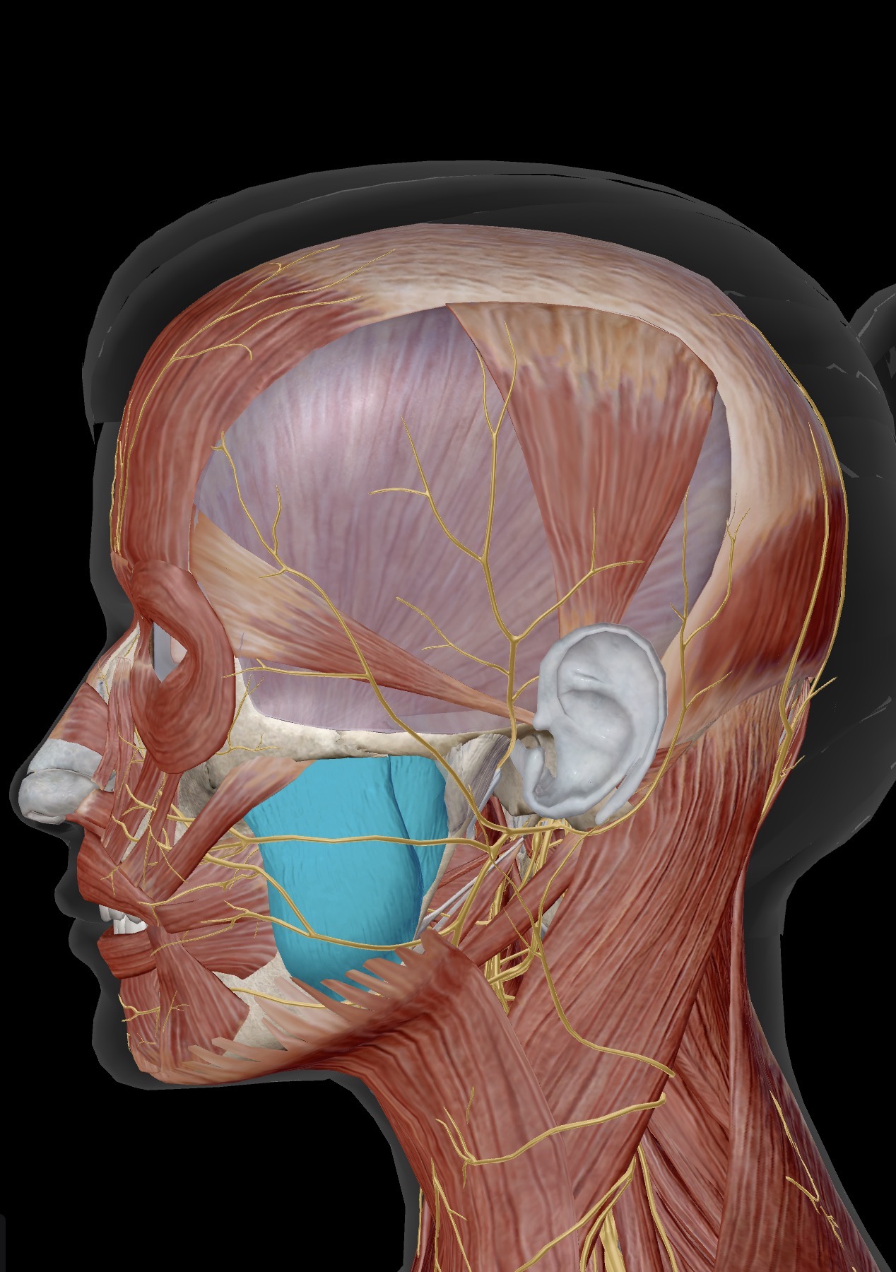
Name the muscle and corresponding letter ( O = origin, I = insertion, A = actions, IN = innervation)
O
I
A
IN
Masseter
O. Zygomatic arch
I. Ramus and angle of the mandible
A. Raises the mandible against the maxilla
IN. Branch of the trigeminal nerve (CN V)
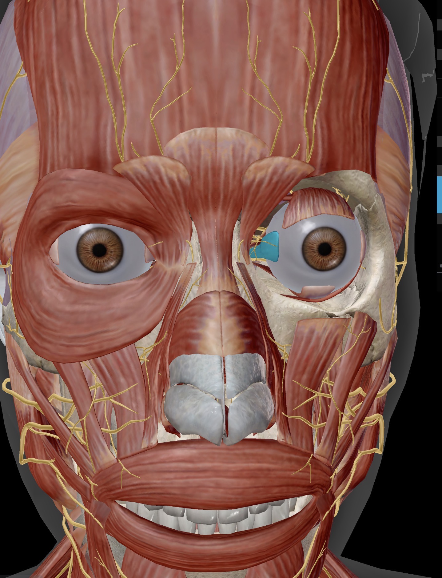
Name the muscle and corresponding letter ( O = origin, I = insertion, A = actions, IN = innervation)
A
IN
Medial rectus:
A. Primary action: abducts eyeball/moves the eyeball medially
IN: oculomotor nerve (CN lll)
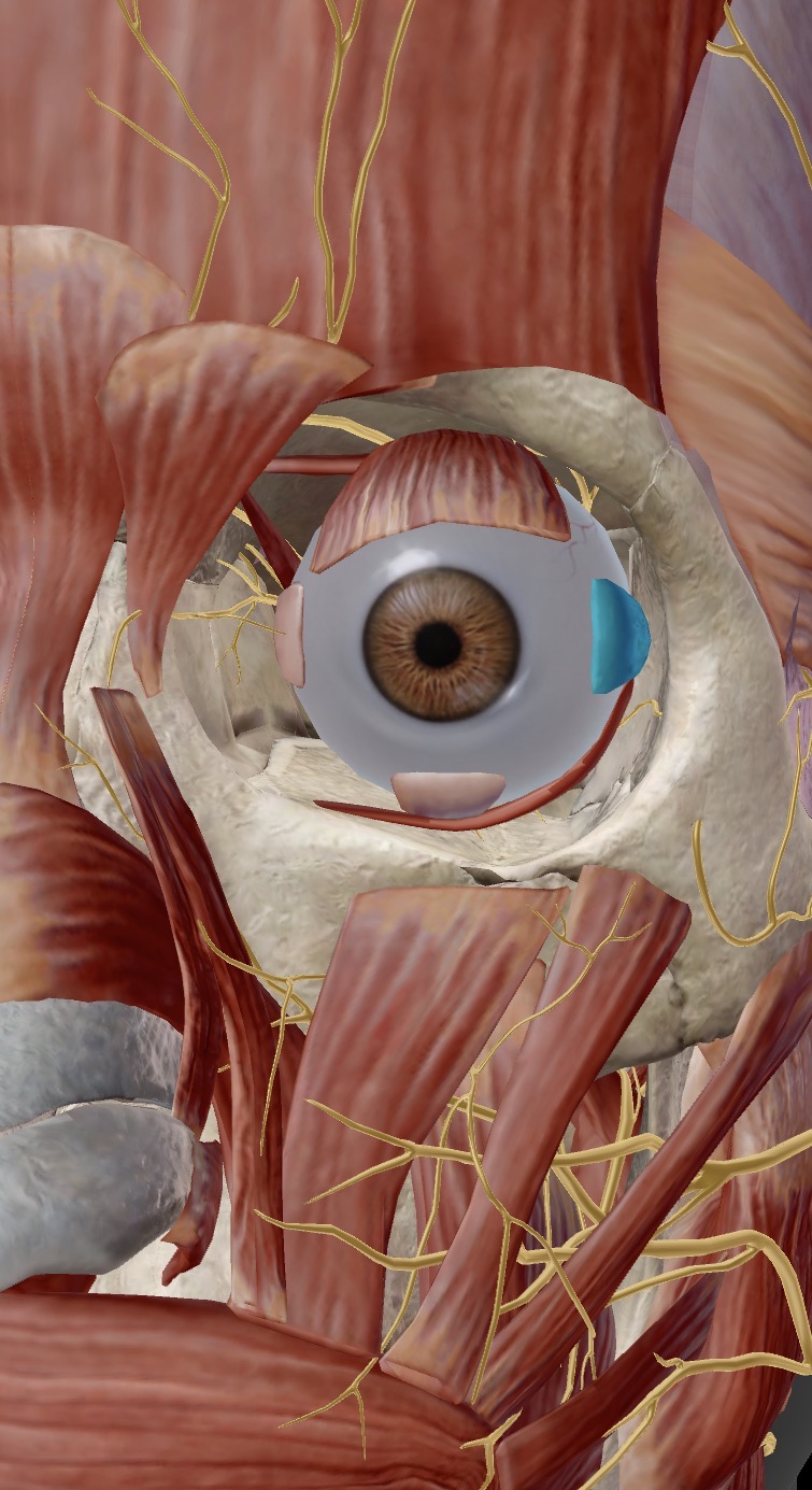
Name the muscle and corresponding letter ( O = origin, I = insertion, A = actions, IN = innervation)
A
IN
Lateral rectus:
A: Primary action: abducts the eyeball/moves laterally
IN. Abducens nerve (CN Vl)
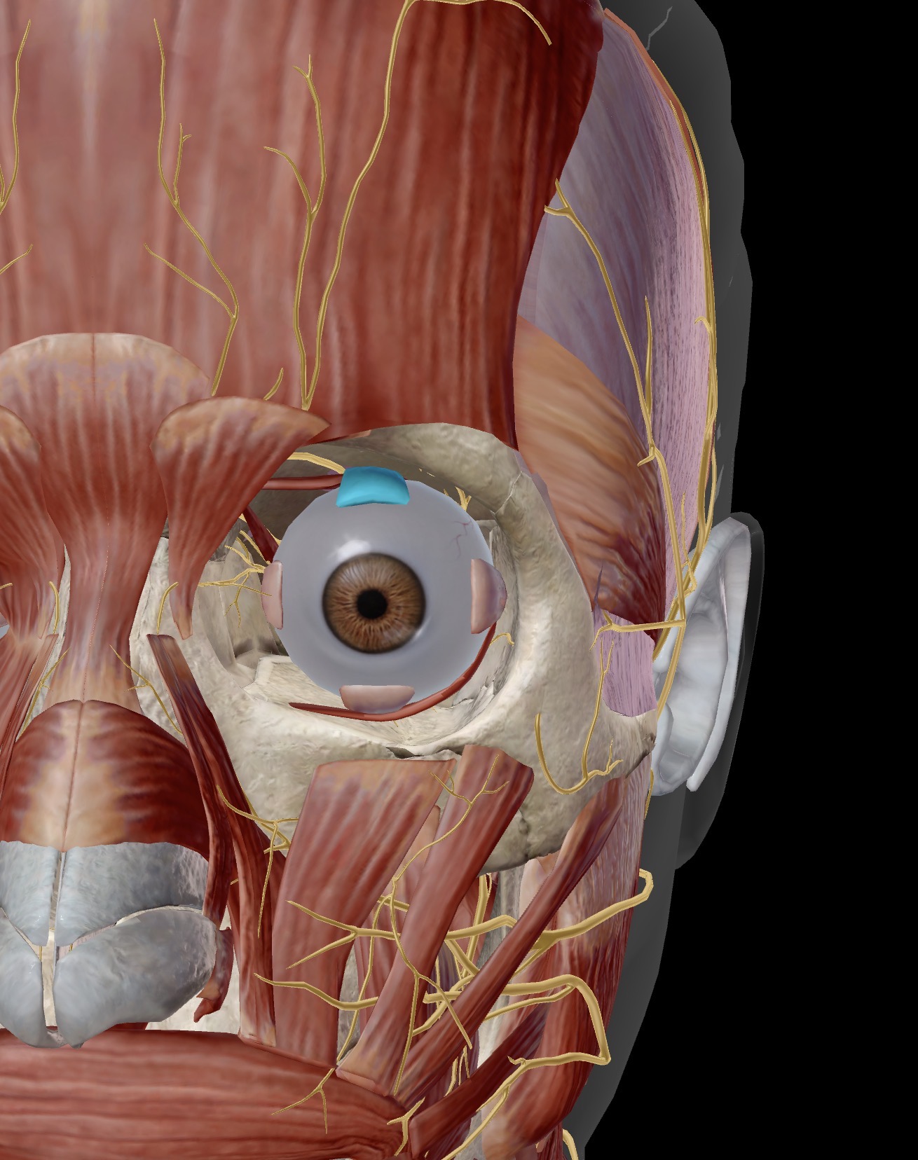
Name the muscle and corresponding letter ( O = origin, I = insertion, A = actions, IN = innervation)
A
IN
Superior rectus:
A primary action: elevates the eyeball
IN. Oculomotor nerve (CN lll)
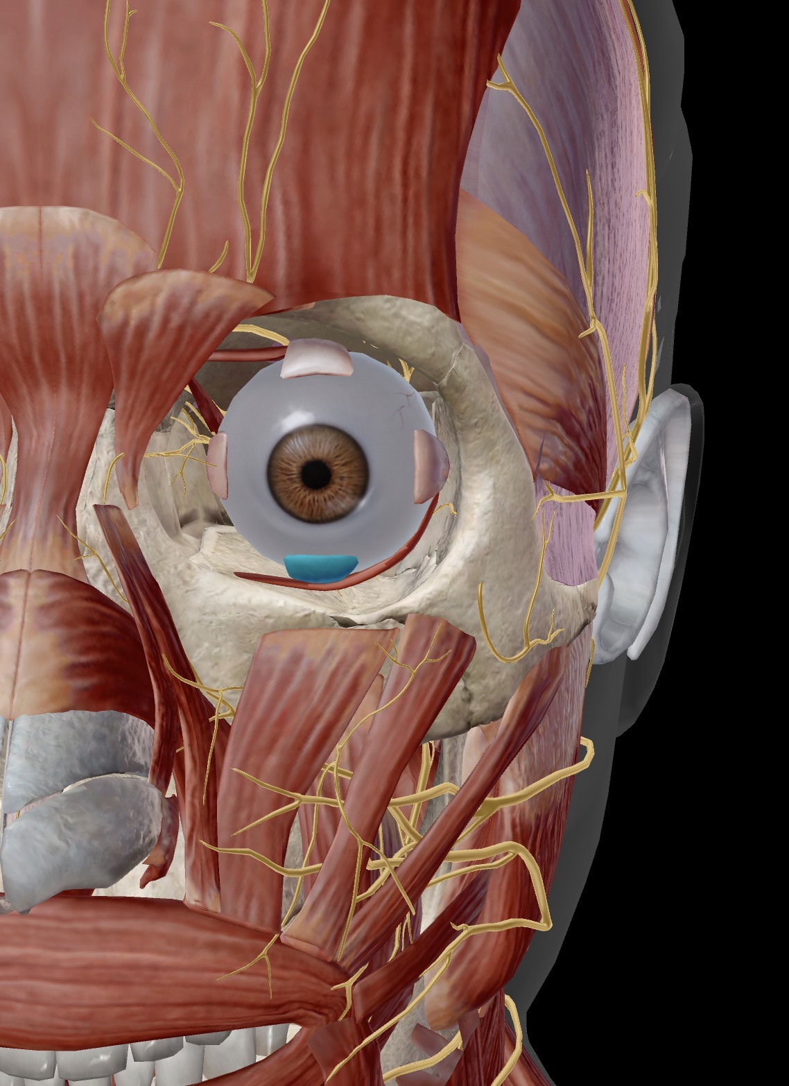
Name the muscle and corresponding letter ( O = origin, I = insertion, A = actions, IN = innervation)
A
IN
Inferior rectus
A. Primary action: depresses the eyeball
IN. Oculomotor nerve
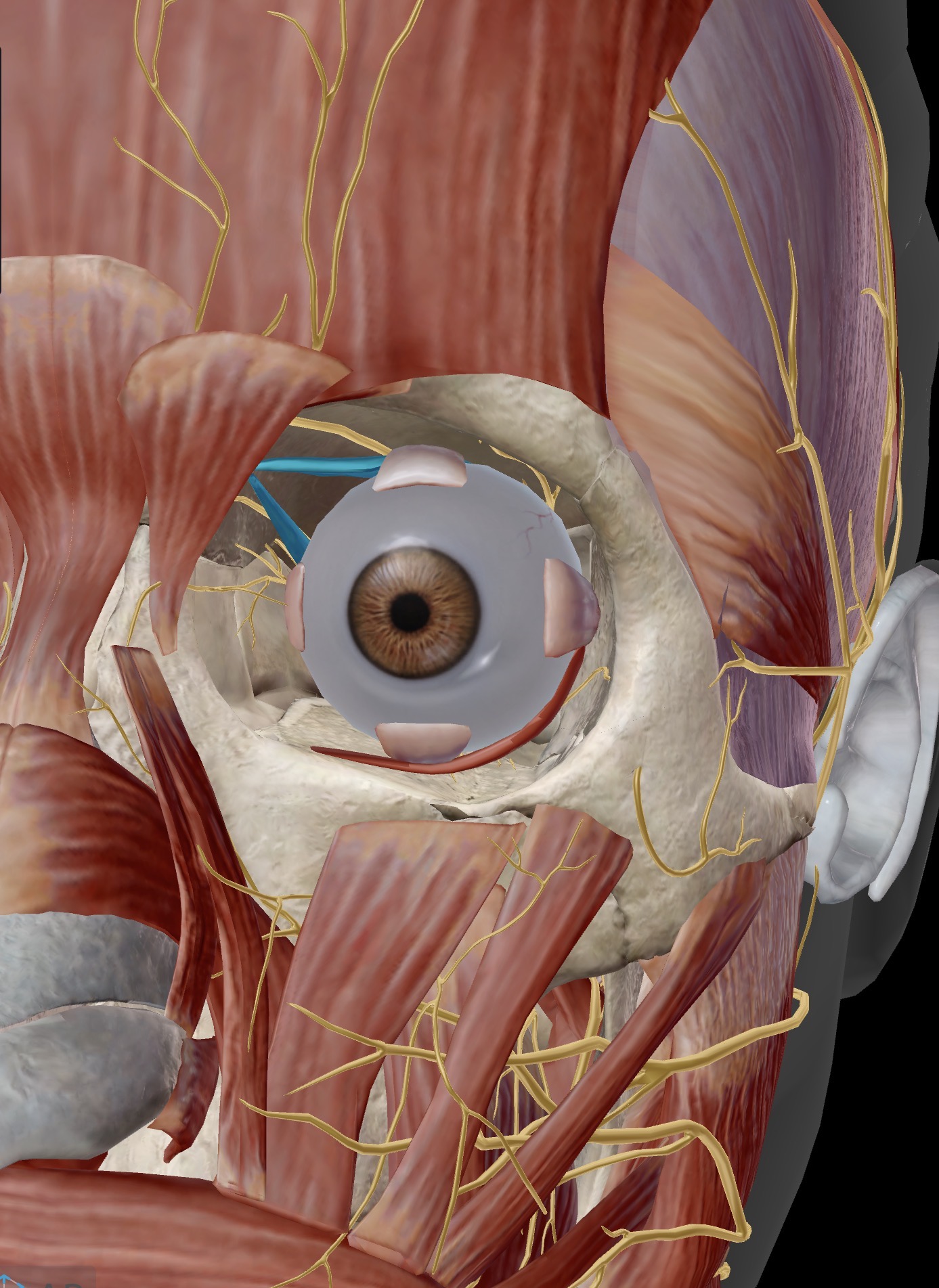
Name the muscle and corresponding letter ( O = origin, I = insertion, A = actions, IN = innervation)
IN
A
Superior oblique:
IN. Innervation: trochlear nerve (CN lV)
A. Assists in eyeball movement
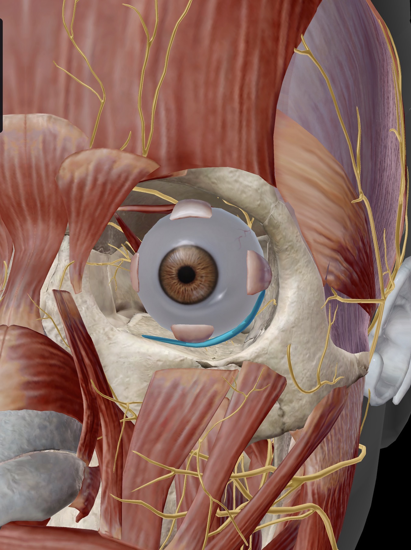
Name the muscle and corresponding letter ( O = origin, I = insertion, A = actions, IN = innervation)
IN
A
Inferior oblique:
IN: oculomotor nerve (CN lll)
A. Assists with eyeball movement
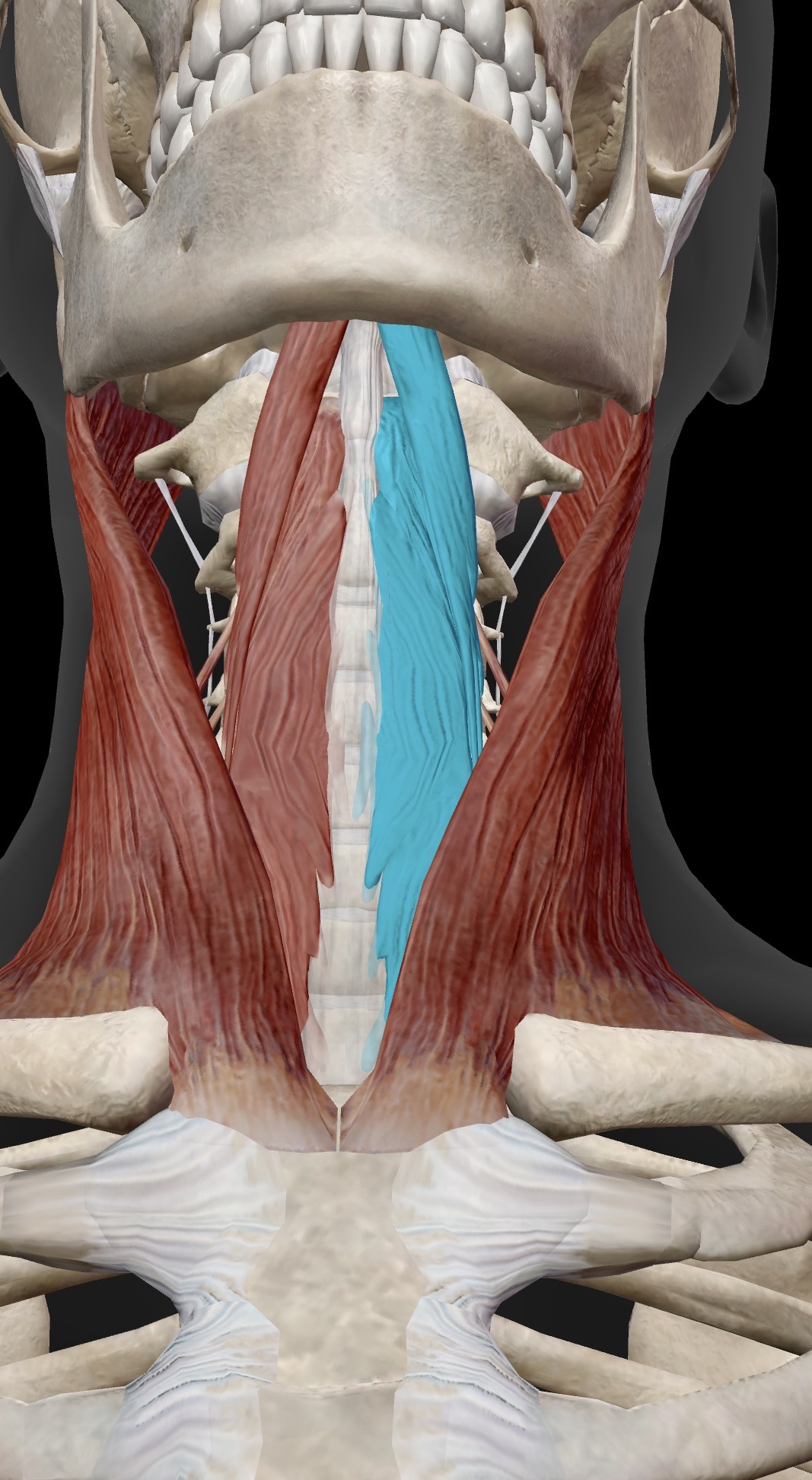
Name the muscle and corresponding letter ( O = origin, I = insertion, A = actions, IN = innervation)
A
Longus colli:
A. Flexes the cervical spine (flexion of the cervical vertebrae at the cervical intervertebral joints)
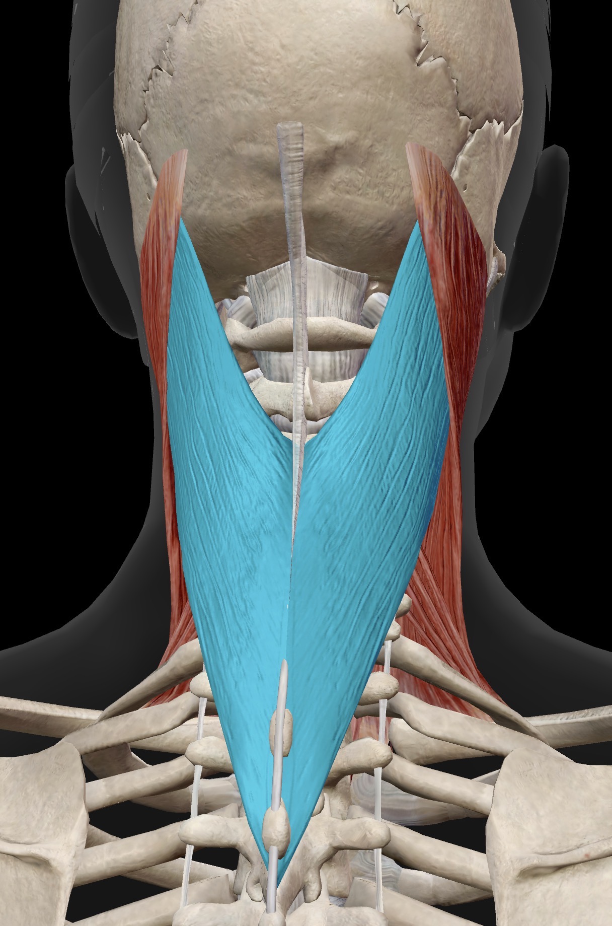
Name the muscle and corresponding letter ( O = origin, I = insertion, A = actions, IN = innervation)
A
Splenius capitius:
A. Acting bilaterally, extends the head.
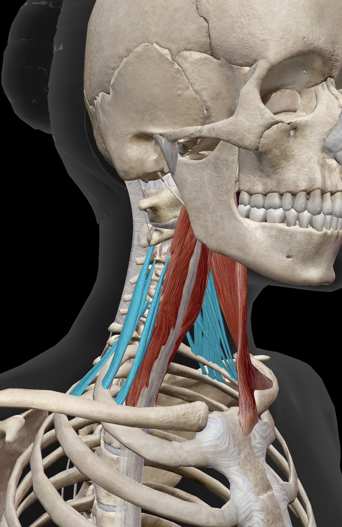
Name the muscle and corresponding letter ( O = origin, I = insertion, A = actions, IN = innervation)
A
Scalenes:
A. Ipsilateral side flexion of the neck (draws the neck into Side flexion on the Same side muscle is situated on)
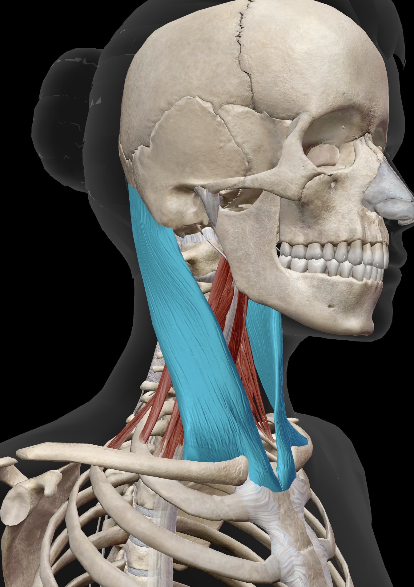
Name the muscle and corresponding letter ( O = origin, I = insertion, A = actions, IN = innervation)
O
I
A
IN
Sternocleidomastoid:
O. Manubrium of sternum and medial 1/3 of clavicle
I. Mastoid process of temporal bone plus part of occipital bone
A: Unilateral contraction : Draws the head towards the shoulder of same side (ipsilateral side flexion of the head) And rotates the head towards the opposite side (contralateral rotation of the head)
Bilateral contraction: Flexion of the head
IN. Accessory nerve (Xl)
Name the movements associated with with chewing / movements of the jaw and tongue
Protrusion: sticking the tongue out
Retraction: pulling the tongue back in the mouth
Depression: lowering the tongue toward the floor of the mouth
Elevation: raising the back or sides of the tongue
Lateral movement: shifting the tongue horizontally
Name the movements of the eyeball
Elevation (closing.) bringing the jaw up to close the mouth.
Depression. (opening) lowering the jaw
Lateral movement
Protrusion
Retraction
Close the eyelids
Close the lips/protrude the lips
Elevate the upper eyelids
Raise the eyebrows
Depress/Lower the mandible at the temporomandibular joints
Raise/elevate the mandible at the temporomandibular joints
Retract the mandible at the temporomandibular joints
Protract/protrude the mandible at the temporomandibular joints
Laterally deviate the mandible at the temporomandibular joints
Medially deviate the mandible at the temporomandibular joints
Elevate the eyeballs
Depress the eyeballs
Abduct the right eyeball
Abduct the left eyeball
Adduct the right eyeball
Adduct the left eyeball
Flex the head/neck at the atlanto-occipital joint and at the cervical intervertebral joints.
Extend the head/neck at the atlanto-occipital joint and at the cervical intervertebral joints.
Right side flex the head/neck at the atlanto-occipital joint and at the cervical intervertebral joints.
Left side flex the head/neck at the atlanto-occipital joint and at the cervical intervertebral joints.
Right rotate the head/neck at the atlanto-axial joint and at the cervical intervertebral joints.
Left rotate the head/neck at the atlanto-axial joint and at the cervical intervertebral joints.
How many cervical, thoracic, lumbar, sacral and coccygeal vertebrae are there?
There are 7 cervical Vertebrae and the associated vertebral discs, 12 Thoracic vertebrae and the associated discs, 5 Lumbar vertebrae and associated discs, 5 fused sacral (discs usually absent but may be some disc “remnants”, and 4 fused coccygeal (discs usually absent)
What are the curvatures of the spine called for each associated area?
Cervical spine - lordosis
Thoracic spine - Kyphosis
Lumbar - lordosis
Sacral and coccyx - Kyphosis


Give the definitions of the following terms:
Foramen
Canal
Process
Facet
Foramen: An opening; in the vertebral column there are several intervertebral foramina that allow for the passage of spinal nerves consisting of neurons that are either travelling toward the spinal cord/brain (sensory neurons) or leaving the brain/spinal cord (motor neurons)
Canal: a tunnel or enclosed pathway; in the vertebral column, there is a vertebral canal that allows for spinal neurons to be continuous with the brain
Process: a projection or outgrowth of bone
Facet: A facet is a flattened ending of a bone where it articulates with another bone at a joint. Although there are several “facets” in the body, the term “facet” usually refers to the facets in the vertebral column

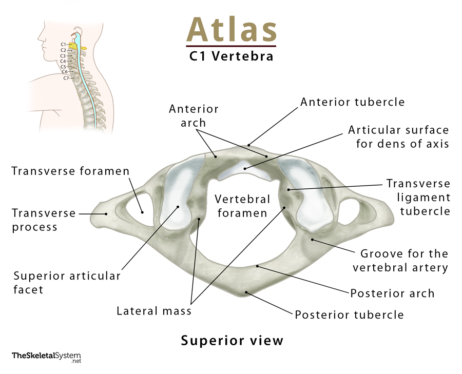
Name the parts of the atlas bone (C1)

Name the parts of the axis (C2)

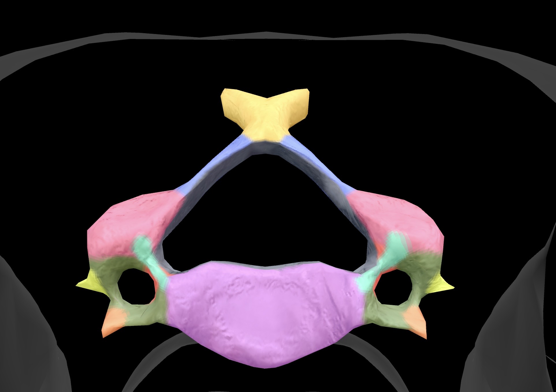

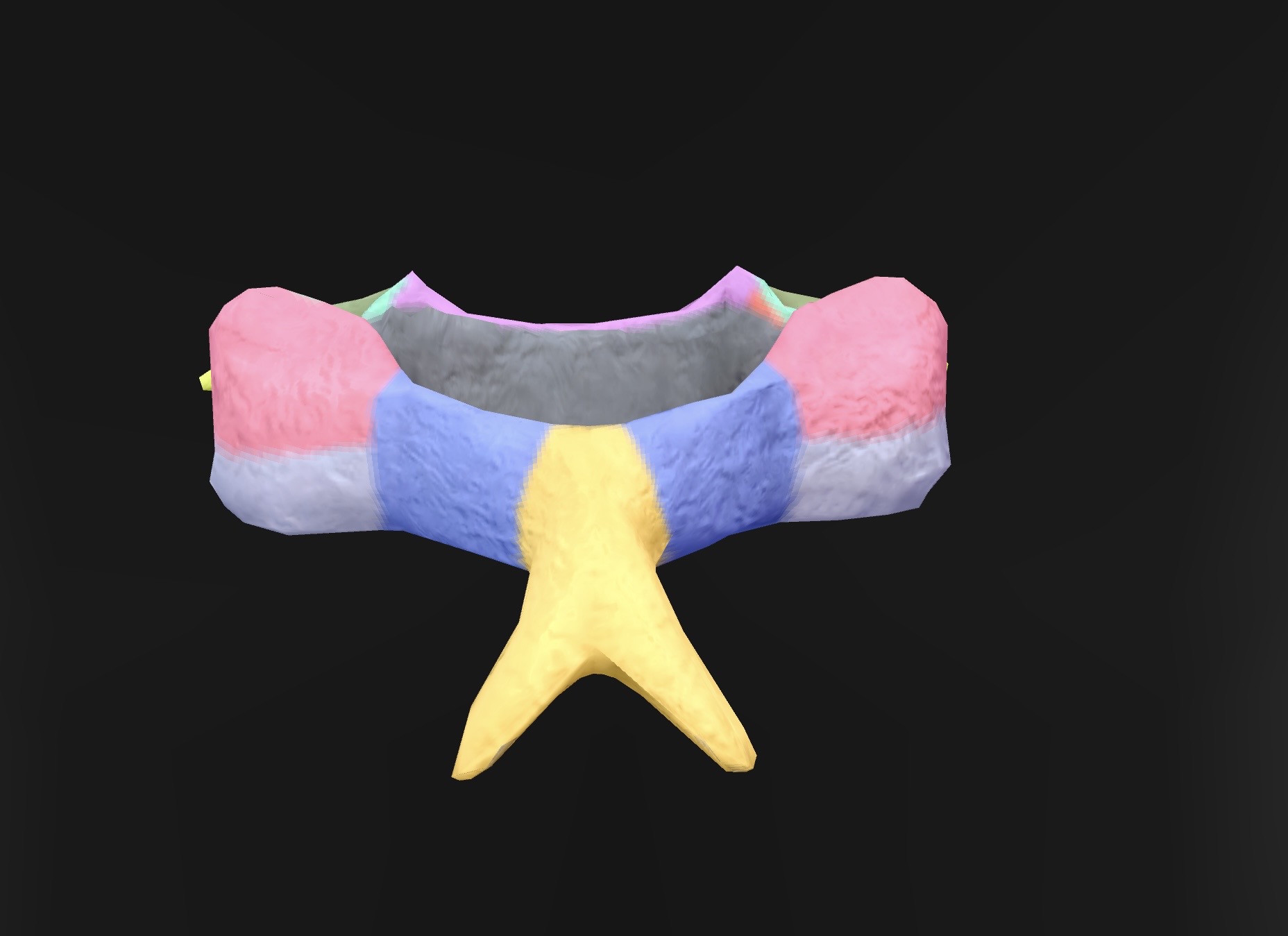
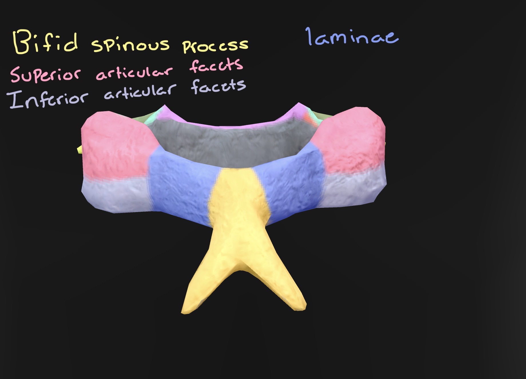

What kind of vertebra is this as well as name the parts
Thoracic vertebrae


What kind of vertebra is this as well as name the parts
Thoracic vertebrae


What kind of vertebra is this as well as name the parts
Thoracic vertebrae


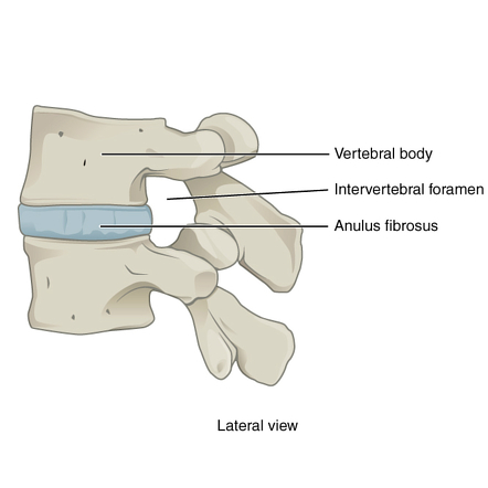

What kind of vertebra is this as well as name the parts
Thoracic vertebrae