ECG
 Call Kai
 Learn
Learn Practice Test
 Spaced Repetition
 Match
Match Flashcards
 Knowt Play
1/55

There's no tags or description
Looks like no tags are added yet.
Name | Mastery | Learn | Test | Matching | Spaced | Call with Kai | Chat |
|---|
No analytics yet
Send a link to your students to track their progress
56 Terms
The spread of excitation through the atria, ventricles and conducting system is brought about by what
local electrical currents
What is an action potential
the abrupt reversal of the membrane potential to a positive value
In the active depolarised region, the interior of the membrane is _______ charged while in the resting zone ahead is ________ charged
positively
negatively
Conduction velocity in atria
0.5 m/s
Conduction velocity in AV node
0.05 m/s
Conduction velocity in Bundle of His, bundle branches
1.0 m/s
Conduction velocity in Purkinje fibres
5.0 m/s
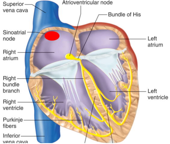
Conduction velocity in Ventricles
0.5 m/s
Depolarisation of atria & ventricles goes in what direction
Top to bottom
Does repolarisation occur spontaneously
yes
In what direction does repolarisation occur in atria & ventricles
Bottom to top
Resting membrane potential cells are _______ charged inside and __________ charged outside
Negatively charged inside
Positively charged outside
During atrial depolarisation cells are _______ charged inside and __________ charged outside
Positively charged inside
Negatively charged outside
During ventricular repolarisation cells are _______ charged inside and __________ charged outside
Negatively charged inside
Positively charged outside
What is an ECG a recording of
a recording of potential changes at the skin surface, resulting from depolarisation and repolarisation of heart muscle
How does cardiac excitation have any effect on currents around the body
The spread of cardiac excitation creates currents in the extracellular fluid.
The currents generate small potential differences across the body surface of around 1mV
What can ECGs be recorded by and on (& at what speed)
Recorded by a sensitive voltmeter connected to metal electrodes on the skin surface
It is recorded on a strip of moving paper or computer screen to produce the ECG trace
The paper speed is set at 25mm/s (1 large division every 0.2 sec)
The magnitude of the skin potential difference depends on what
the mass of the myocardium that is activated
Does the surface ECG detect activity of the SA and AV nodes
No - just activity of atria and ventricles
The electrical current set up by atrial and ventricular depolarisation is a _____
vector (has both magnitude & direction)
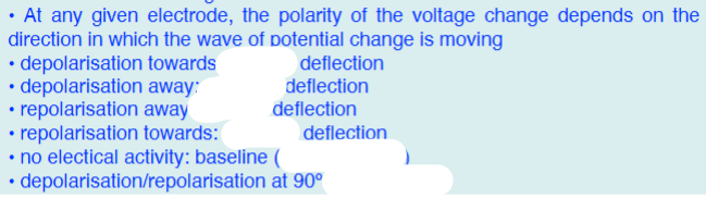
Positive deflection by depolarisation towards = R
Positive deflection by depolarisation away = T
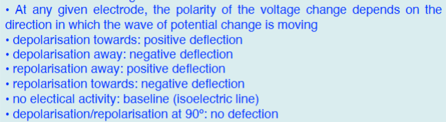
What part of the cardiac cycle do we not see on an ECG? Why?
atrial repolarisation
It is hidden by the QRS complex
The overall direction of depolarisation changes between going towards / away from the detecting electrode which is placed on the leg. Depolarisation in different parts of the heart goes in different directions. In what direction does it go from:
The SA node:
Ventral septum:
Ventricle:
Ventricle last portion:
Repolarisation of heart:
The SA node: Towards AV node - towards electrode - positive deflection (p wave)
Ventral septum: Away from electrode - small negative deflection (q wave)
Ventricle: Towards electrode - large positive deflection (r wave)
Ventricle last portion: Away from electrode - small negative deflection (s wave)
Repolarisation of heart: Away from electrode - positive deflection (t wave)
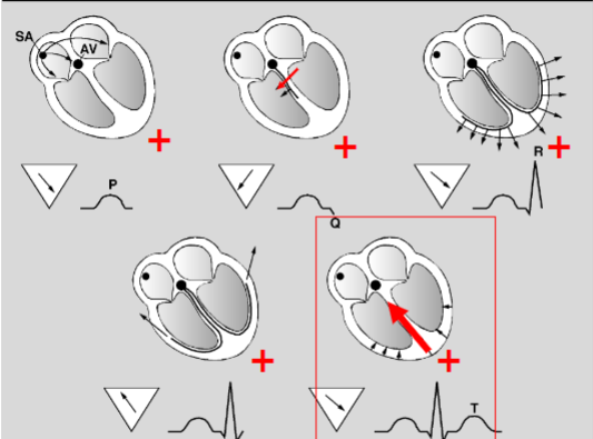
Why is atrial repolarisation not seen
It occurs at the same time as the QRS
It’s too small to have an effect
What did Einthoven invent
The original electrocardiographic lead system
How did the original electrocardiographic lead system work
Recording electrodes are placed on:
• left arm (LA)
• right arm (RA)
• left leg (LL)
• a fourth electrode on the right leg acts as an electrical earth
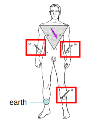
A bipolar lead was used in the original electrocardiographic lead system. What does this mean
Recordings are made between 2 electrodes
There are 3 limb leads used in the original electrocardiographic lead system. What was the function of each
Lead I : records potential difference between LA and RA (LA is positive, RA is negative)
Lead II : records potential difference between LL and RA (LL is positive, RA is negative)
Lead III : records potential difference between LL and LA (LL is positive, LA is negative)
Mean QRS axis
The average direction in which the dipole causing the QRS complex is oriented
Usually lies within the 0 º to 90 º quadrant down and to the left.
It corresponds to the anatomical pathway the wave of depolarisation takes
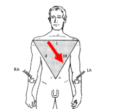
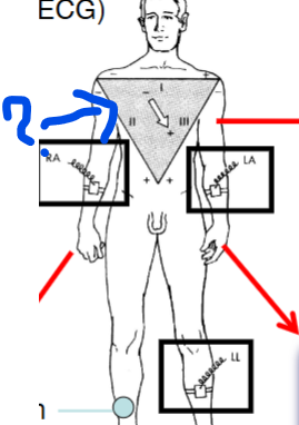
What is that called
Einthoven triangle
oriented in the frontal plane of the body (2 dimensional)
What does the Einthoven triangle represent
2 negative electrodes (RA & LA) and 1 positive electrode are placed on the person’s 2 arms and left leg. By swapping the position of the positive electrode, the direction of the ECG vector changes to go between:
+ 90º with + on leg (aVF - augmented voltage left foot)
- 30º with + on left arm (aVL - augmented voltage left arm)
- 150º with + on right arm (aVR - augmented voltage right arm)
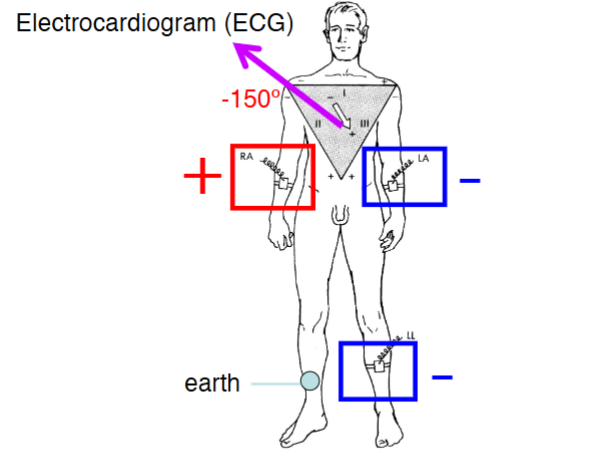
The 3 augmented limb leads aVR, aVL, aVF are unipolar/bipolar
unipolar
What is the hexaxial reference system
The unipolar (aV) and bipolar limb leads (standard) record electrical activity of the heart in the frontal plane of the body
The main deflection is positive for all leads except the aVR
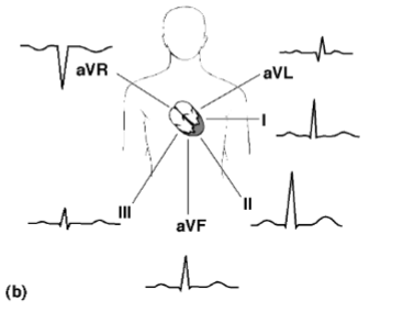
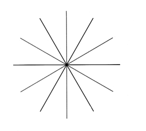
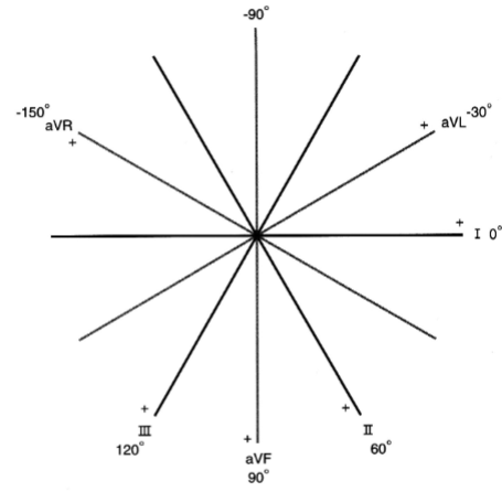
Place the right leads on the right lines using the hexaxial system
- lead I : 0º
- lead II : +60º
- aVF: +90º
- lead III: +120º
- aVR: -150º
- aVL: -30º
When an ECG is taken using electrodes on the chest, how many leads are there and what are they called
6
Called V1, V2, V3, V4, V5, V6,
There are still electrodes on the La, Ra & LL & right foot is earthed
The chest leads examine the heart in what plane
horizontal
Do chest leads give large/small ECG deflections
large
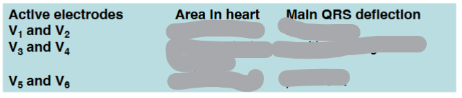
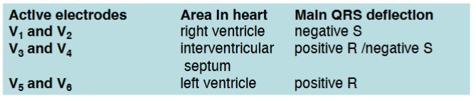
V1 versus V6
V1: negative wave - S wave
V6: positive wave - R wave
The depolarisation of the left ventricle is far greater than in the right ventricle
Being a vector, the LV depolarisation cancels out the RV depolarisation
Both V1 and V6 only see a wave of depolarisation heading towards V6 (V1 is somewhat cancelled out)
V1 lead: RS
V6: QR
ECG is recorded at what speed
25mm/s (5 large boxes, 25 small squares)
Spaces between waves are called?
segments
Most important segment and why?
ST segment
Used to test for ischemic activity in the heart
What does the PR interval represent
time from initial depolarisation of the atria to initial depolarisation of the ventricle
Which interval correlates with conduction time through the AV node
PR interval
PR interval length
0.12 - 0.2 s
Sinus arrhythmia
the physiological slowing of firing by the SA node during expiration and acceleration during inspiration
A phasic rise in vagal activity occurs during expiration which is initiated by lung stretch receptors which slows the heart rate
During inspiration vagal activity is reduced and heart rate increases
(this is normal)
first degree AV-nodal block effect on PR interval
the conduction velocity through the AV node is slowed (due to injury), the PR interval is lengthened (>0.20 s)
second degree AV-nodal block * frequently asked
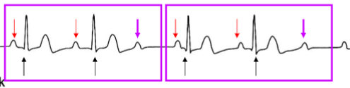
the AV node conducts only some of the supraventricular depolarisations:
- some P waves are followed by QRS complexes
- other P waves are not followed by a QRS complex
In this example there are 3 P waves for every 2 QRS complexes (3:2 ratio)
- other ratios are possible (e.g, 2:1)
Caused by increased injury to the AV node
third degree AV-nodal block
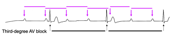
There is complete electrical block between the atria and the ventricles
The AV node fails to conduct any signals
P waves occur regularly
QRS complexes occur regularly but with low frequency (secondary pacemaker)
PVC
Premature ventricular contraction
The heartbeat doesn’t originate within the atria (SA node) like normal. It originates within the ventricle
These ventricular beats disturb the normal rhythm of the heart (arrhythmias)
PVC on an ECG
The PVC is not preceded by a P wave
The next QRS would have been expected at the arrow
The shape, amplitude and duration of the PVC are abnormal
The wave of depolarisation is not conducted by the normal conducting pathways
The diastolic interval after the PVC is long this compensatory pause allows for increased ventricular filling before the next beat
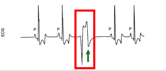
What’s an ectopic beat
A PVC
How does injured heart tissue affect depolarisation
In a normal heart at point d the 2 currents cancel each other out. In an injured heart, the conduction is interrupted.
Depolarisation passes back through injured area and area G is induced to depolarisation again but too early!
An ectopic beat can occur
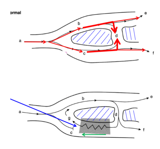
Ventricular fibrillation
Re-entry can occur causing a series of rapid uncoordinated excitations
These rapid un-coordinated excitations are collectively referred to as ventricular fibrillation (VF)
There is no cardiac output during VF
Death follows within minutes
(Normally, an electrical impulse travels through the heart tissue once, then stops (because the cells behind it are refractory). In re-entry, the impulse doesn't stop — it circles back into areas that are no longer refractory and keeps re-exciting the tissue.)
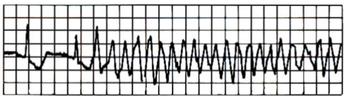
What can cause Ventricular fibrillation
Ventricular fibrillation is often a fatal consequence to myocardial ischaemia (loss of blood flow to a potion of the muscle in the heart), anaesthetic overdose or electrocution
During ischaemia, ventricular ectopics can occur
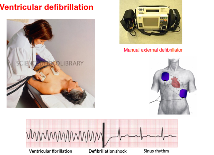
How is ventricular fibrillation treated
An electrical shock is delivered through pads or paddles placed on the chest