C. Chapter 3 - Plasma Membrane, Cytoplasm, Nucleus
1/117
There's no tags or description
Looks like no tags are added yet.
Name | Mastery | Learn | Test | Matching | Spaced | Call with Kai |
|---|
No analytics yet
Send a link to your students to track their progress
118 Terms
Cell theory
– A cell is the structural and functional unit of life
– How well the entire organism functions depends on individual and combined activities of cells
– Biochemical functions of cells are dictated by shape of cell and specific subcellular structures
– Continuity of life has cellular basis
– Cells can arise only from other preexisting cells
Generalized cell
All cells have some common structures and functions
Human cells have three basic parts:
1. Plasma membrane: flexible outer boundary
2. Cytoplasm: intracellular fluid containing organelles
3. Nucleus: DNA containing control center
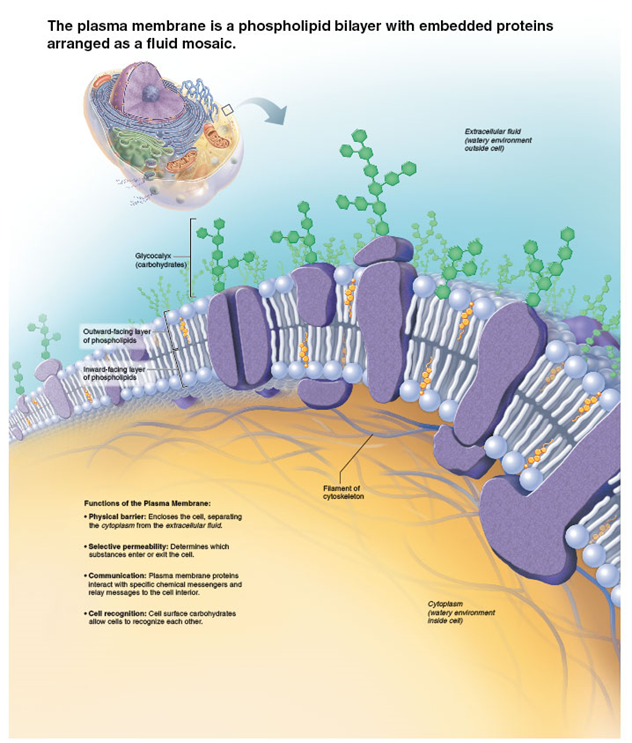
Plasma Membrane
acts as an active selectively permeable barrier that separates intracellular fluid from extracellular fluid (ex. role in cellular activity and controls what enters/leaves cell)
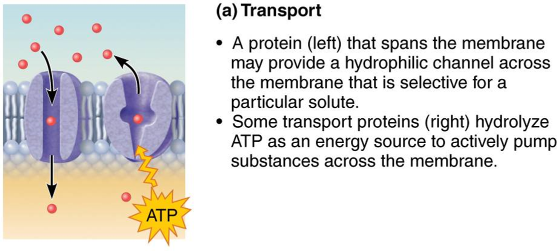
Transport (membrane protein)
hydrolyze ATP as an energy source to actively pump substances across a membrane
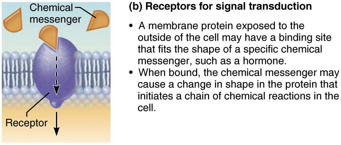
Receptors (membrane protein)
membrane protein exposed to outside of cell may have biting site for chemical messengers (hormones)
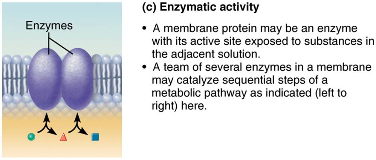
Enzymatic activity (membrane protein)
active site exposed to substances in the adjacent solution (may catalyze sequential steps of a metabolic pathway)
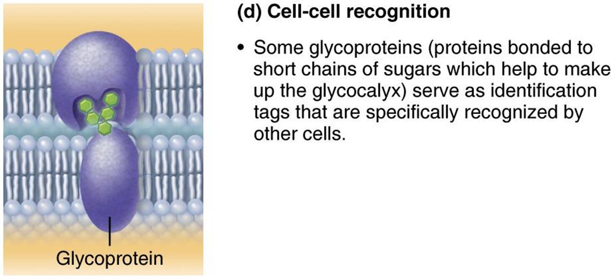
Cell-cell Recognition (membrane protein)
ex. some glycoproteins serve as identification tags that are specifically recognized by other cells
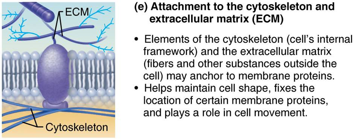
Attachment to ECM/ cytoskeleton (membrane protein)
elements of the cytoskeleton and extracellular matrix may anchor to membrane proteins (helps maintain cell shape, cell movement)
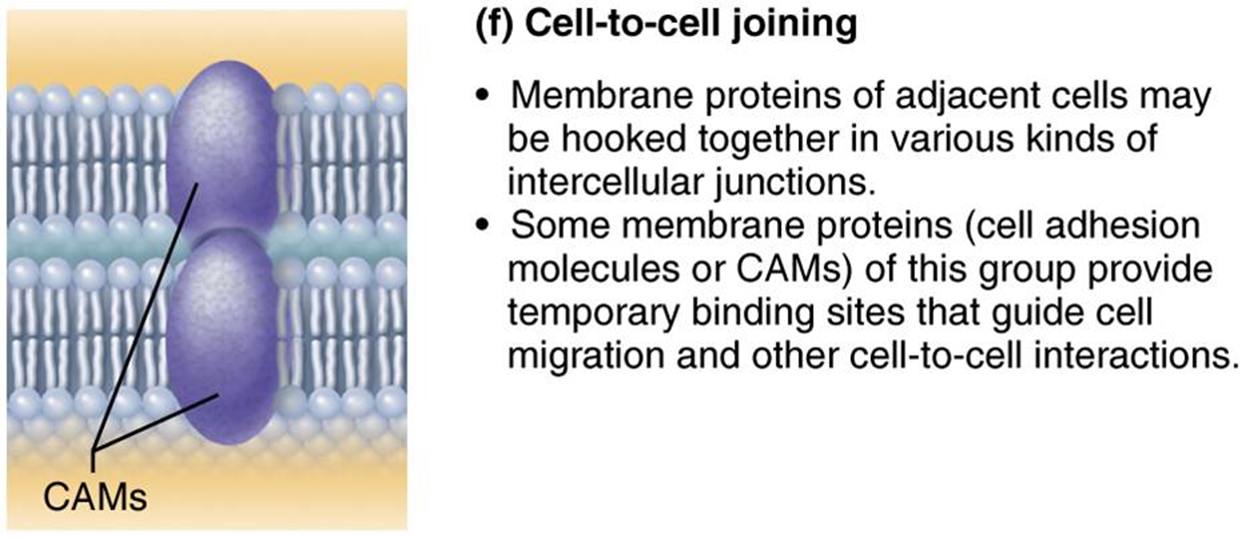
Cell-to-cell Joining (membrane protein)
membrane proteins hooked together in intercellular junctions
Cell Junctions
cells bound together to form tissues and organs
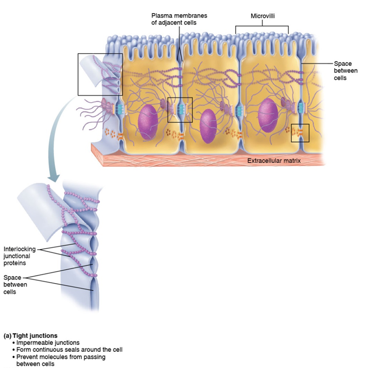
Tight Junction
impermeable junction that encircles the cell, prevent molecules from passing between cells
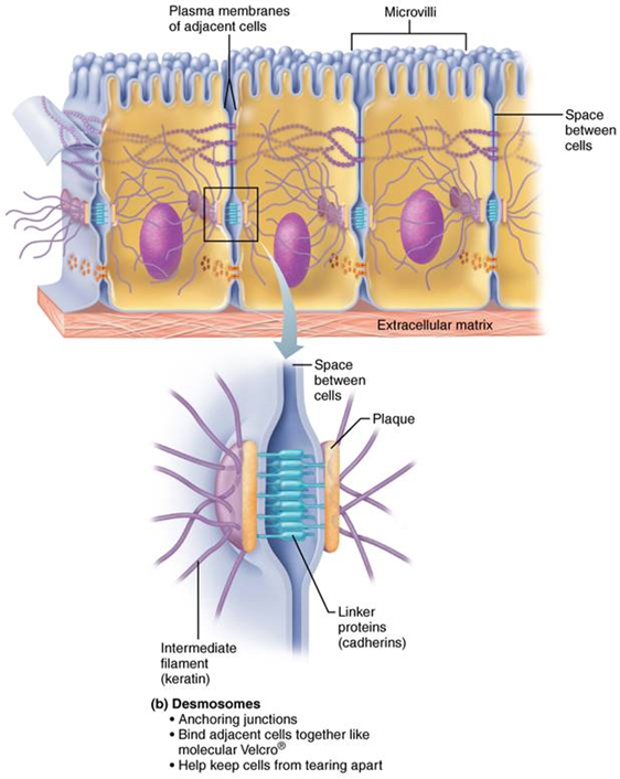
Desmosomes
anchoring junctions, bind cells together and keep cells from tearing apart
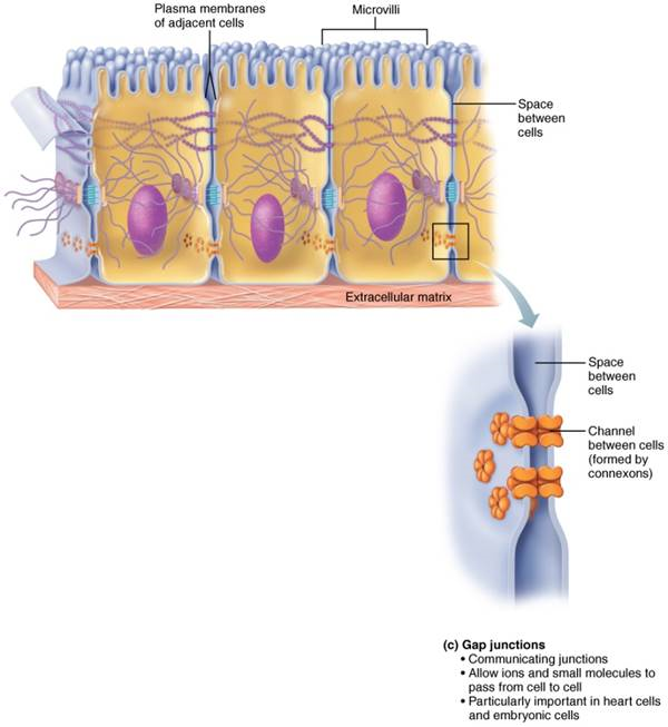
Gap Junctions
communicating junctions that allow ions/small molecules to pass from cell to cell (important in heart and embryonic cells)
Passive Transport
No energy required; transport of small and medium materials across the plasma membrane (Osmosis, Diffusion, and Facilitated Diffusion)
Diffusion
natural movement of molecules from areas of high concentration to areas of low (moves down concentration gradient)
Speed of Diffusion (passive transport)
influenced by 3 factors: concentration - the greater the difference, the faster the diffusion, molecular size - smaller molecules diffuse faster, and temp - higher temps increase kinetic energy which results in faster diffusion
Equilibrium
this is reached when there is no net movement of directions in one direction only
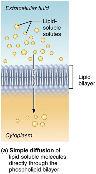
Simple Diffusion (passive transport)
Nonpolar lipid-soluble hydrophobic substances diffuse directly through the phospholipid bilayer (ex. O2, CO2, steroid hormones, fatty acids, and small polar substances like H2O)
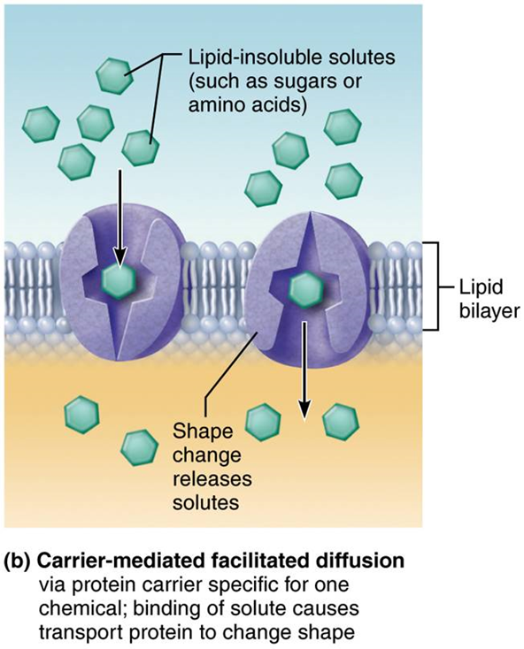
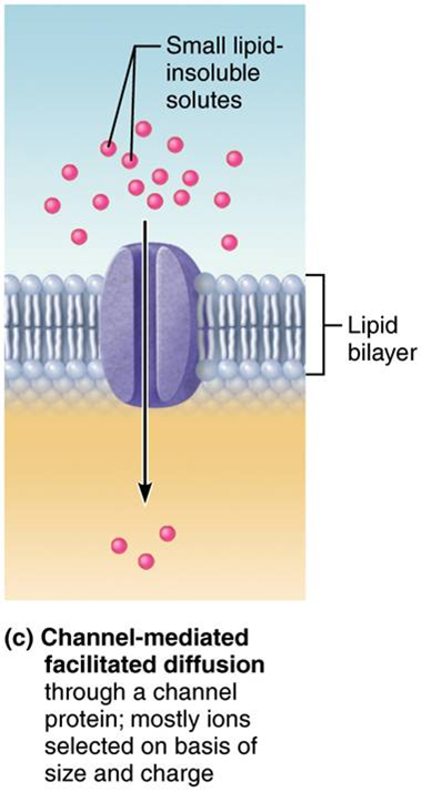
Facilitated Diffusion (passive transport)
Movement of specific molecules across cell membranes through protein channels (ex. channel-mediated facilitated diffusion through a protein channel: ions selected based on size/charge)
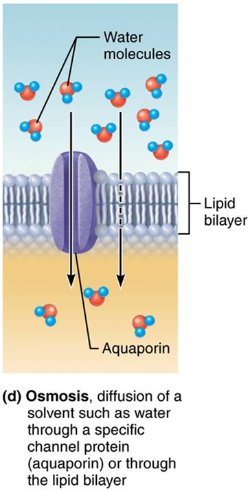
Osmosis (passive transport)
movement of water across a semipermeable membrane by specific protein channels - aquaporin or through lipid bilayer from areas of low solute (high water) to areas of high solute (low water)
Osmolarity (passive transport)
measure the concentration of the total number of solute particles in solvent
– Water moves by osmosis from areas of low solute (high water) concentration to high areas of solute (low water) concentration
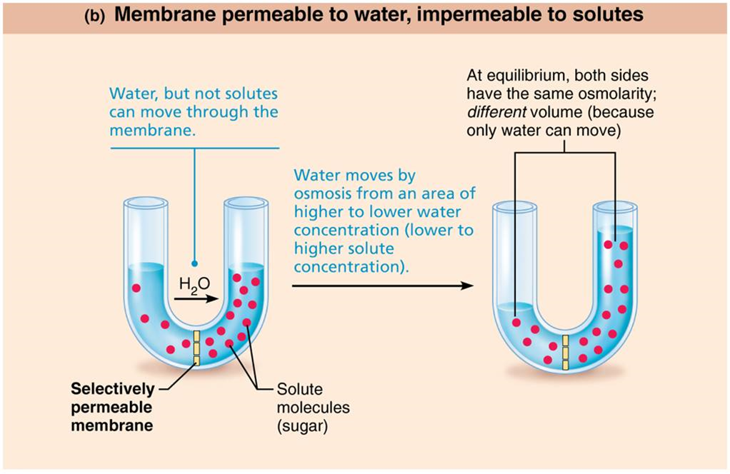
Water Concentration
varies with number of solute particles in solvent (when solute concentration goes up, water concentration goes down vice vera)
Tonicity
Ability of a solution to change the shape or tone of cells by altering the cells' internal water volume
Isotonic Solution
has same osmolarity as inside the cell, so volume remains unchanged (equal)
Hypertonic Solution
has higher osmolarity than inside cell, so water flows OUT of cell, resulting in cell shrinking
Hypotonic Solution
has lower osmolarity than inside cell, so water flows INTO cell, resulting in cell swelling
Lysing
effect of hypotonic solution that can cause the cell to burst
Active Membrane Transport
Energy-requiring process requires ATP to moves solutes across concentration gradient because solute is too big for channels and is not lipid soluble - two types: active and vesicular transport
Active Transport (active membrane transport)
requires carrier proteins called solute pumps, bind specifically and reversibly with moving substance
– Some carriers transport more than one substance; antiporters and symporters
–Moves solutes against their concentration gradient (from low to high), this requires energy (ATP)
Antiporters (active carrier transporters)
transport one substance into cell while transporting a different substance out of cell
Symporters (active carrier transporters)
transport two different substances in the same direction
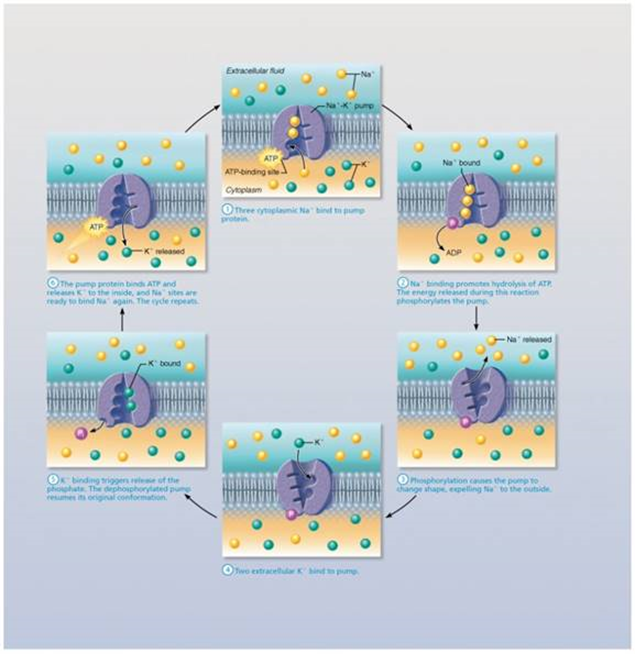
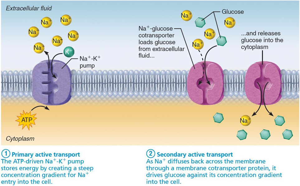
Primary Active Transport
required energy comes directly from ATP hydrolysis and this energy causes change in shape of transport protein (shape change causes ions bound to protein to be pumped across membrane ex of pumps: calcium, Na - k pumps)
Secondary Active Transport
Required energy is obtained indirectly from ionic gradients created by primary active transport (mostly Na+)
Vesicular Transport (active membrane transport)
involves transport of large particles, macromolecules and fluids across membrane in membranous sacs called vesicles
Vesicular Trafficking (vesicular transport)
transport from one area or organelle in cell to another
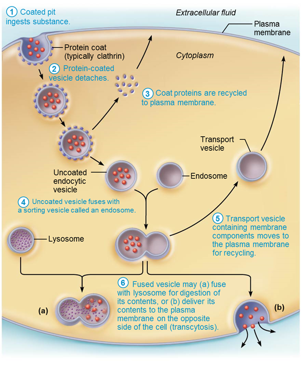
Endocytosis (vesicular transport)
transport into cell and involves formation of protein-coated vesicles
– Except for pinocytosis, usually involves receptors making it very selective; substance being pulled in must be able to bind to its unique receptor
– Once vesicle is pulled inside cell, it may fuse with a lysosome
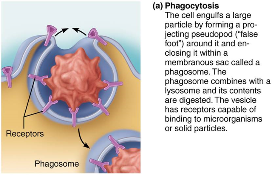
Phagocytosis (endocytosis)
large molecules, bacteria, or viruses are ingested by the cell to be broken down
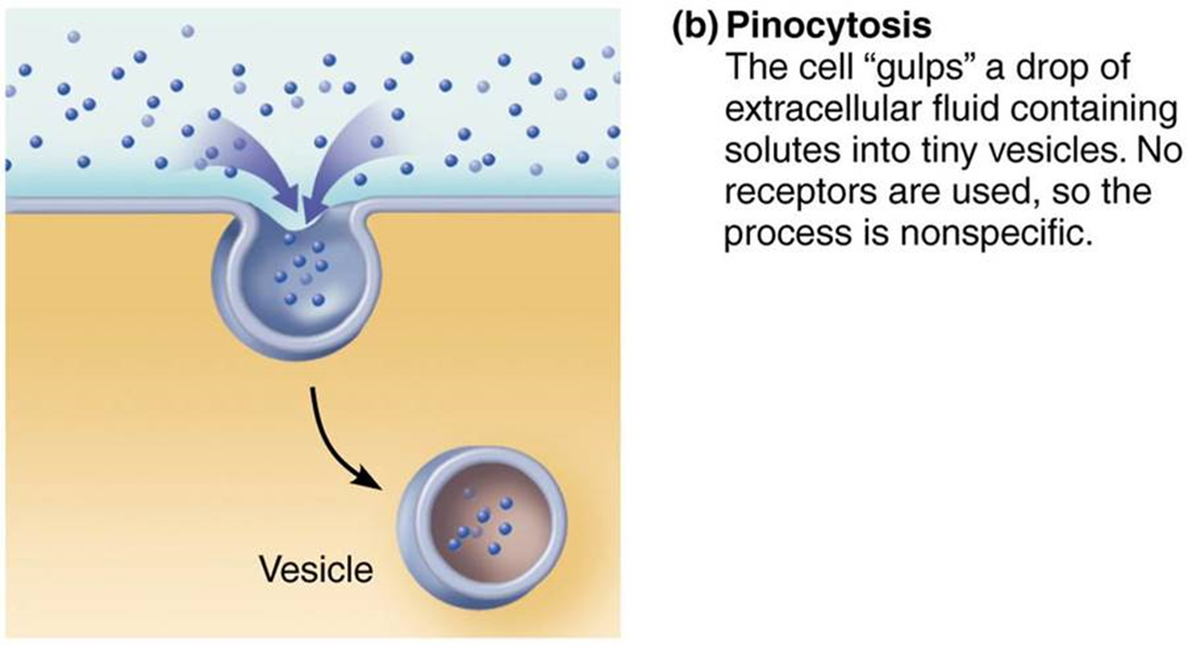
Pinocytosis (endocytosis)
cell 'gulps' a drop of extracellular fluid containing solutes into tiny vesicles
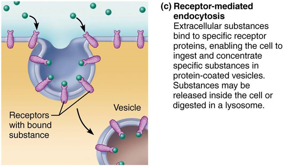
Receptor-mediated endocytosis
Extracellular substances bind to specific receptor proteins, enabling the cell to ingest and concentrate specific substances (ligands) in protein coated vesicles. Ligands may simply be released inside the cell, or combined with a lysosome to digest contents.v
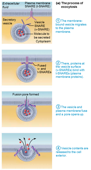
Exocytosis (endocytosis)
transport out of cell where materials enclosed in secretory vesicle are ejected out (ex. hormones, neurotransmitters, mucus, wastes)
– Protein on vesicle called v-SNARE finds and hooks up to target t-SNARE proteins on membrane
– Docking process triggers exocytosis
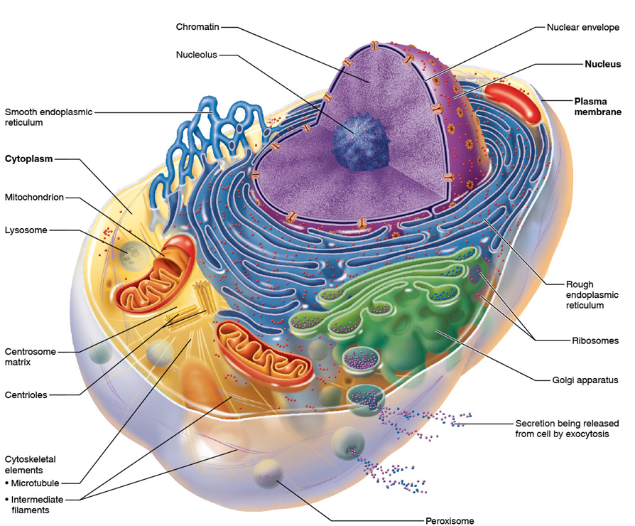
Cytoplasm
A jellylike fluid inside the cell in which the organelles are suspended (between plasma membrane and nucleus); made of cytosol, inclusions, and organelles
Cytosol
gel-like solution made up of water and soluble molecules such as proteins, salts, sugars, etc.
Inclusions
insoluble molecules; vary with cell type (examples: glycogen granules, pigments, lipid droplets)
Organelles
metabolic machinery structures of cell; each with specialized function; either membranous or nonmembranous
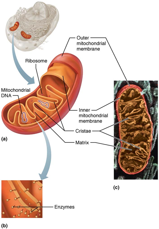
Mitochondria (Membranous)
Powerhouse of the cell because it produces most of cell's ATP molecules through aerobic cellular respiration; contain their own DNA, RNA, and ribosomes
Endoplasmic Reticulum (ER) (Membranous)
Consists of several series of parallel, interconnected cisterns
2 types - rough (site of protein synthesis) and smooth (contain enzymes)
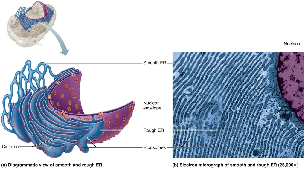
Rough ER (Membranous)
External surface appears rough because it is studded with attached ribosomes
Site of synthesis of proteins that will be secreted from cell
Site of synthesis of many plasma membrane proteins and phospholipids
Proteins enter cisterns as they are synthesized and are modified as they wind through fluid-filled tubes
Final protein is enclosed in vesicle and sent to Golgi apparatus for further processing
Smooth ER (Membranous)
Network of looped tubules continuous with rough ER
Enzymes found in its plasma membrane (integral proteins) function in:
Lipid metabolism; cholesterol and steroid-based hormone synthesis; making lipids for lipoproteins
Absorption, synthesis, and transport of fats
Detoxification of certain chemicals (drugs, pesticides, etc.)
Converting of glycogen to free glucose
Storage and release of calcium
Sarcoplasmic reticulum is specialized smooth ER found in skeletal and cardiac muscle cells
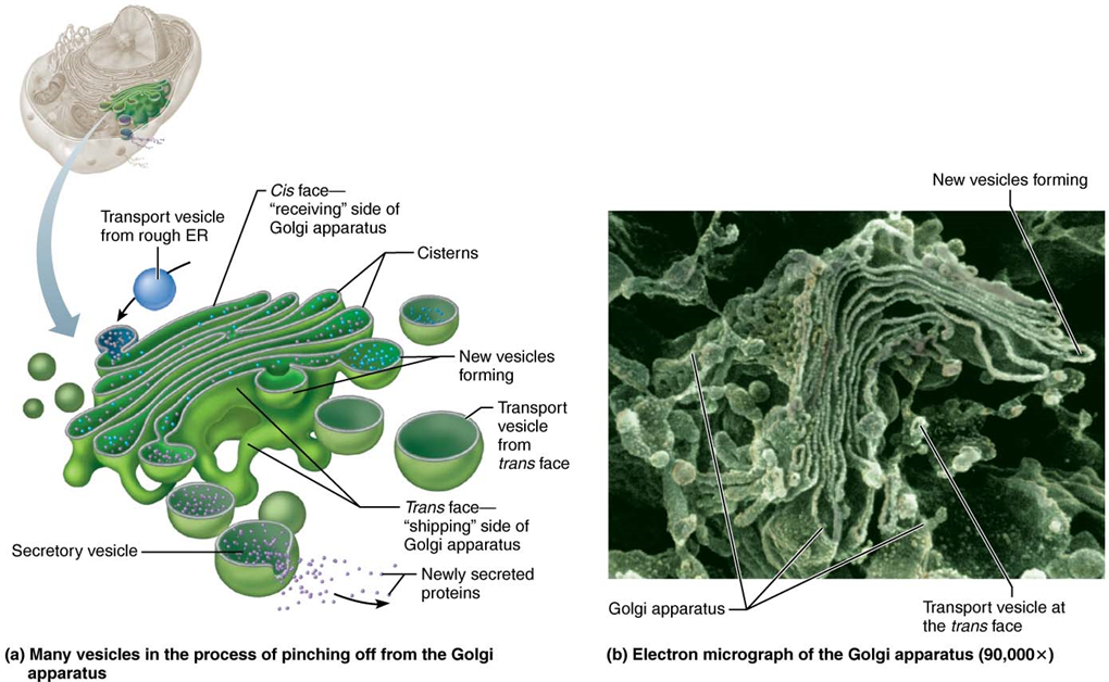
Golgi Apparatus (Membranous)
Modifies, sorts, and packages proteins and lipids for storage or transport out of the cell
Golgi Apparatus - Pathway A
Secretory vesicles containing proteins to be used outside of cell fuse with plasma membrane and exocytosis contents

Golgi Apparatus - Pathway B
Vesicles containing lipids or transmembrane proteins fuse with plasma membrane or organelle membrane, inserting contents directly into destination membrane
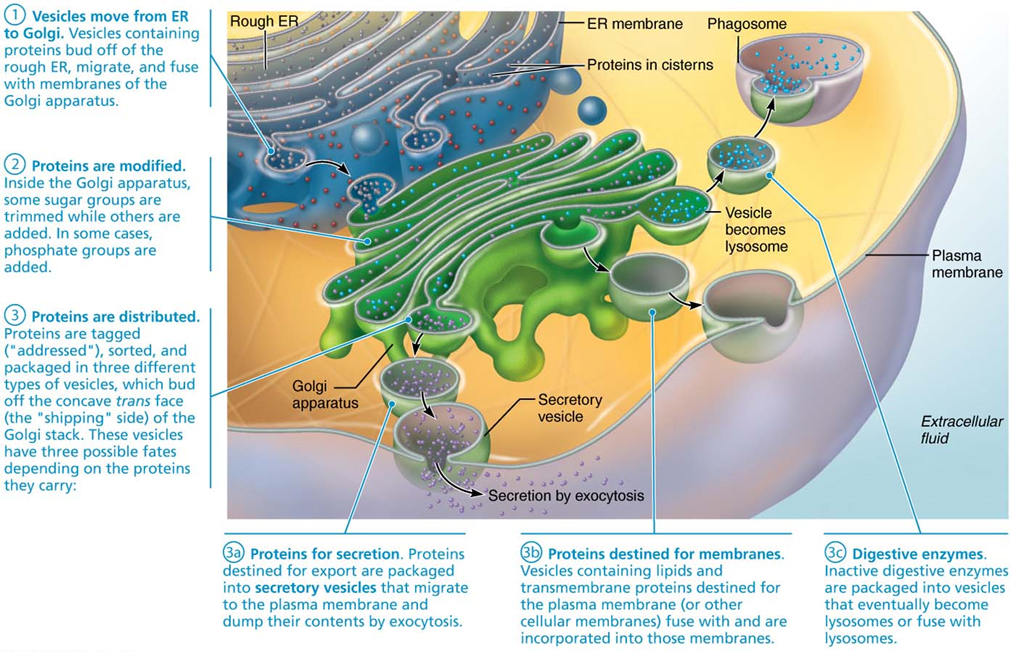
Golgi Apparatus - Pathway C
Lysosomes containing digestive enzymes remain in cell, holding contents in vesicle until needed

Peroxisomes (Membranous)
Membranous sacs containing enzymes used to break down free radicals (highly toxic and reactive molecules)
– Free radicals: toxic, highly reactive molecules that are natural by-products of cellular metabolism; can cause havoc to cell if not detoxified
– Two main detoxifiers: oxidase uses oxygen to convert toxins to hydrogen peroxide (H2O2), which is itself toxic; however, peroxisome also contains catalase, which converts H2O2 to harmless water
Lysosomes (Membranous)
Spherical membranous bags containing digestive enzymes; digest ingested bacteria, viruses, and toxins
– Considered “safe” sites because they isolate potentially harmful intracellular digestion from rest of cell
– Degrade nonfunctional organelles
– Metabolic functions: break down and release glycogen; break down and release Ca2+ from bone
Tay-Sachs Disease
Condition where patient lacks lysosomal enzyme needed to break down glycolipids in brain cells
Endomembrane System
Consists of membranous organelles in cytoplasm, nuclear/plasma membranes that work together to produce, store, degrade, and export biological molecules
Ribosomes (Non-membranous)
Site of protein synthesis
Free ribosomes
free floating; site of synthesis of soluble proteins that function in cytosol or other organelles
Membrane-bound ribosomes
attached to membrane of endoplasmic reticulum (ER); site of synthesis of proteins to be incorporated into membranes or lysosomes, or exported from cell
Cytoskeleton (Non-membranous)
Elaborate network of rods that run through cytosol
3 types - microfilaments, intermediate filaments, and microtubules
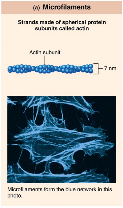
Microfilaments
Strands made of spherical protein units called actin
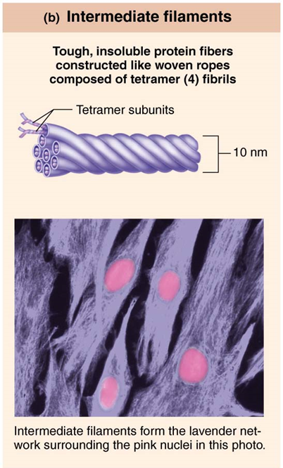
Intermediate Filaments
Tough, insoluble protein fibers constructed like woven ropes composed of tetramer (4) fibrils
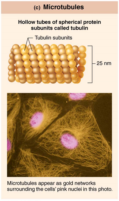
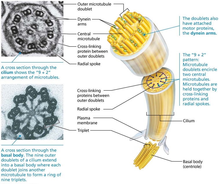
Microtubules
Hollow tubes of spherical protein subunits called tubulin
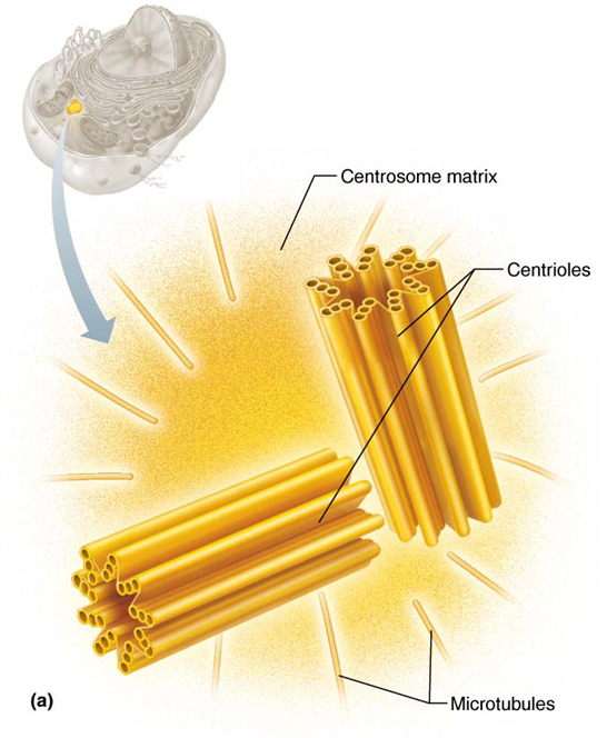
Centrioles (Non-membranous)
Forms basis of cilia and flagella
Cilia and Flagella
Aid in the movement of the cell or of materials across the surface of the cell
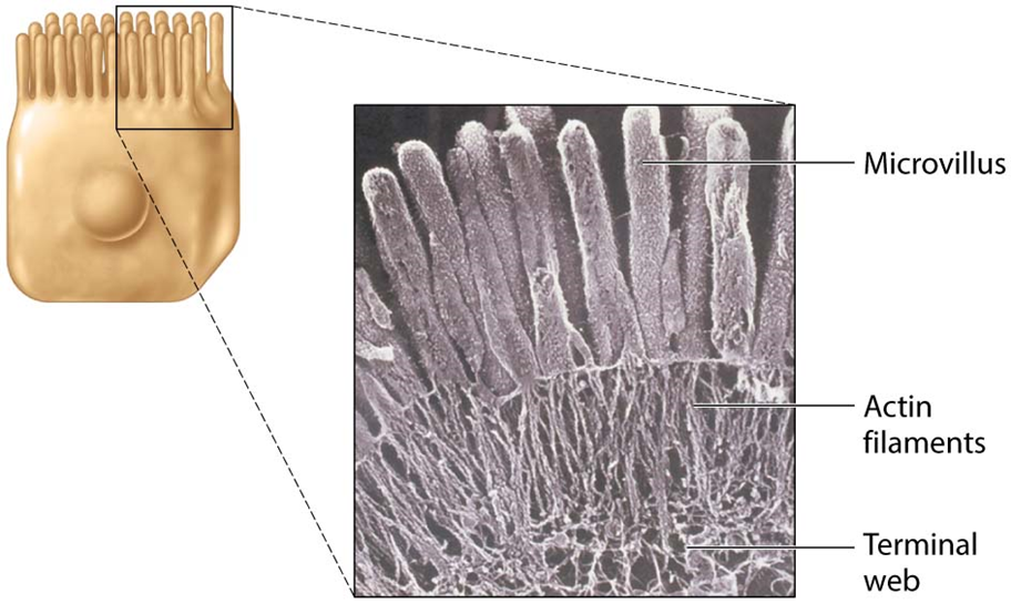
Microvilli
Finger-like projections that increase surface area of cell
Nucleus
Contains genetic library of blueprints for synthesis of nearly all cellular proteins
The nucleus has three main structures:
– Nuclear envelope
– Nucleoli
– Chromatin
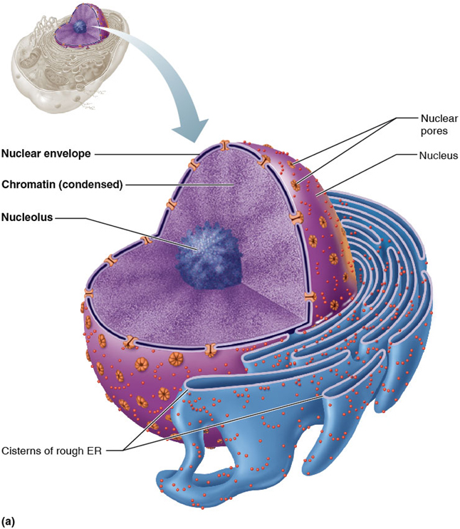
Nuclear Envelope
A double membrane that surrounds the nucleus in the cell
– Outer layer is continuous with rough ER and, like the rough ER, is studded with ribosomes
– Inner layer, called nuclear lamina, is a network mesh of proteins that maintains nuclear shape and acts as scaffolding for DNA
Nuclear Pores
Allow substances to pass into and out of nucleus; they are guarded by the nuclear pore complex, which regulates transport of specific large molecules
Nucleoli
Dark-staining spherical bodies within the nucleus
Involved in ribosomal RNA (rRNA) synthesis and ribosome subunit assembly
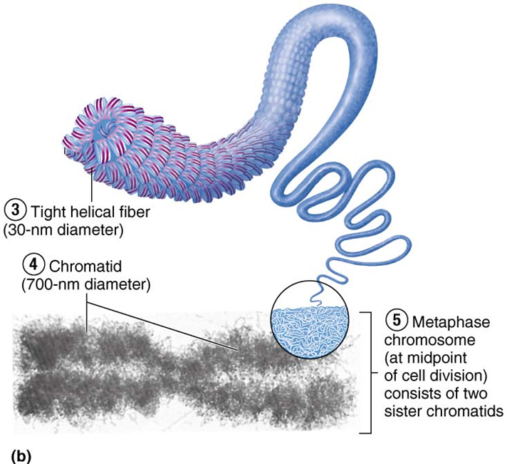
Chromatin
Consists of 30% threadlike strands of DNA, 60% histone proteins, and 10% RNA
Arranged in fundamental units called nucleosomes, which consist of DNA wrapped around histones
Histones help regulate gene expression
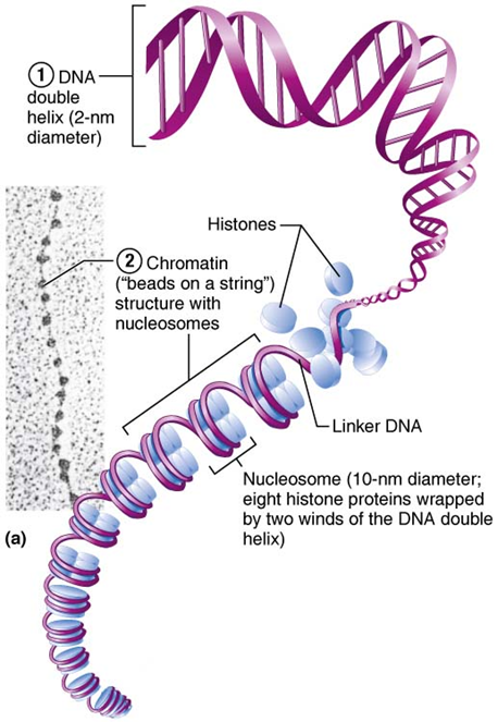
Chromosome
is condensed chromatin
Condensed state helps protect fragile chromatin threads during cell division
Cell Cycle
Series of changes a cell undergoes from the time it is formed until it reproduces
Two major periods of cell cycle:
– Interphase
– Cell division (mitotic phase)
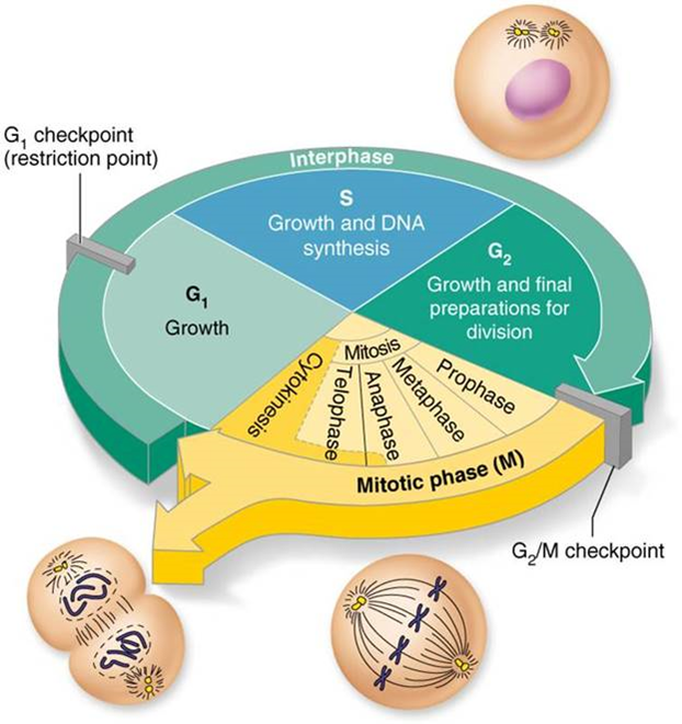
Interphase
Period from cell formation to cell division, in which cell carries out its routine activities and prepares for division
During interphase, nuclear material is in uncondensed chromatin state
Interphase consists of subphases, which include the process of DNA replication
G1 Phase
Vigorous growth, protein synthesis, and metabolism
S Phase
DNA replication occurs
G2 Phase
Preparation for division
DNA Replication
Prior to division, the cell makes a copy of DNA
Double-stranded DNA helices unwind and unzip
Each strand acts as a template for a new complementary strand
RNA starts replication by laying down short strand that acts as a primer
A polymerase attaches to primer and begins adding nucleotides to form new strand
§DNA polymerase synthesizes both new strands at one time (one leading and one lagging strand)
DNA polymerase works only in one direction, so leading strand is synthesized continuously; however, because lagging strand is “backwards,” it is synthesized discontinuously into segments
Another enzyme, DNA ligase, then splices short segments of discontinuous lagging strand together
End result: two identical “daughter” DNA molecules are formed from the original
During mitotic cell division, one complete copy will be given to new cell while one is retained in original cell
Process is called semiconservative replication because each new double-stranded DNA is composed of one old strand and one new strand
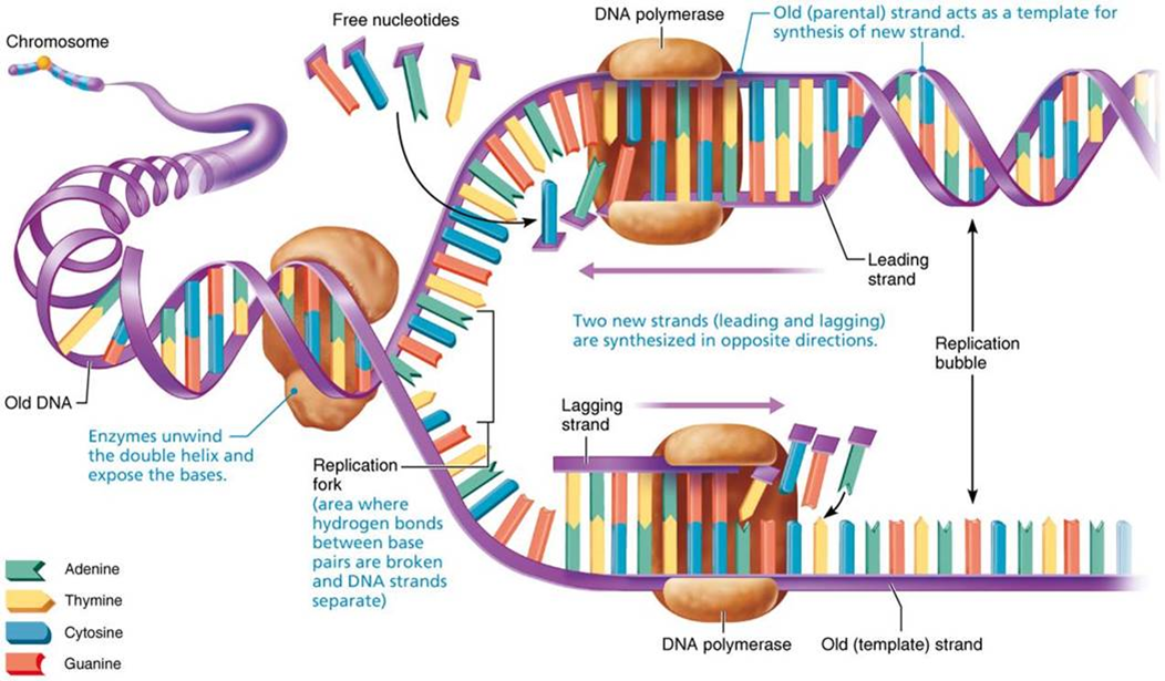
Replication fork
point where strands separate
Replication bubble
active area of replication
M (mitotic) Phase
division occurs; consists of 2 distinct events:
– Mitosis
– Cytokinesis
Mitosis
Division of nucleus, in which replicated DNA is distributed to new daughter cells (4 stages)
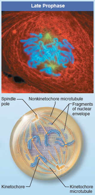
Prophase
Chromatin fibres coil up to become chromosomes (2 chromatids joined at the centromere)
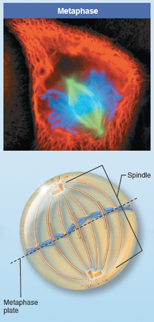
Metaphase
Mitotic spindle is formed and chromosomes are lined up in middle of the 2 spindle poles
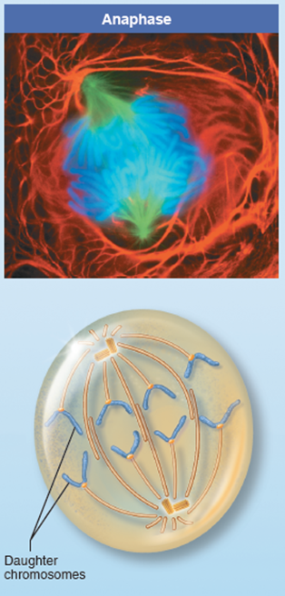
Anaphase
Centromeres come apart and 2 sister chromatids are pulled to opposite poles
Telophase
Nuclear envelopes form around identical sets of chromosomes at 2 poles
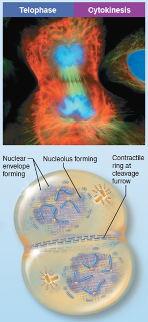
Cytokinesis
Splits cytoplasm and separates 2 daughter cells

Control of Cell Division
“Go” and “Stop” signals direct when a cell should and should not divide
Go signals include:
– Critical surface-to-volume ratio of cell, when area of membrane becomes inadequate for exchange
– Chemicals (example: growth factors, hormones)
Stop signals include:
– Availability of space; normal cells stop dividing when they come into contact with other cells
– Referred to as contact inhibition
S Phase Proteins
Two groups of proteins are crucial to cell’s ability to accomplish S phase and enter mitosis:
– Cyclins: regulatory proteins that accumulate during interphase
– Cdks (Cyclin-dependent kinases) that activate cyclins when they bind to them
– Cyclin-Cdk complex in turn activates enzyme cascades that prepare cell for division
– Cyclins are destroyed after mitotic cell division, and process begins again
Checkpoints
key events In the cell cycle where cell division processes are checked and, if faulty, stopped until repairs are made
– G1 checkpoint (restriction point) is the most important of the three major checkpoints
– If cell does not pass, it enters G0, in which no further division occurs
Gene
segment of DNA that holds the code for one polypeptide
The code is determined by the specific order of nitrogen bases (Adenine, Guanine, Thymine, and Cytosine) in the gene
– Code consists of three sequential bases (triplet code)
– Each triplet specifies the code for a particular amino acid
– Genes are composed of exons and introns
Exons
part of gene that actually codes for amino acids
Introns
non-coding segments interspersed amongst exons
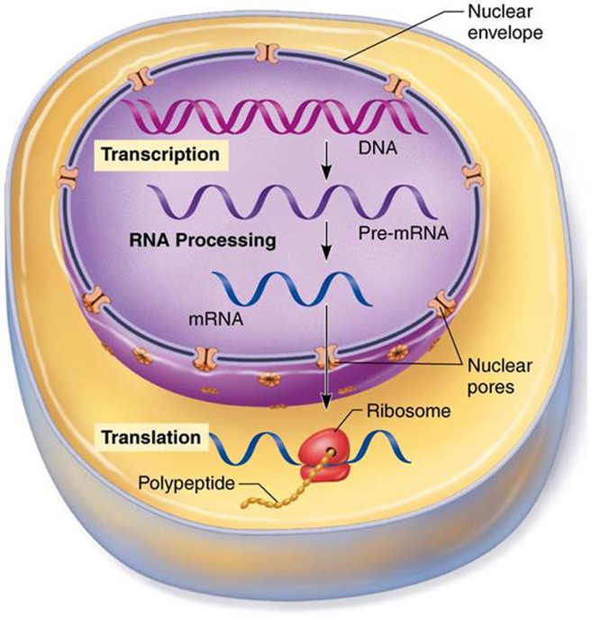
Role of RNA
RNA is the “go-between” molecule that links DNA to proteins
RNA copies the DNA code in nucleus, then carries it into cytoplasm to ribosomes
All RNA is formed in nucleus
RNA differs from DNA
– Uracil is substituted for thymine in RNA
– RNA has ribose instead of deoxyribose sugar
Three types of RNA:
– Messenger RNA (mRNA)
– Ribosomal RNA (rRNA)
– Transfer RNA (tRNA)
Messenger RNA (mRNA)
Single stranded
Code from DNA template strand is copied with complementary base pairs, resulting in a strand of mRNA
– Process is referred to as transcription
– mRNA maintains the triplet code (codon) from DNA
Codon
3 bases of mRNA that code for a single amino acid
Ribosomal RNA (rRNA)
Structural component of ribosomes, the organelle where protein synthesis occurs
Along with tRNA, helps to translate message from mRNA into polypeptide
Transfer RNA (tRNA)
Carrier of amino acid
Have special areas that contain a specific triplet code (anticodon) that allows each tRNA to carry only a specific amino acid
Anticodon of tRNA will complementary base-pair with codon of mRNA at ribosome, adding its specific amino acid to growing polypeptide chain
– Process is referred to as translation
Anticodon
3 bases of tRNA that code for a single amino acid
Transcription
DNA information coded in mRNA
Has 3 phases:
– Initiation
– Elongation
– Termination