BIOL 1510 Chapter 4
 Call Kai
 Learn
Learn Practice Test
 Spaced Repetition
 Match
Match Flashcards
 Knowt Play
1/46

There's no tags or description
Looks like no tags are added yet.
Name | Mastery | Learn | Test | Matching | Spaced | Call with Kai | Chat |
|---|
No analytics yet
Send a link to your students to track their progress
47 Terms
Why are peptide bonds planar?
Because of resonance, which gives them a partial double bond character

Can peptide bonds rotate?
No! Rotation is restricted by partial double bond character
What bonds can rotate in the peptide backbone?
N-C and C-C bonds that are no involved in the peptide bond itself

Is bond rotation easy in peptides?
No, most rotation angles are restricted by steric hinderance
Ramachandran plot
Depicts which rotation angles are allowed and which ones aren’t
darker color=allowed
lighter color=disallowed
How are alpha helices formed?
Hydrogen bonds between main chain atoms (O-H)
How many residues are present in each alpha helix?
3.6 residues
Where are the R groups positioned in an alpha helix?
Point outwards
What direction does an alpha helix wind in?
right-handed helix
Dipole moment in alpha helix
Dipole forms from N terminus to C terminus
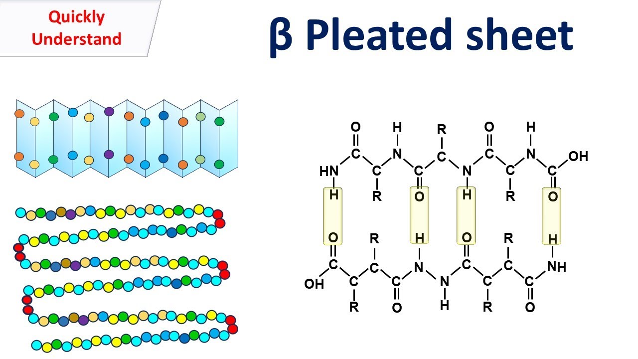
Beta strand
Formed by hydrogen bonds between main chain O and H atoms
Beta sheet
When beta strands hydrogen bond together
Parallel beta sheets
N terminus lined up with N terminus and vice versa

Antiparallel beta sheet
N terminus lined up with C terminus
Which form of beta sheet (parallel or antiparallel) is stronger and why?
Antiparallel is more stable because the hydrogen bonds between beta strands are head on, leading to stronger interactions
Beta turn
Rounding of a corner that connects two strands of an antiparallel beta sheet
two types: type I and type II
What amino acids are involved in a beta turn?
glycine or proline
Why is glycine good for beta turns?
small and flexible amino acid
Why is proline good for beta turns?
Bulky but can assume a cis or trans configuration
Position of R groups in a beta sheet
R groups protrude in opposite direction from the zig zag pattern

Fibrous proteins
long stringy proteins that are often important for structure
Fibrous protein structure
Usually made of tight alpha helices
can have repeat sequences of glycine, proline, hydroxy-proline
Globular protein
Balled up and compact proteins often important for cellular functions
Ways to represent protein structure
ribbon representation, surface model, ball and stick model, space-filling model
Ribbon representation
Mostly secondary structures

Surface model
This thing

Ball and stick model
Can clearly see atoms and functional groups
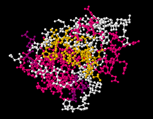
Space filling model
Similar to surface model

Beta-alpha loop
A loop formed by a repeating pattern of
beta strand
alpha helix
beta strand
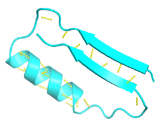
Alpha-beta barrel
A type of fold when multiple beta-alpha loops come together
Supersecondary structure
3D structure consistent of multiple secondary structures
Motif
Any simple or complex distinct folding patter
supersecondary structure
Domain
A distinct structural unit that may have a separate function
Do all proteins have ordered secondary structures?
No!
Intrinsically disordered domains
Parts of a protein that don’t have a set protein structure
What is the purpose of intrinsically disordered domains?
Allows for morphing/conformational change
Proteostasis
homeostasis but for proteins
Chaperone proteins
proteins that create favorable microenvironments that allow naive proteins to fold properly
Chaperonins and heat shock proteins
helper proteins that interject during protein misfolding and help proteins refold into the correct form
Anfinsen’s classical experiment
denatured and reduced ribonuclease A—>denaturing and reducing agent added
Both agents were slowly removed
Ribonuclease A activity was measure and found that it rose over time
Conclusion of Anfinsen classical experiment
Protein refolded itself!
Techniques to determine protein structure
circular dichroism
NMR
X-ray crystallography
cryo-EM
Circular dichroism
A spectroscopy technique that allows you to determine the fraction alpha helices and beta sheets
NMR spectroscopy
3D NMR used to determine small protein structure by placing it in magnetic field
X-ray crystallography
protein crystals bombarded with x-rays to create a diffraction pattern. Then, the pattern is analyzed to generate a model
cryo-EM
determines structure of large complexes
What secondary structure is an extended conformation of the polypeptide chain?
beta sheets