Basic ECG interpretation
 Call Kai
 Learn
Learn Practice Test
 Spaced Repetition
 Match
Match Flashcards
 Knowt Play
1/63

There's no tags or description
Looks like no tags are added yet.
Name | Mastery | Learn | Test | Matching | Spaced | Call with Kai | Chat |
|---|
No analytics yet
Send a link to your students to track their progress
64 Terms
The components of the conduction system comprises of:
The Sino-Atrial (SA) node
The Atrio-Ventricular (AV)
The bundle of His
The right bundle branch
The left bundle branch
Purkinje Fibres
What is the primary pacemaker of the heart?
The SA node
Where is the SA node located?
Top right of the atrium → its electrical impulses initiates the cascade of depolarization through the heart
What is the rate of the SA node
Fires an electrical impulse of 60-100 per minute
What are the 3 internodal tracts of the right atrium ?
Anterior internodal tract
Middle internodal tract
Posterior internodal tract
The internodal tracts of the right atrium conduct the impulse from the SA node through to the AV node in the right atrium.
What conducts the impulse to the left atrium?
Bachmann’s Bundle
Where is the AV node located?
Located at the base of the right atrium
What are the 3 functions of the AV node?
Conduct impulses from atria to ventricles
AV delays: if atrial rate is too fast the AV node will block some of the impulses so that the ventricular contraction rate is slower that atrial rate e.g. in AF with an atrial rate of 300bpm the ventricular rate can be 150bpm =2:1 conduction
Secondary pacemaker of the heart when SA node fails. It has the intrinsic ability to generate a junctional impulse at a rate of 40-60bpm
Where is the bundle of His located?
Base of the AV node
What is the bundle of His?
It is a short bundle of fibres loacted at the base of the AV node, from here the impulse bifurcates into both the left and right bundle branches
What is the role of the Right bundle branch?
To conduct the impulse to the right ventricle via the Purkinje fibres
Where is the RBB located?
To the right of the inter ventricular septum towards the apex and only has one fasicle
What are the purkinje fibres and where are they located?
Loacted at the end of the bundle branches. They conduct the impulse to the ventricle muscle cells. They are the last backup pacemaker and usually generate a ventricular rate of 20-40bpm
What is the role of the left bundle branch block ?
To conduct impulse to the left ventricle via the Purkinje fibres
Where is the LBB located?
To the left of the interventricular septum and splits into two fasicles
What two fascicles does the LBB split into?
Anterior Fascicle
Posterior Fascicle
Cardiac action potential phases
Phase 4 (Resting): The cell rests at about -90 mV. K+ maintains resting potential.
Phase 0 (Depolarisation): Fast sodium (Na⁺) channels open. Sodium rushes in. The cell turns positive (up to +30 mV).
Phase 1 (Initial Repolarisation): Sodium channels close. Potassium (K⁺) starts to leak out, causing a tiny drop in voltage.
Phase 2 (Plateau): Calcium (Ca²⁺) channels open. Calcium flows in while potassium keeps leaking out. This balance holds the charge steady and causes the muscle to contract.
Phase 3 (Repolarisation) Calcium channels close. Potassium keeps leaving. The cell returns to its negative resting state
Phase 4: Returns to resting membrane potential. K+
What is depolarization of the atria characterised by?
P wave
What does the PR interval measure?
The time required for an impulse to travel across the atria and through the AV node
What is the depolarisation of the ventricles characterised by?
QRS complex
What is the Q wave?
It is an initial negative deflection and should be <0.03sec and <25% of R wave amplitude
What is the first positive deflection?
The R wave
What is a negative deflection following an R wave?
S wave
What is generally unseen and is hidden in the QRS complex?
The repolarisation of the atria (the recovery phase where the upper chambers of the heart (atria) electrical charge resets and the muscle relaxes after contraction)
What is the repolarisation of the ventricles characterised by?
T wave
Why is it called a 12 lead ECG?
Because it looks at 12 different views of the heart
Why are their only 10 electrodes placed?
Are used in combination to produce the 12 leads. Each electrode has a positive or negative pole and the electrical current moves from negative to positive.
What leads is an ECG comprised of?
3 bipolar leads (I, II and III)
9 unipolar leads (aVR, aVL, aVF, V1-6)
Basic law of electrophysiology
If the average flow of impulses are traveling toward a positive pole then the deflection on the ECG will be positive
If the impulses are traveling away from the positive pole then the deflection will be negative
If the impulses are at right angels to the positive pole the deflection will be equiphasic
If no electrical activity is detected then no deflection occurs
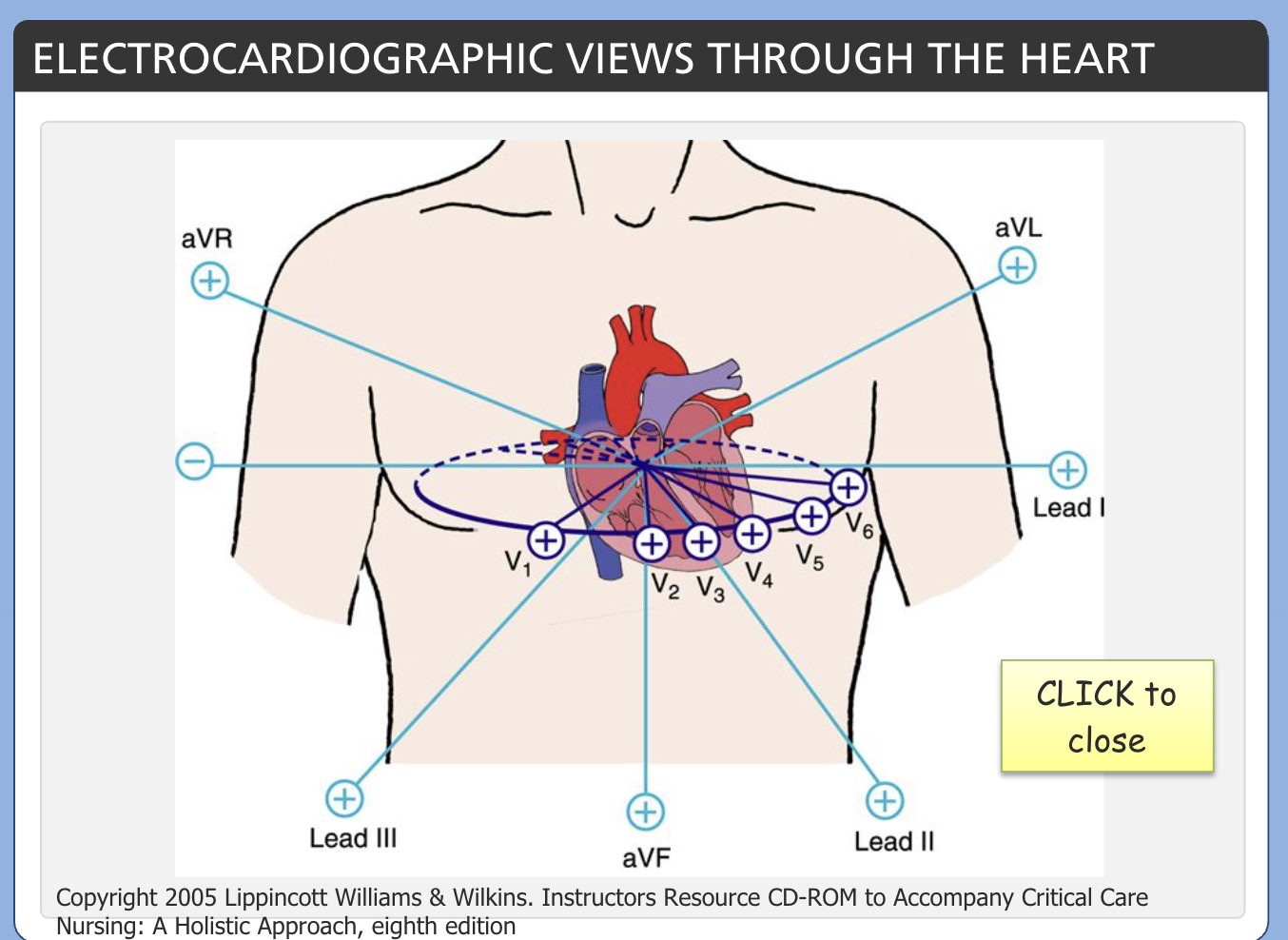
Electrocardiographic views through the heart
Refer to picture.
Leads I, II and III use the three main electrodes. Right Arm, Left Arm, Left Leg to gain capture. The electrodes form an imaginary triangle called Einthoven’s Triangle. It is an equilateral triangle which has the heart at its centre.
What is the standard voltage for an ECG?
1mv (10mm)
→ each small 1mm x 1mm square equals 0.1mv or 0.04 sec
→ each large 5mm x 5mm square equals 0.5mv or 0.2 sec
What is the standard speed for an ECG?
25mm per sec
What do 1, 5, 15 and 30 large squares represent on an ECG paper?
1 = 0.2 sec
5 = 1 sec
15 = 3 sec
30 = 6 sec
How to calculate rate using the ECG paper?
To count how many QRS complexes are within 6 seconds (30 large squares), then times by 10 to calculate heart rate per minute
What is the isoelectric line?
This is the flat base line that all waveforms arise from. It is important to have a level isoelectrical line so that the following waves, intervals and segments can be accurately measured.
A wandering baseline is commonly caused by chest movement related to respirations.
What is the P Wave?
This is atrial depolarisation
→ should be <0.2mv in amplitude and <0.12 sec in duration
→ P Mitrale or ‘m’ shaped P waves with a duration >0.12 sec may indicated left atrial dilation
→ P Pulmonale or notched/peaked P waves with an amplitude >0.3mv may indicate right atrial dilation
What are Q waves?
Q waves are deemed to be pathological if:
→ >2mm (0.2mv) deep or
→ >1mm (0.04sec) wide or
→ more than 25% of the height of the following R wave
They usually appear after an ST elevation Myocardial infarction (STEMI) and indicate myocardial damage
What is the QRS complex?
This is ventricular depolarisation
→ Should be 0.04-0.10 sec
→ Measured from the point it first leaves the baseline to the end of the last wave.
→ If >0.12 sec, indicates intraventricular conduction delay
→ Amplitude in any one lead should not exceed 25mm (2.5mv) or 35mm (3.5mv)
→ If the amplitude of the S wave of V1 or V2 is added to the R wave of V5 or V6 and the total is over 35mm (7 large squares), it may indicate left ventricular hypertrophy
What is the T wave?
This is Ventricular repolarisation
→ Amplitude <5mm (0.5mv) in limb leads and <10mm (1mv) chest leads
→ Tall T waves may be a sign of hyperkalemia or early MI
→ T wave inversion may be a sign of ischemia or non-STEMI in combination with other leads
→ T wave inversion may also be seen when repolarisation of the ventricle is affected, as in bundle branch block or hypertrophy
What is the PR interval?
→ The time impulses take to cross the atria and through the AV node
→ Measured from the beginning of the P wave to the beginning of the QRS complex
→ Normal PR interval is 0.12-2.0 sec
→ A consistently prolonged PR interval represents 1st degree AV block
What is the QT interval?
→ Duration of ventricular depolarisation and repolarisation
→ QT should be <0.45 sec. QT interval may be prolonged if bradycardic
→ QTc (corrected QT interval) is the QT divided by the square root of the R-R interval
→ A prolonged QTc may be caused by electrolyte imbalance, drug toxicity or bradyarrhythmias. This may cause Torsades de Pointe ventricular tachycardia
What is the ST segment?
→ End of ventricular depolarisation and the beginning of repolarisation
→ Elevation above the isoelectric line >1mm (0.1mv) in limb leads and >2mm (0.2mv) in chest leads is consistent with MI (infarction)
→ other causes of ST elevation include paced rhythm, LBBB, pericarditis, ventricular aneurism and dissection of the aortic root and coronary arteries.
→ Depression below the isoelectric line may be consistent with myocardial ischemia
What is the R-R interval?
→ The distance from the top of the QRS complex on a rhythym strip to the next QRS
→ Measuring the R-R interval is especially useful for counting HR or identifying regularity of a heart rhythm
Analysing the ECG and questions to ask myself
Rate - is the atrial and ventricular rate normal (60-100bpm)
Rhythm - is it regular with no dropped beats
P waves - does it have a normal shape and size
P-R interval - is there a constant distance and is it 0.12-0.20 sec (2-5 small squares)
QRS - does it have a normal QRS complex, with a duration 0.06-0.10 sec and amplitude <3.0mv?
ST segment - do the wave forms return to isoelectric line?
Conduction - is the sinus impulse always conducted to the ventricles? Is there a P wave for every QRS compelx
What are the characteristics of Junctional Rhythym?
Absence of P waves
40-60 bpm
QRS less than 0.12 sec
Regular R-R complex
The time impulses take to cross the atria and through the AV node is called the ?
P-R interval
What is the arrhythmia with varying R-R intervals which is associated with the changes in thoracic pressure is?
Sinus arrhythmia (often seen in children, young adults and dogs)
Areas of the heart and their back-up pacemaker rates
Atria - 60-100bpm
A-V junction - 40-60bpm
Ventricles - 20-40bpm
What is the arrhythmia characterised by irregular QRS complexes and fibrillatory P waves
Atrial fibrillation
True or False. Q waves can indicate, among other conditions, previous MI
True. They represent necrosis or death of the myocardial tissue
Torsades de Pointe ventricular tachycardia may be caused by?
Prolonged QTc
True or False. Asystole is a shockable rhythm
False
What is the correct standardised speed for ECG recording paper?
25mm/sec
Notched or peaked P waves with an amplitude >0.3mv may indicate right atrial dilation and are called?
P Pulmonale
The arrhythmia characterised by a prolonged PR interval (>0.20sec) is:
First degree AV block