exam - basic extra and intra oral anatomy
 Call Kai
 Learn
Learn Practice Test
 Spaced Repetition
 Match
Match Flashcards
 Knowt Play
1/103

There's no tags or description
Looks like no tags are added yet.
Name | Mastery | Learn | Test | Matching | Spaced | Call with Kai | Chat |
|---|
No analytics yet
Send a link to your students to track their progress
104 Terms
Which anatomical landmark is normally seen on intraoral radiographs?
Maxillary sinuses
When labeling radiographs, what should be included?
Date of exposure
Which radiographic exposure produces a radiograph that shows the crowns of both the maxillary and mandibular teeth?
Bitewing
Panoramic images are used to
Assess growth and development
Proper care of phosphor plates includes
Disinfection according to the instructions for use
What are common parts of a dental X-ray machine?
Control panel, extension arm, tubehead
A quality assurance policy for dental radiographs should include a
Written quality assurance (QA) program
What should you ask a patient to do before you begin exposing intramural radiographs?
Remove any removable oral appliance and objects from the head and neck area.
When exposing radiographs on an edentulous patient, compared to a dentyolous patient, the exposure setting should:
Be reduce (decreased)
What is the result of primary, secondary and scatter radiation?
Flip fog (image fogging)
To reduce cross contamination during dental radiography
Wear appropriate PPE and cover the control unit and tubehead with plastic barriers
The paralleling technique is recommended for which radiographic exposure?
Periodical radiographs
Joker smile appearance
Panoramic radiograph when pts chin is too low
PSP receptors should always be handled by the edges to prevent
Scratches
PSP
Photostimulable phosphor - type of digital image receptor
Which structure appears radiopaque on a dental radiograph?
Latina dura
What is the maximum annual permissible dose (mod) of ionizing radiation for a member of the public?
0.1 REM per year L1 msg o 0.001 Sv)
Ionization occurs when a X-ray strikes an acorn and the atom..
Loses or gains an electron, becoming charged (an ion)
Radiolucent
The dark, or black, portions of image • less dense structures
Radiopaque
The light, or white portions of the images
Radiolucent
Periodontal ligaments, pulpal tissues • soft tissue and air space
Radiopaque
Dentil, enamel, filling
Condole of the mandible
Rounded upper back of the lower jawbone. Radiopaque
Maxilla
Two bones that form the upper jaw and part of the maxillary structure •radiopaque
Ramus
Vertical portion at each end of the mandible that are perpendicular•radiopaque
Plural of ramus
Rami
Body of the mandible
Curve, horizontal part of the mandible that extended from one ramus to the other. Radiopaque
Mandible
Single bond lower jaw and oly movable bone in the skull. Made of condoled process, ramus, alveolar
Condoled process
Thin rectangular bone
Alveolar
Compact bone lining the tooth sock
Midsagittal plane (midline)
Imaginary vertical line that equally separate left & right
Orbit
Eye cavity, appears on ceph & pano • radiolucent
Frankfort plane
Imaginary line from the bottom of the eye socket to the ear canal.
Landmark
Specific recognizable feature or point on a bone or body structure to identify its location. To not confuse them with diseases.
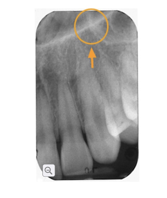
What image is this?
Later al way of the nasal fossa and the wall of maxillary sinus. Located on the maxillary canine in the premolar. Radioplaque
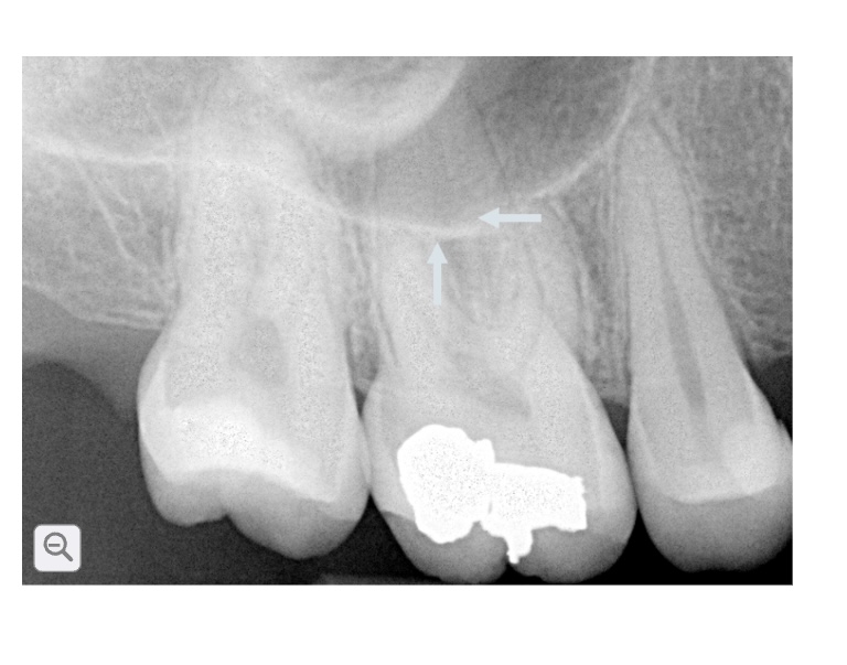
This is the?
Air filled cavity located above premolars and molars on each side. Appears radiolucent with a thin radioplaque line
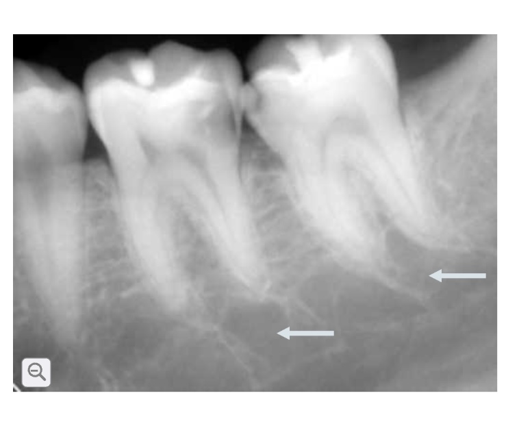
This is?
Pulp tissue once they enter the tooth. Small channels on the jaw bone - blood vessel nerves , supple to the teeth and bone. Radiolucent lines.
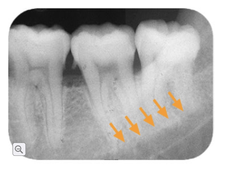
This is?
Bony ridge inside of the lower jaw. Extending from molar toward the front. Radiopaque below molar root.
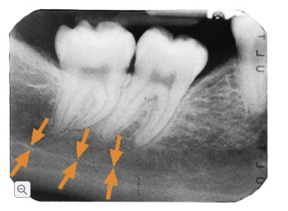
This is?
Passageway inside lower jaw carrier the inferior alveolar and blood vessel. Appears outlined by two thin radioplaque.
Cortical bone
Dense and lines the outside layers of the maxilla and mandible.
Concellous bone
Spongy or very porous and forms the inner portion of the bone
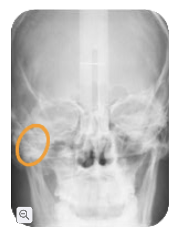
Zygoma
Cheek bone. Appears as radiopaque on maxillary posterior.
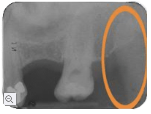
This is?
Coronoid process
Coronoid process
Triangular bony protection on the front part of the mandibular ramus (lower jaw) radioplaque
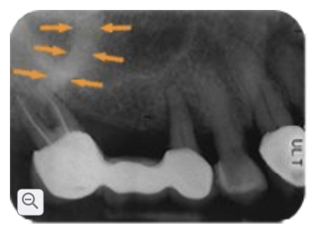
This is?
Zygomatic process of the maxilla.
Zygomatic process of the maxilla
Upper jaw connects with the cheek bone
Appears as a 5 or u-shape radio plaque on the periodical maxillary posterior
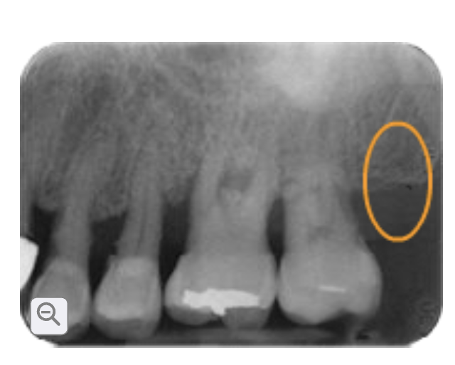
This is?
Maxillary tuberosity
Maxillary tuberosity
Located behind the last upper molar
Blood vessels and nerves enter through the maxilla. Radioplaque
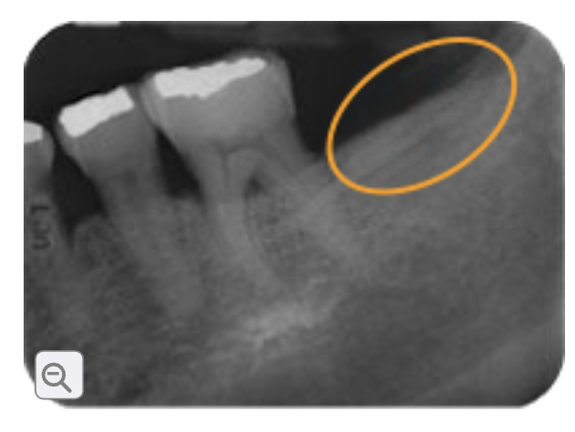
This is?
External oblique ridge or external oblique line
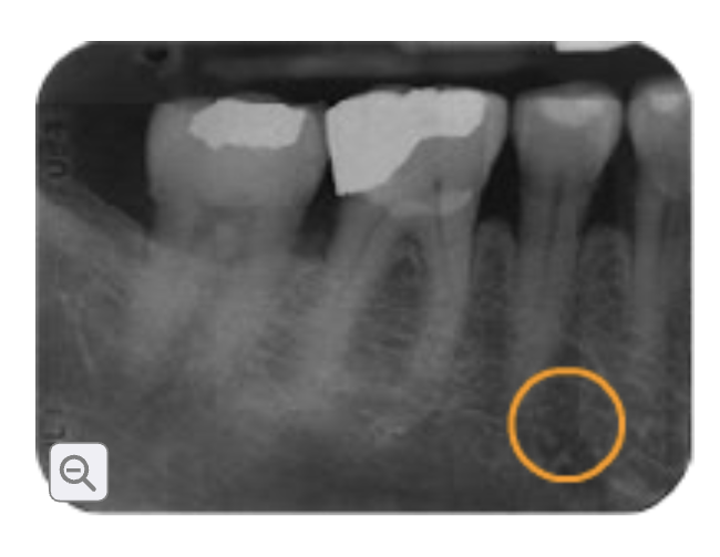
This is?
Mental foremen
External oblique ridge or external oblique line
Outer surface of the lower jaw
Front ramus to molar radioplaque
Mental foramen
Opening located on the outside lower jaw apices of premolars. Allows nerve and blood vessel to pass through
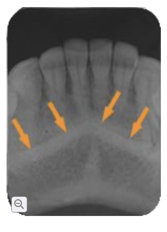
This is?
Mental process or ridge
Mental process or ridge
Bony prominence located front of lower jaw in chin area. Radioplaque V SHAPE
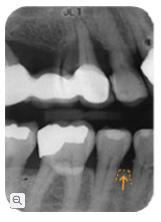
This is?
Alveolar process
Alveolar process
Upper and lower jawbone that holds teeth on the sock •radioplaque
Alveolar ridge
Bony ridge that contains the tooth socket
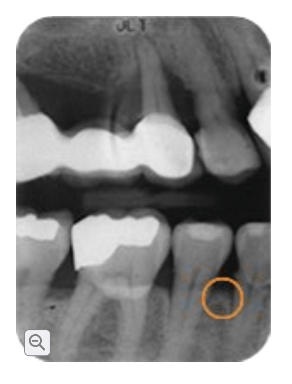
This is?
alveolar ridge
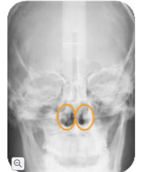
This is?
Nasal fossa ( nasal cavity)
Nasal fossa ( nasal cavity)
Two large air space above upper front teeth. Radiolucent
Nasal septum
Bone structure that divides the left & right nasal. Visible on intraoral X-rays. Radiopaque.
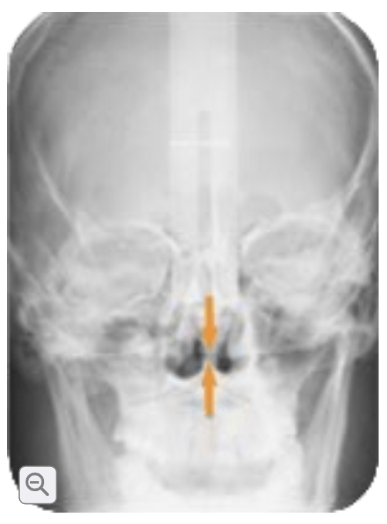
This is?
Nasal septum
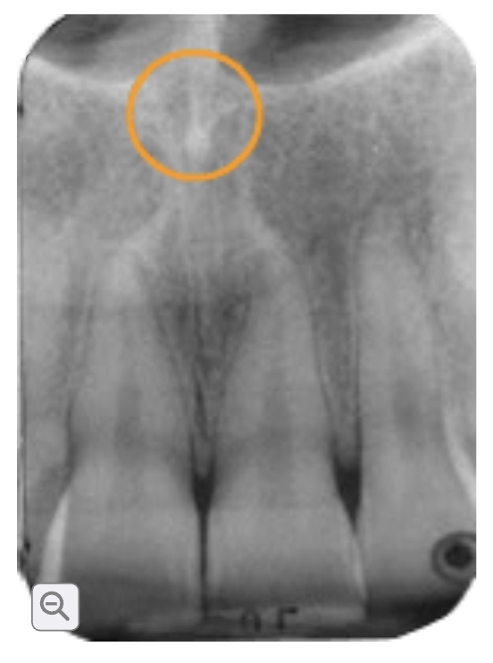
This is?
Anterior nasal spine
Anterior nasal spine
Triangle projection from the floor of the nasal fossa located above the maxillary central incision. Appears as a V SHAPE Radioplaque
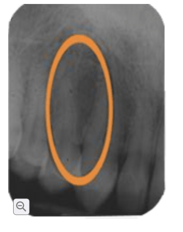
This is?
Lateral fossa /canine fossa
Lateral fossa /canine fossa
Shallow depression in the maxilla near the canine area. Radiolucent
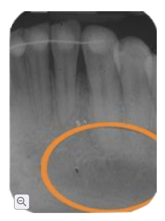
This is?
Mental fossa
Mental fossa
Shallow depression in the front of mandible. Radiolucent
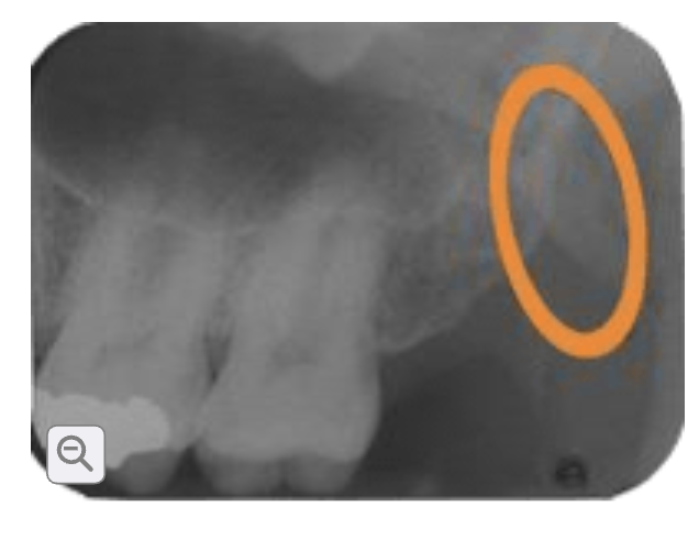
This is?
Hamulus / humular process
Hamulus / humular process
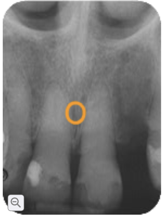
This is?
Incisive foreman
Incisive foreman
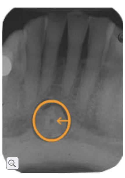
This is?
Lingual foreman
Lingual foreman
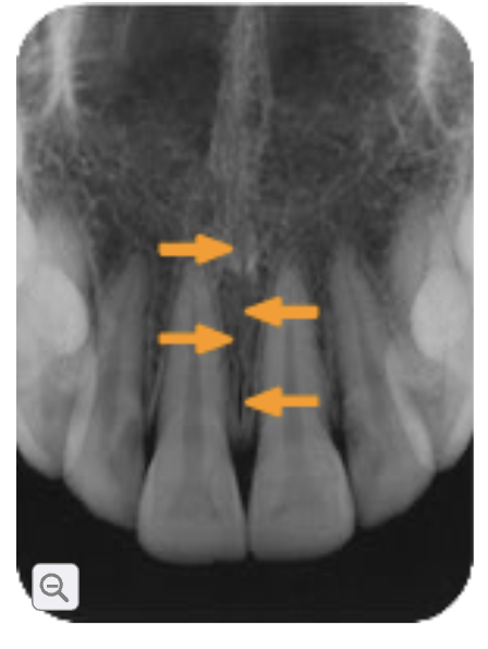
This is?
Median palatial suture
Median palatial suture
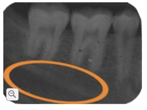
This is?
Mylohyoid ridge
Mylohyoid ridge
Submandibular fossa
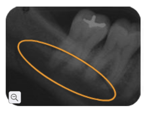
This is?
Submandibular fossa
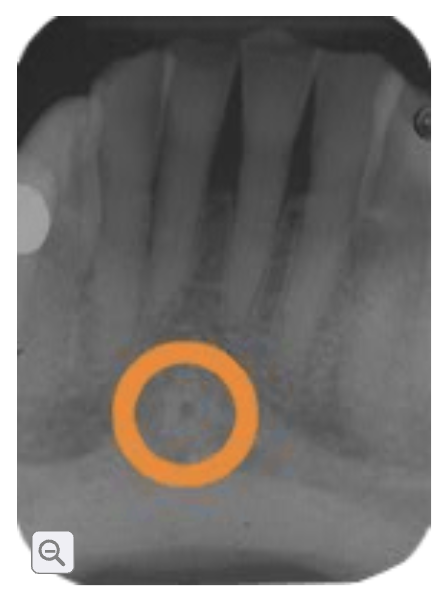
This is?
Genial tubercles
Genial tubercles
Small bony bump on the inner surfaces of the mandible near the midline.
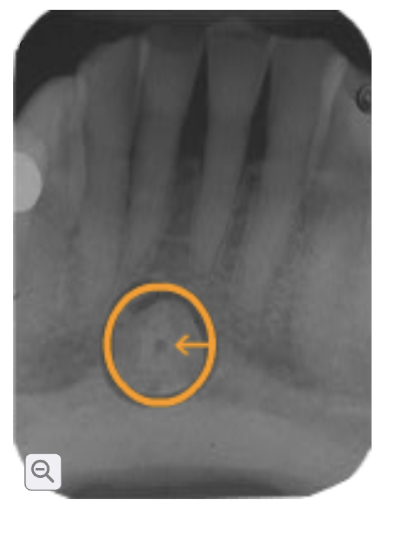
This is?
Lingual foramen
Lingual foreman
A small opening on the inner surface of the mandible, near the midline and surrounded by the genial turbercles. Allows a small blood vessel to pass through. Small radiolucent dot .
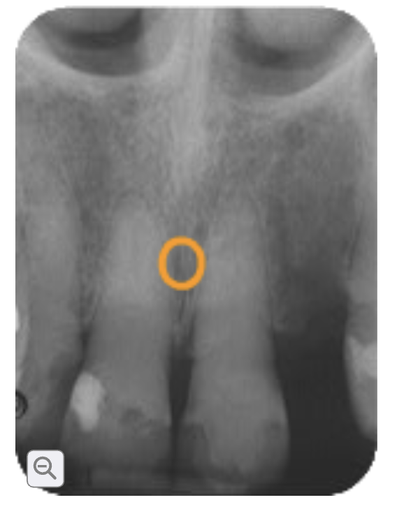
This is?
Incisive foramen
Incisive foramen
A pear shape small opening in the anterior hard palate, behind the upper central incisors. It allows nerves and blood vessels to pass through. Round, radiolucent between the roots of the upper central incisors.
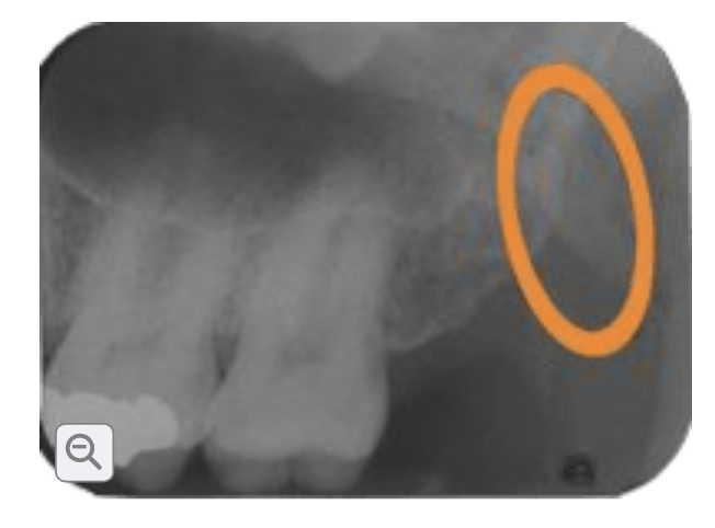
This is?
Hamulus / humular process
Hamulus / humular process
A small, hook-shaped portion of bone extending located behind the maxillary tuberosity. It provides attachment for muscles and ligaments involved in swallowing and movement of the soft palate. Radiopaque
Enamel
The hard, outer covering of the tooth’s crown. It protects the tooth from wear and decay and is the hardest substance in the human body. Appears radioplaque.
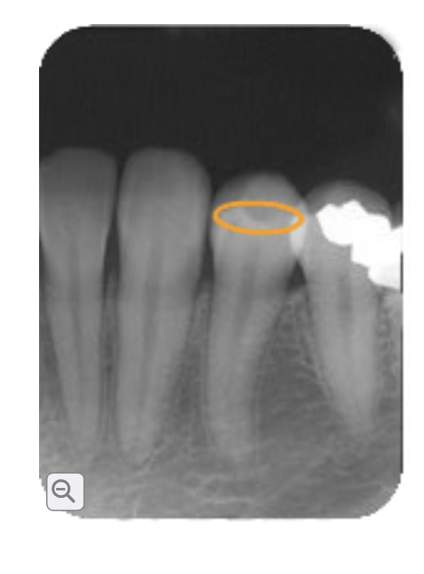
Enamel
Dentin
The hard tissue that forms most of the tooth and protects the pulp. It is found under the enamel and cementum. Appears radiopaque but slightly darker.
Dentinoenamel junction
Strong, wavy connection that holds the enamel and dentin together and helps prevent cracks from spreading through the tooth. Radiopaque line.
Pulp
Interior of the tooth, which contains nerves, blood vessels, and lymphatic tissue. Appears radiolucent.
Periodontal ligament
Is a thin layer of connective tissue between the tooth root and surrounding alveolar bone. Holds the booth in its socket, absorbs pressure,
Apical foreman
Small tip at the tip of a tooth's root where nerves and blood vessels enters and leave the tooth. Not easily distinguishable on X-rays
Apical foreman
Small tip at the tip of a tooth's root where nerves and blood vessels enters and leave the tooth. Not easily distinguishable on X-rays
Apex or apices
General term for the tip of the root of the tooth. Continuation or the radioplaque.
Latina dura
Bony wall of the loot socket that surrounds the root. Tin radioplaque around the root.
Cementum
Thin, hard layer covering the tooth's root • helps attach the tooth to the surroundings through the periodontal ligament. Usually not distinguish on X-rays.
Alveolar crest.
The highest part of the jawbone between the teeth. Supports and holds the teeth in place. Appears as radiopaque.