calcaneus and ankle procedures
 Call Kai
 Learn
Learn Practice Test
 Spaced Repetition
 Match
Match Flashcards
 Knowt Play
1/85

There's no tags or description
Looks like no tags are added yet.
Name | Mastery | Learn | Test | Matching | Spaced | Call with Kai | Chat |
|---|
No analytics yet
Send a link to your students to track their progress
86 Terms
A radiographic image of the ankle is obtained in the lateral projection. Dorsiflexion of the foot is required to accurately demonstrate:
superior tibiotalar joint
A radiographic image of the ankle is obtained in the AP oblique projection. In order to maximize the opening of the tibiofibular joint space the leg should be rotated:
45 degrees medially
In the 45 degree oblique of the ankle the medial mortise will be ____ and the tibiofibular joint will be partially ___.
closed, open
A patient is brought into the radiography department to rule out a trimalleolar fracture. The radiographic projection that is most commonly obtained to demonstrate this condition is the:
AP projection of ankle joint
A radiographic image of the ankle is obtained in the AP oblique projection. In order to maximize the opening of the mortise joint, the ankle and leg should be rotated:
15-20 degrees medially
A radiographic image of the calcaneus is obtained in the axial (plantodorsal) projection. The CR should be directed at an angle of:
40 degrees to the long axis of the foot
Should the sinus tarsi be completely open on the 45 degree oblique ankle?
no
A radiographic image of the ankle is obtained in the AP projection. The CR should be directed:
vertically to the midpoint between the malleoli
Which view of the ankle demonstrates the talofibular joint space in profile?
AP, internal rotation 15 degrees
What is the proper movement of the foot for a lateral projection of the ankle?
dorsiflexion
The fibula will be superimposed on the posterior half of the tibia on which projection?
lateral(mediolateral)
The method that will best demonstrate the talofibular joint best is:
15-20 degrees medially
The lateral mortise is the ___ joint.
talofibular joint
The medial mortise is the ___ joint.
tibiotalar joint
The intermalleolar plane is parallel to the IR in the ___ projection of the ankle. Which view of the ankle demonstrates the talofibular joint space in profile?
Ap internal rotation 15 degrees
In the Oblique projection of the ankle if the calcaneus is superimposed on the fibula, what error was made?
foot was not dorsiflexed
A radiographic image of the calcaneus is obtained in the lateral projection. The CR is directed to enter perpendicular to a point:
1st distal to the medial malleolus
Which of the following is NOT TRUE concerning the AP projection of the ankle?
calcaneus is well demonstrated free of other structures
A radiographic image of the calcaneus is obtained in the axial plantodorsal projection. The CR should be directed at a cephalad angle of 40 degrees to enter the plantar surface at the level of the:
base of the third metatarsal
What is the proper central ray angulation for the AP projection of the ankle?
perpendicular
In which of the following positions/projections will the talocalcaneal joint be visualized?
lateral calcaneus
Which of these are true for the oblique ankle when the foot is dorsiflexed?
the fibular will not overlap the calcaneus
When a radiographic image is obtained of the ankle in the lateral projection, which position will place the joint closest to the IR?
lateromedial position
If the lateral malleolus is slightly superimposed over the talus and the lateral joint space is not open in the AP mortise oblique projection, what is the most likely cause?
insufficient medial rotation of the foot and ankle
How should the patient be positioned on the AP, mortise oblique, and routinue AP oblique of the ankle?
supine with leg extended
How should the body part be positioned on the AP projection of the ankle?
slight dorsiflexion of the foot and plantar surface to the IR
on the AP projection of the ankle, how should the CR be directed?
perpendicular to the Ir midway between the malleoli at the ankle joint. 1/2-3/4 inches superior to the tips of the malleoli
What structures are shown on the AP projection of the ankle?
1. distal tibia and fibula
2. proximal talus
3. medial and lateral malleoli
4. ankle joint
on the AP projection of the ankle, what are the criterias?
1. tibia should be slightly superimposed on the fibula
2.superior tibiotalar joint shuld be open
3. medial tibiotalar joint should be open
4. talus should be slight superimposed on the fibula-lateral joint space is not open
5. foot should be dorsiflexed and calcaneus not seen
How should the body part be positioned on the mortise oblique (medial oblique)?
1. the leg should be turned medially 15-20 degrees so the intermalleolar plane is parallel to the IR.
2. malleoli are in profile
3. foot slightly dorsiflexed
how should the CR be directed on the mortise oblique (medial oblique)?
perpendicular to IR midway between malleoli at the ankle joint
What structures are shown on the mortise oblique (medial oblique)?
mortise joint of the ankle, talofibular joint, (best method to see this joint)
What criterias are shown on the mortise oblique (medial oblique)?
1. the foot should be dorsiflexed
2. distal fibula should be demonstrated without talar superimposition
3. lateral mortise (talofibular joint) should be open
4. medial tibiotalar joint should be open
5. medial mortise should be open
6. the lateral and medial malleoli should be demonstrated in profile
7. tibia should be very slightly superimposed on the fibula
How should the body part be positioned on the AP medial oblique?
turn the leg medially 45 degrees with the foot slightly dorsiflexed
how should the CR be directed on the AP medial oblique?
perpendicular to IR midway between malleoli at the ankle joint
What structures are shown on the AP medial oblique?
distal fibula-lateral malleolus free of superimposition, distal tibia, talus, distal tibiofibular joint open
What criterias are shown on the AP medial oblique?
1. medial mortise should be closed; lateral mortise is slight closed
2. fibula should be demonstrated without tibial superimposition
3. distal tibiofibular joint is open.
4. calcaneus should be demonstrated distal to the lateral mortise and fibula
5. sinus tarsi should be slightly seen
How should the patient be positioned on the lateral projection (mediolateral)?
lateral recumbent
How should the body part be positioned on the lateral projection (mediolateral)?
leg and foot in true lateral position with the lateral aspect of the ankle adjacent to the IR. foot is dorsiflexed into a right angle with the leg
how should the CR be directed on the lateral projection (mediolateral)?
perpendicular to the medial malleolus
What structures are shown on the lateral projection (mediolateral)?
tuberosity of the 5th metatarsal, distal 1/3 of leg, superior talofibular joint, talus, calcaneus, navicular, cuboid, ankle joint, and anterior and posterior displacement of fractures
What criterias are shown on the lateral projection (mediolateral)?
1. long axis of the foot is 90 degrees to the lower leg
2. superior talotibial joint is open
3. distal fibula is superimposed over the posterior half of the tibia
4. domes of the talus are superimposed
5. pretalar fat pad is seen
How should the patient be positioned on the AP weight bearing?
AP position- patient standing with weight evenly distributed, heels adjacent to IR. CR perpendicular
How should the patient be positioned on the lateral weight bearing?
lateral- patient standing with affected side adjacent to IR. CR
What structures are shown on the the weight bearing projections?
demonstrates ankle joint space narrowing with weightbearing
How should the patient be positioned on the Ap axial projection?
supine or seated with leg extended (can also be weight bearing)
How should the body part be positioned on the Ap axial projection?
ankle is dorsiflexed; plantar surface is perpendicular to IR
How should the CR be directed on the Ap axial projection?
angled 40 degrees cephalad- toward long axis of the plantar surface at the base of the 3rd metatarsal
what structures should be seen on the Ap axial projection?
calcaneus and talocalcaneal joint
How should the patient be positioned on the lateral projection of the calcaneus?
lateral recumbent
How should the body part be positioned on the lateral projection of the calcaneus?
leg should be rotated laterally and foot dorsiflexed. plantar surface perpendicular to the IR
How should the CR be directed on the lateral projection of the calcaneus?
perpendicular 1 inch inferior to the medial malleolus
what structures should be seen on the lateral projection of the calcaneus?
entire calcaneus in profile (perpendicular to the IR), ankle joint, subtalar joint
what is another name for the ap axial of the calcaneus
plantodorsal or inferior-superior projection or the Harris method
why is the CR angled at 40 degrees on the AP axial?
to elongate the calcaneus
when is it appropriate to use the dorsoplantar or superoinferior projection?
if the calcaneus cannot be rested on the IR
what is the medial mortise?
medial tibiotalar joint
what projection best demonstrates the medial malleolus and the talus?
15 degree oblique projection
what is the superior mortise?
superior tibotalar joint (ankle joint)
what projections are best used to demonstrate the superior tibiotalar joint?
AP, mortise oblique and lateral
what is the lateral mortise?
talofibular joint
what projection is used to best demonstrate the lateral malleolus (fibula) and the talus?
15 degree oblique projection
what projection is used to best demonstrate the distal tibiofibular joint?
45 degree oblique projection
Ap ankle error: if the ankle is not in true AP, too much of the fibula is superimposed on the tibia and the tibiotalar (medial mortise) joint is not seen. the leg is slightly externally rotated. how do we correct it?
rotate slightly internally to place the leg in true AP position
AP ankle error: the foot is plantar flexed- this will cause the calcaneus to be too close to the fibula and will obscure the talofibular joint (lateral mortise). how do we fix it?
dorsiflex the foot
oblique mortise ankle error: the leg is not rotated enough- this will cause the talofibular joint not to open enough and the lateral malleoli are not equal distance from the IR. how do we correct this?
rotate the lower leg internally (medially) until the malleoli are equal distance from the IR
oblique mortise ankle error: the foot is plantar flexed- this will cause the calcaneus to be too close to the fibula and will obscure the talofibular joint. what do we do to correct this?
dorsiflex the foot
medial mortise oblique ankle error: oblique in the wrong direction, leg oblique laterally instead of medially. foot not dorsiflex. how to correct it?
rotate the leg 15-20 degrees medially dorsiflex the foot and center IR correctly
oblique 45 degree ankle error: over rotation- the fibular will be superimposed on the talus and the sinus tarsi will be completely open. what is needed to correct this?
dorsiflex the foot and decrease the rotation of leg to 45 degrees
oblique 45 degree ankle error- under rotation and foot is not dorisflexed, the sinus tarsi is not seen, and the fibula is superimposed on the calcaneus. how is this corrected?
increase rotation of leg to 45 degree
lateral ankle error- leg is externally rotated, the fibula is too far posterior, the talus is rotated. how is this corrected?
internally rotate the leg
situation: a radiograph of a AP mortise projection of the ankle reveals that the lateral malleolus is slightly superimposed over the talus and the lateral joint is not open.
solution: insufficient medial rotation of the foot and ankle was most likely to cause for this radiographic outcome
situation: a radiograph of a plantodorsal (axial) projection of the calcneus reveals foreshortening. the technologist used a 30 degree cephalad angle from the long axis of the foot
solution: increasing the CR cephalad angulation to increase angle to 40 degrees will produce a more diagnostic image of the calcaneus
AP ankle

AP mortise oblique
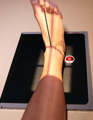
AP oblique ankle

lateral ankle

axial projection- harris method

axial projection- dorsoplantar projection

lateral calcaneus

standing ap ankle

axial projection-harris method

lateral calcaneus xray image

standing lateral ankle
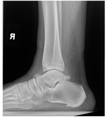
CT calcaneus

standing AP ankle
