Lecture 15: Pericardial Disease & Cardiac Tumors
 Call Kai
 Learn
Learn Practice Test
 Spaced Repetition
 Match
Match Flashcards
 Knowt Play
1/39

There's no tags or description
Looks like no tags are added yet.
Name | Mastery | Learn | Test | Matching | Spaced | Call with Kai | Chat |
|---|
No analytics yet
Send a link to your students to track their progress
40 Terms
What are the 2 main layers of the pericardium?
Fibrous pericardium (outer layer):
• Tough connective tissue
• Anchors heart to diaphragm & sternum
• Prevents overdistension of heart
Serous pericardium (inner layer):
• Parietal layer: lines inner surface of fibrous pericardium
• Visceral layer (epicardium): adheres tightly to heart surface
• Pericardial cavity is between layers (0.5-2 mls lubricating serous fluid)
What is a peritoneopericardial diaphragmatic hernia (PPDH)?
• Incomplete development of diaphragm & caudal pleuropericardial membrane
• Abdominal contents herniated into the pericardial space
• Liver, stomach, omentum, small intestines, spleen, rarely colon
• Pericardial sac distends
• May be associated with sternal deformities or pectus excavatum
• Maybe be associated with ductal plate malformations
What are the clinical signs of PPDH?
• Often long asymptomatic period
• GI signs – Vomiting, anorexia, weight loss, diarrhea
• Respiratory signs – Tachypnea, cough, shortness of breath
• Cardiovascular signs less common
What are the typical PE findings of a PPDH?
• Muffled heart sounds
• Absent or displaced cardiac sounds
• Absent or displaced apex beat
• Tachypnea
• Relative absence of abdominal organs on palpation
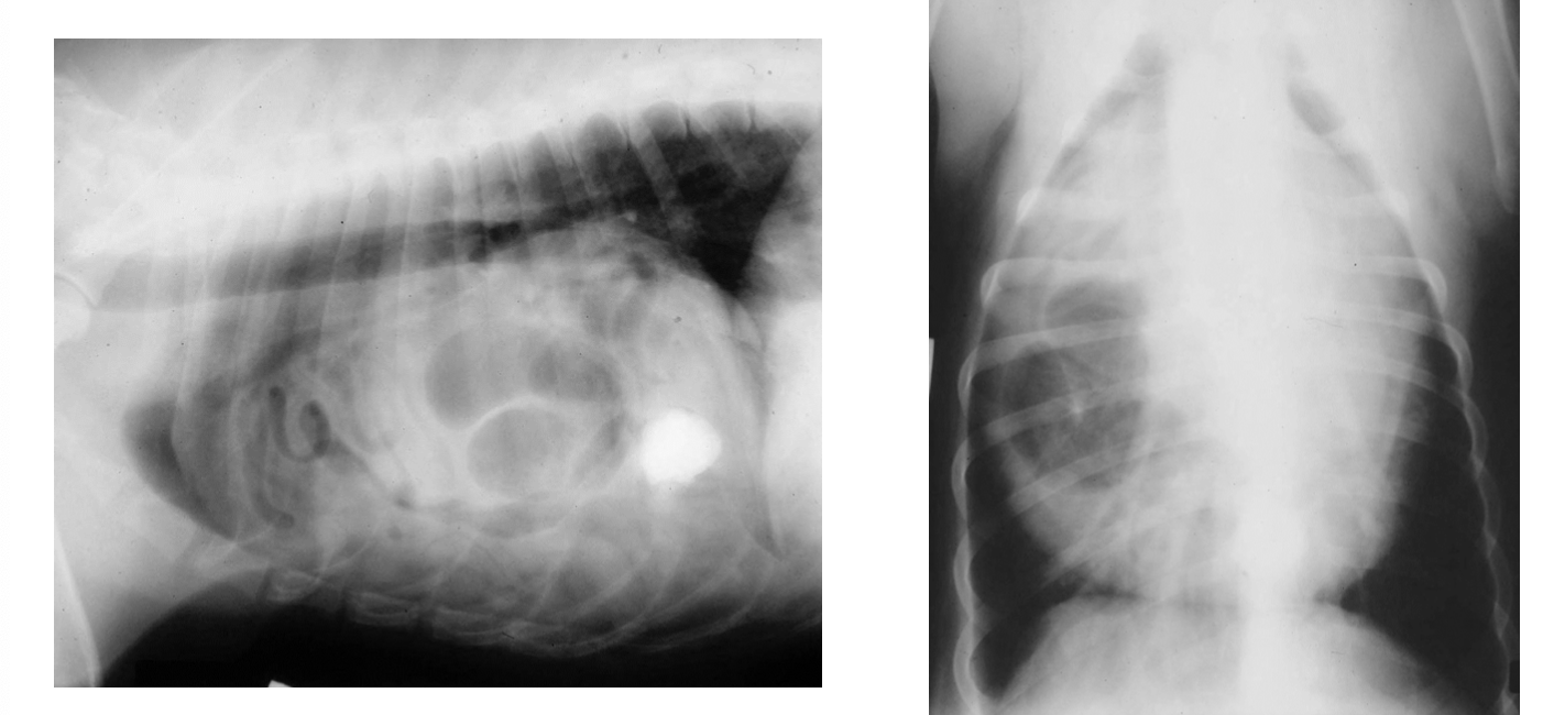
What is seen on this rad?
PPDH
What is the treatment and prognosis of PPDH?
• Surgical correction for large hernias or symptomatic patients
• Medical management considered for asymptomatic or minimally symptomatic patients, especially if older animal with higher surgical risk → Close monitoring, Avoid strenuous activity
• Prognosis generally excellent with surgical correction provided no liver necrosis, GI strangulation, or pericardial compromise
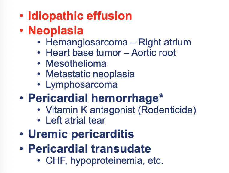
What are the typical causes of canine pericardial effusion?
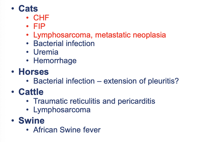
What are the typical causes of feline, equine, bovine, and porcine pericardial effusion?

What is on this right atrium?
right atrial hemangiosarcoma
What tumor of malignant endothelial cells is most often found in the right atrium or auricle?
hemangiosarcoma
What are the effects of HSA?
• Fragile, abnormal tumor vasculature has spontaneous rupture or oozing of blood into pericardial space
• Bleeding can be acute and massive or slow and recurrent
• Pericardium’s limited stretch causes rapid pressure rise → tamponade
• Fluid usually hemorrhagic
• May clot if acute, won’t if more chronic
What neuroendocrine tumor at the heart base near aorta/pulmonary artery puts external compression on the great vessels causing increased venous pressure and transudate fluid into pericardial sac?
chemodectoma (aortic body tumor)
How does mesothelioma cause pericardial effusion?
• Malignant tumor of mesothelial cells lining serous membranes → Often diffuse and tumor sheets coat the pericardial surface
• Tumor cells infiltrate the pericardial lining causing inflammatory exudate → Can invade or compress pericardial lymphatic vessels impairing lymphatic drainage → Can invade small pericardial vessels causing low grade or episodic bleeding
• Extensive tumor surface area continuously produces abnormal fluid leading to rapid recurrence even after pericardiocentesis
• Overall pericardial effusion is often serosanguinous or hemorrhagic, sometimes with high protein and mixed-cell populations
What type of tumor infiltrates the pericardium causing increased vascular permeability leading to an inflammatory exudate?
lymphoma (cardiac lymphoid tissue or secondary metastasis)
What type of tumor involves functional thyroid or parathyroid tissue at the heart base that causes external compression of great vessels increasing venous pressure and transudate fluid into the pericardial sac?
ectopic thyroid or parathyroid carcinoma
What is the common presenting history for pericardial effusion?
• Medium to large breed dogs: GSD, Golden retriever, Lab, Rottweiler
• Brachycephalic (heart base tumor)
• Middle age or older (> 4 years)
• Male > female
• Often history of acute collapse
• Abdominal distention
• Dyspnea
• Syncope
What are the clinical signs and PE findings of pericardial effusion?
• Lethargy, weakness, exercise intolerance
• Muffled heart sounds
• Jugular vein distension
• Tachycardia
• Pulsus paradoxus
• Ascites/hepatomegaly
• Dyspnea, muffled lungs ventrally
• MM color muddy / CRT delayed
What is pulsus paradoxus?
• Exaggerated drop in systolic blood pressure during inspiration due to pericardial effusion/cardiac tamponade
• Exaggeration of a normal physiologic change
• Alternating weak/normal femoral pulses
What is seen on thoracic rads of pericardial effusion?
• Generalized cardiomegaly
• Globoid heart
• Enlarged caudal vena cava
• Variable pulmonary vessel findings; often small
• Some pleural effusion common
• Search for pulmonary metastatic disease

What is seen on these rads?
pericardial effusion
What is seen on an ECG with pericardial effusion?
• Sinus tachycardia
• Electrical alternans
• Low-voltage QRS complexes
• ST segment elevation
• Ventricular premature depolarizations common (esp. post-tap)
What bloodwork should be performed in pericardial effusion cases?
• CBC: Anemia, thrombocytopenia, RBC changes
• Chemistry profile: Pre-renal azotemia, increased LE’s, low sodium & chloride if ascites
• FIP & FeLV testing in cats
• Cardiac troponin I: Elevated in many dogs; highest with hemangiosarcoma in dogs
• NT-proBNP: Usually NOT elevated (heart is not stretched)
• Central venous pressure (CVP): Elevated
What is typically seen on echocardiography of pericardial effusion?
• Anechoic fluid between peri- & epicardium (pericardial effusion)
• RA +/- RV diastolic collapse (cardiac tamponade)
• Heart swinging within pericardial sac
• Small LV size
• Search for cardiac mass
What is the treatment for pericardial effusion?
Pericardiocentesis:
• Emergency procedure to relieve tamponade
• Performed from right side, under ultrasound guidance
Pericardiectomy:
• Surgical removal of part of the pericardium
• Indicated in recurrent effusion or neoplasia
Specific therapies:
• Neoplasia: Chemo (e.g., doxorubicin for hemangiosarcoma)
• Infectious: Antibiotics, anti-fungals (rare)
• CHF: Address underlying cardiac disease in cats
What are the key signs of a pericardial effusion emergency?
Muffled heart sounds
Jugular venous distension
Weak pulses, pulsus paradoxus
Tachycardia
Ascites (if chronic)
Collapse or shock in severe cases
What are the immediate goals of an emergency pericardial effusion case?
1. Relieve pericardial pressure on heart
2. Maintain perfusion until definitive therapy (pericardiocentesis)
3. Avoid interventions that worsen cardiac filling
What are the initial stabilization steps of a pericardial effusion emergency?
• Positioning: Keep patient in sternal recumbency or sitting (orthopneic) to maximize venous return and lung expansion
• Oxygen Therapy: Provide supplemental oxygen (mask, flow-by, oxygen cage) to address hypoxia and reduce myocardial workload
• Fluid Therapy: Modest boluses (5-10 mls/kg) to help maintain preload until fluid is drained
What drugs should be avoided if pericardial effusion is present?
• Diuretics: Reduce preload further and worsen hypotension
• Vasodilators: Drop systemic vascular resistance and compromise coronary perfusion
What are possible complications of pericardiocentesis?
• Serious complications & risks (10-15%)
• Sudden death (2-5%)
• Exsanguination or rebleed into pleural or pericardial space (3-5%)
• Coronary artery laceration (rare but a problem)
• Serious arrhythmias (10-12%)
What is pericardectomy?
• Surgical removal of part or all of the pericardium to prevent recurrent fluid accumulation in the pericardial sac & relieve cardiac compression
• Effusion drains into thoracic cavity where it is absorbed by pleural surfaces
• Indicated for recurrent or refractory pericardial effusion after pericardiocentesis
What are the three types of pericardectomies?
• Subtotal pericardectomy – via median sternotomy/thoracotomy; removes most of pericardium
• Pericardial Window (Partial Pericardectomy) – via thoracotomy or thoracoscopy; excises a section of pericardium ventrally
• Thoracoscopic Pericardectomy – minimally invasive; reduces pain & recovery time
What is the permanent solution for idiopathic pericardial effusion that prevents recurrence?
pericardectomy
What are the risk/complications of pericardectomy?
• Hemorrhage (especially with neoplastic masses near coronary vessels)
• Arrhythmias (perioperative)
• Pneumothorax
• Infection
• Incomplete resolution if insufficient tissue is removed
What is the prognosis for pericardial effusion?
Idiopathic effusion (dogs): Single pericardiocentesis curative in 30-50% of cases, Good with pericardiectomy
Neoplastic effusion: Poor prognosis, especially hemangiosarcoma
Infectious: Variable, depends on pathogen and response
Cats with CHF or FIP-related effusion: Prognosis tied to primary disease
Rapid recognition and drainage of pericardial effusion is life-saving
Prognosis depends heavily on the underlying cause
What is constrictive pericarditis?
• Chronic condition
• Thickened, fibrotic, & sometimes calcified pericardium
• Reduced pericardial compliance, restricted diastolic filling & eventual right-sided heart failure
What causes constrictive pericarditis?
• Chronic pericarditis (idiopathic, infectious, neoplastic)
• Recurrent pericardial effusion
• Post-surgical or traumatic pericardial injury
• Infectious agents (rare; ie, bacterial, fungal)
• Neoplasia involving pericardium
What are the clinical signs of constrictive pericarditis?
• Ascites
• Jugular venous distension
• Hepatomegaly
• Lethargy, exercise intolerance
• Muffled heart sounds
• Weak pulses
• Pleural effusion (sometimes)
How is constrictive pericarditis diagnosed?
• Echocardiography needed
• Thoracic radiographs: pericardial calcification (sometimes)
• CT/MRI: detailed pericardial anatomy
• Cardiac catheterization: equalization of diastolic pressures
• Rule out restrictive cardiomyopathy
How is constrictive pericarditis treated?
• Surgical pericardectomy (subtotal or total) = treatment of choice
• Stabilize with diuretics before surgery if significant congestion
• Address underlying cause if identified
• Complications more frequent than with surgery for effusion alone
What is the prognosis of constrictive pericarditis?
• Good if idiopathic and treated surgically before severe myocardial damage
• Guarded to poor if associated with neoplasia or advanced myocardial disease
• Early recognition and intervention improve outcomes