Cerebral Hemispheres and Cortex
1/54
Earn XP
Description and Tags
VS 6110 Lab
Name | Mastery | Learn | Test | Matching | Spaced | Call with Kai |
|---|
No analytics yet
Send a link to your students to track their progress
55 Terms
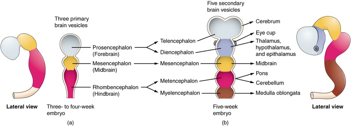
How does the nervous system start out as?
A hollow tube
What happens for the nervous system tube to develop a cranial and caudal end?
3 vesicles
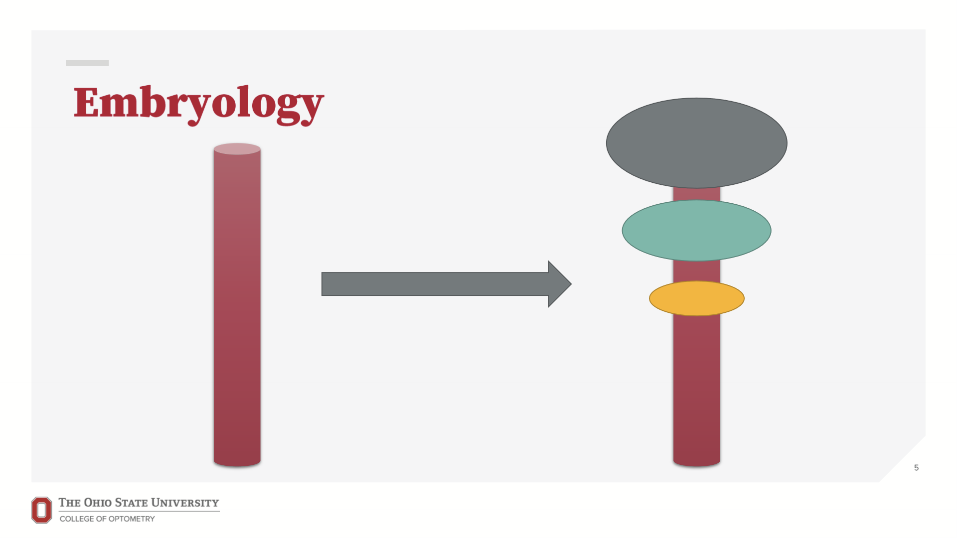
What are the three Vesicles called, cranial to caudal?
Prosencephalon (forebrain), Mesencephalon (midbrain), Rhombencephalon (hindbrain)
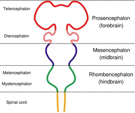
What does the Prosencephalon differentiate into?
Telencephalon and Diencephalon
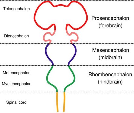
What does the Rhombencephalon differentiate into?
Metencephalon and Myelencephalon
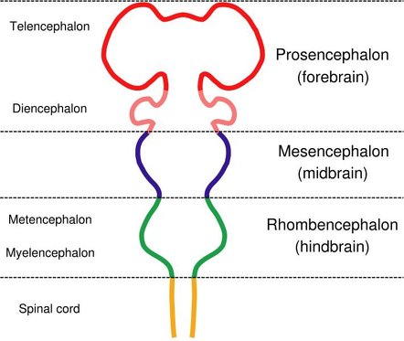
What does the telencephalon become?
Cerebral hemispheres
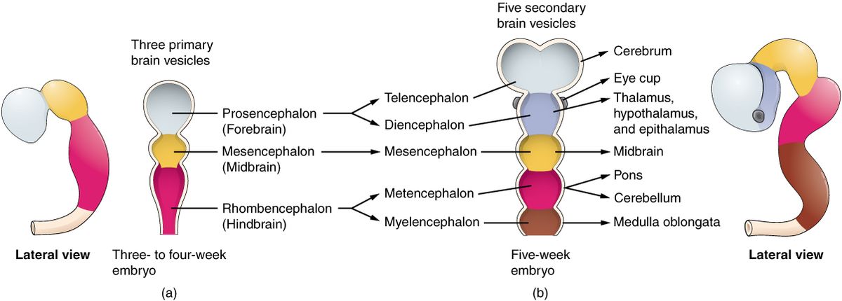
What does the diencephalon become?
Thalamus, Epithalamus, and Hypothalamus
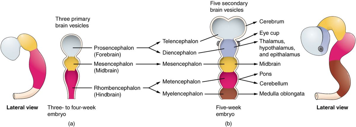
What does the mesencephalon become?
It stays as mesencephalon, but bending starts here called mesencephalic flexion
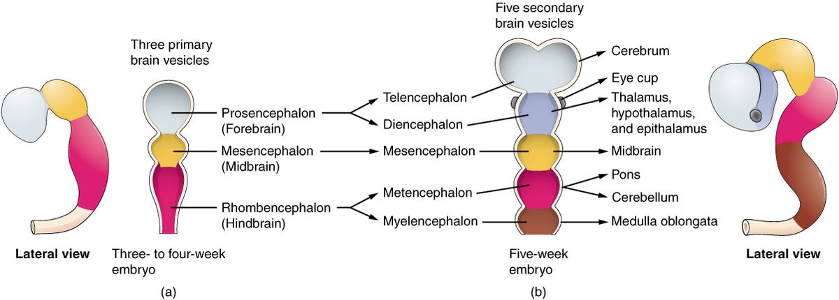
What does the metencephalon become?
Pons and Cerebellum
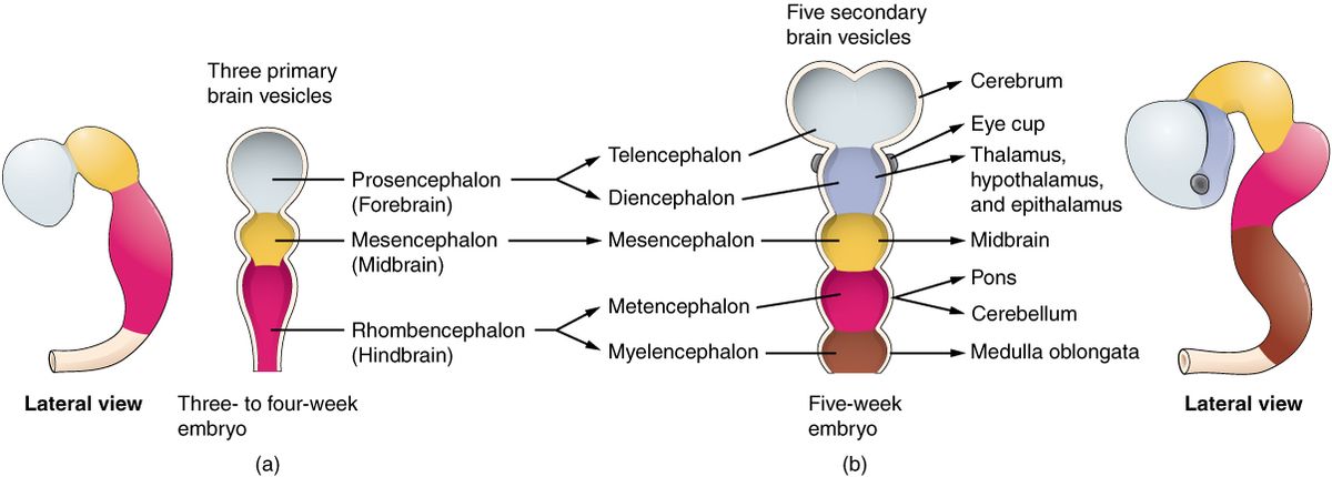
What does the myelencephalon become?
Medulla oblongata
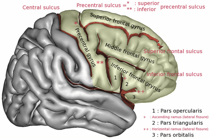
What gyruses is the frontal lobe divided into?
Precentral Gyrus
Superior Frontal Gyrus
Middle Frontal Gyrus
Inferior Frontal Gyrus
Pars Opercular
Pars Triangular
Orbital Gyrus
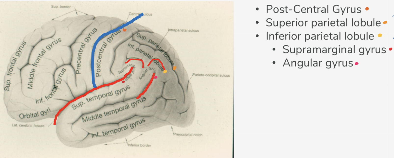
What are the divisions of the parietal lobe?
Post-central gyrus
Superior parietal lobule
Inferior parietal lobule
Supramarginal gyrus
Angular gyrus
What parts of the parietal lobes are used in perceiving depth and location?
Superior and Inferior parietal lobule
What is the functions of the parietal lobe?
Primary sensory cortex and processing location and distance from vision
What are the divisions of the temporal lobe from the lateral view?
Superior temporal sulci
Inferior temporal sulci
Superior temporal gyrus
Middle temporal gyrus
Inferior temporal gyrus
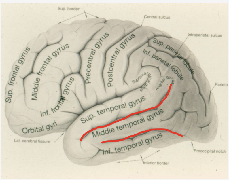
What are the divisions of the temporal lobe from the inferior view?
Inferior temporal gyrus
Lateral occipitotemporal gyrus
Medial occipitotemporal gyrus
Parahippocampal gyrus
Uncus
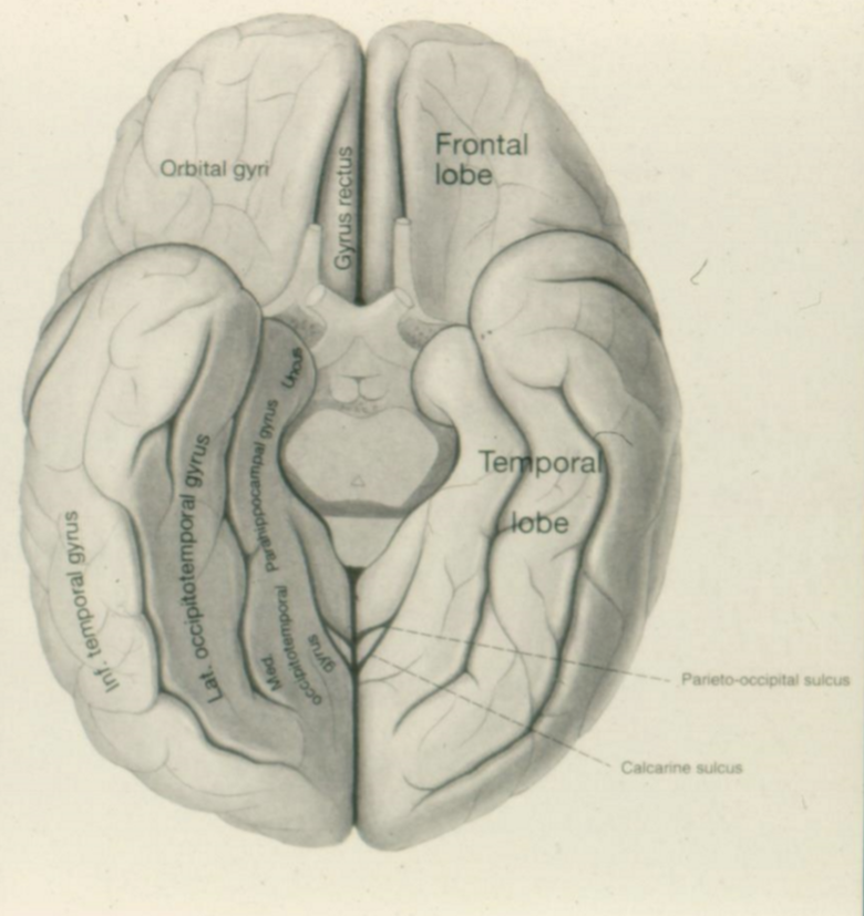
What is the function of the temporal lobe?
Language, hearing, and visual identification
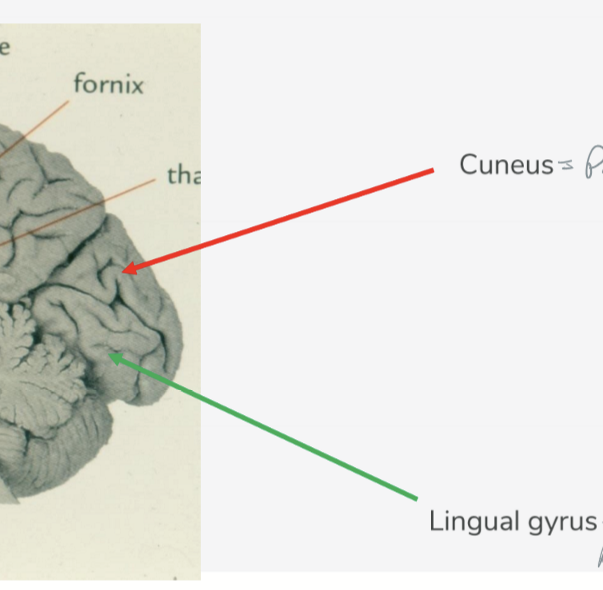
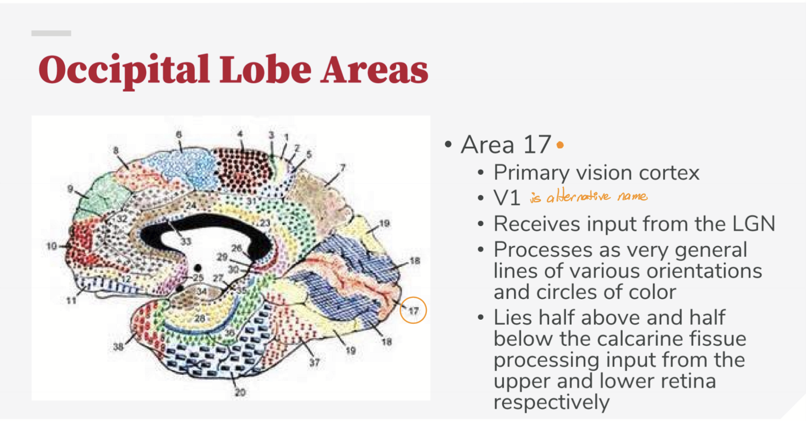
What are the divisions of the occipital lobe?
Parieto-occipital sulcus
Calcarine sulcus
Forms Cuneus above
Forms Lingual gyrus below
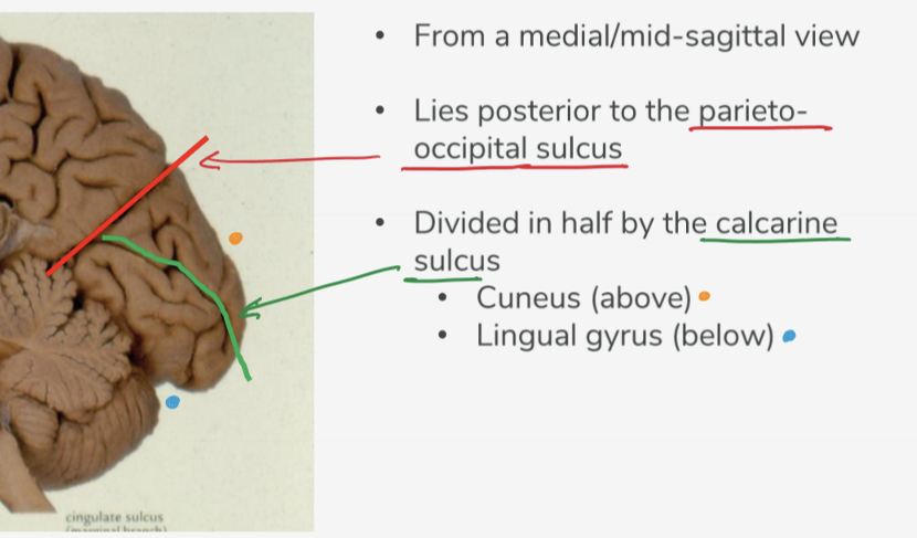
Function of Cuneus is what?
Process visual information, particularly from the contralateral lower visual field; More basic visual processing, like spatial orientation and visual attention
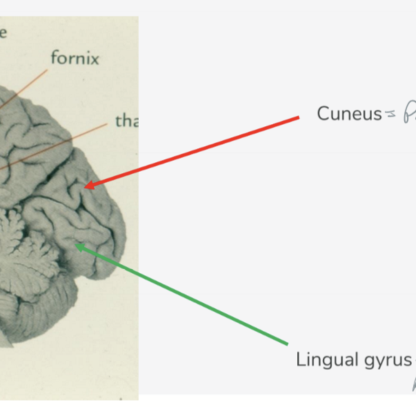
Function of Lingual Gyrus is what?
Process visual information, particularly from the contralateral upper visual field; More complex visual processing, including color perception and object recognition
How many distinct histological layers is the cortex divided into?
6 layers
What are the two primary cell types in the cortex?
Stellate and Pyramidal
Names of the layers of the cortex
Molecular layer
External granular layer
External pyramidal layer
Internal granular layer
Internal pyramidal/Ganglionic layer
Multiform layer
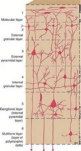
What is the function of the molecular layer (layer 1)?
May be functioning as a coordinating center where layers can communicate action
Every layer sends dendrites back to layer 1
May serve as the water cooler of the brain
What is the function of the external granular layer (layer 2)?
Receives input from other cortical regions, so synapses with axons of cell bodies in layer 3, (external pyramidal layer).
What is the function of the external pyramidal layer (layer 3)?
Sends output to other cortical areas, so the axons of the cells synapse with cells in layer 2
What is the function of the internal granular layer (layer 4)?
Receives input from the Thalamus and other brainstem areas. Very thick with SENSORY areas of cortex (striated cortex). Deals with vision, auditory, and somatosensory information.
What is the function of the internal pyramidal layer (layer 5)?
Sends axons to brainstem (corticobulbar) and spinal cord (corticospinal). Very thick in motor areas of cortex.
What is the function of the Multiform Layer (layer 6)?
Sends axons back to the thalamus via corticogeniculate fibers. Works to modulate what information thalamus sends to cortex (filters out information). It allows the cortex to make a prior decisions with corticogeniculate fibers.
What does TBI have to do with the Multiform Layer (6)?
Damage to the Multiform layer removes individual’s ability to filter and modulate incoming information, thus have trouble processing sensory information, focusing attention, or prioritizing certain sensory inputs.
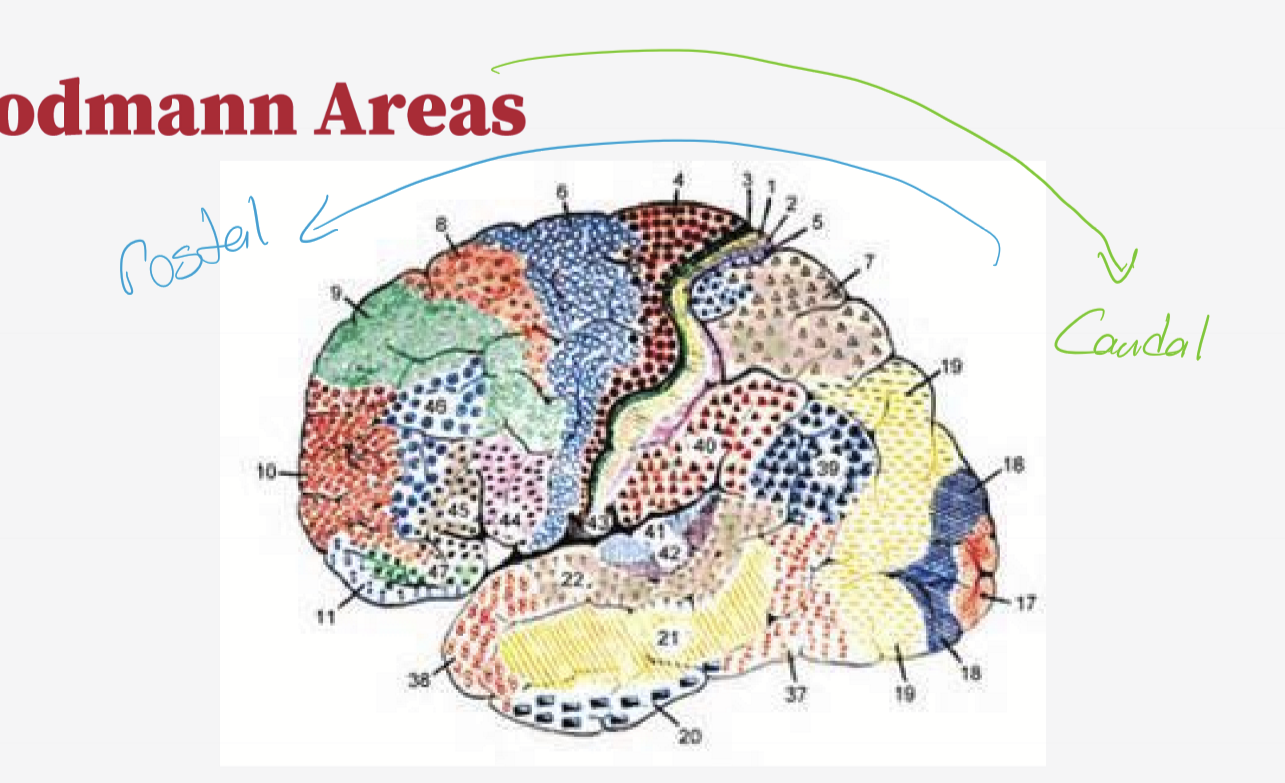
What is BA 4 responsible for?
Primary Motor, thick layer 2
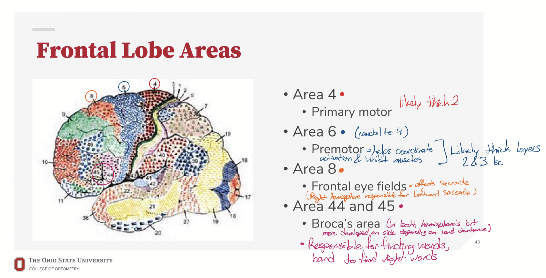
What is BA 6 responsible for?
Premotor, helps coordinate activation and inhibition of muscle, thick layer 2&3
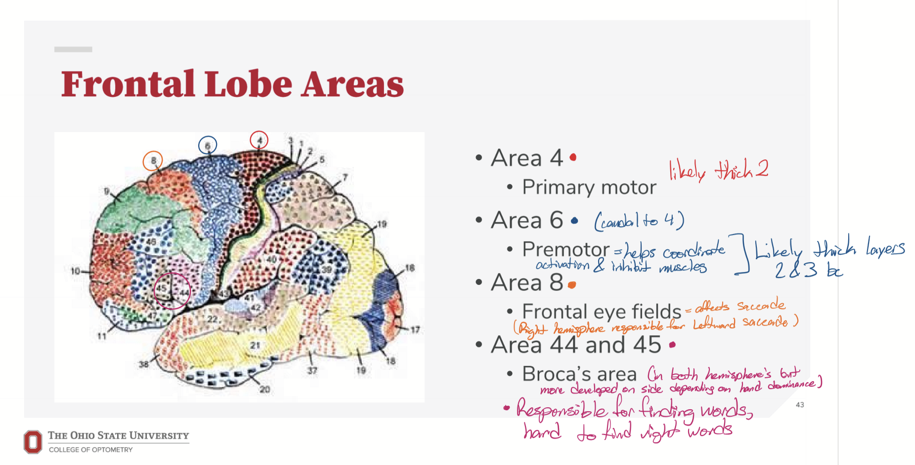
What is BA 8 responsible for?
Frontal Eye Fields, affects saccade, right hemisphere for leftward saccade.
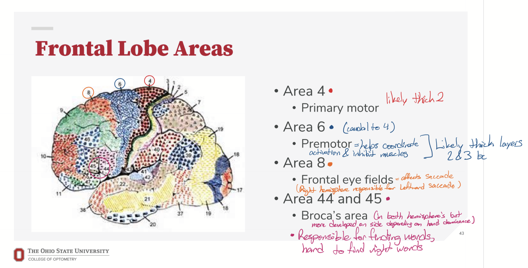
What is BA 44 and 45 responsible for?
It is Broca’s area and is responsible for finding words
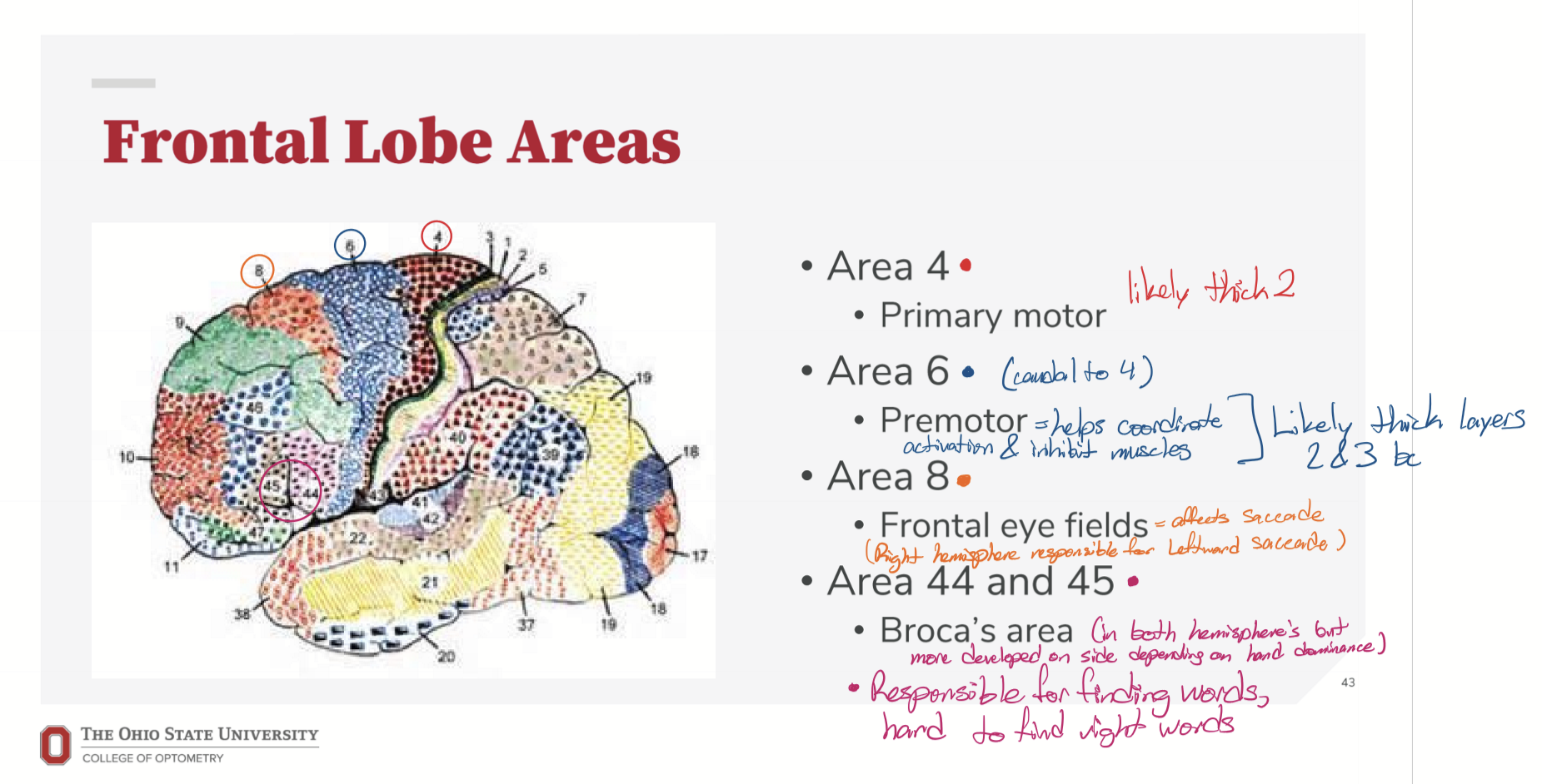
Where is the primary motor cortex?
Right infront of the central sulcus
What happens if the primary motor cortex is damaged?
Spastic paralysis, then over time, flaccid paralysis.
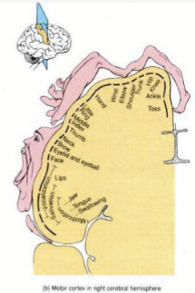
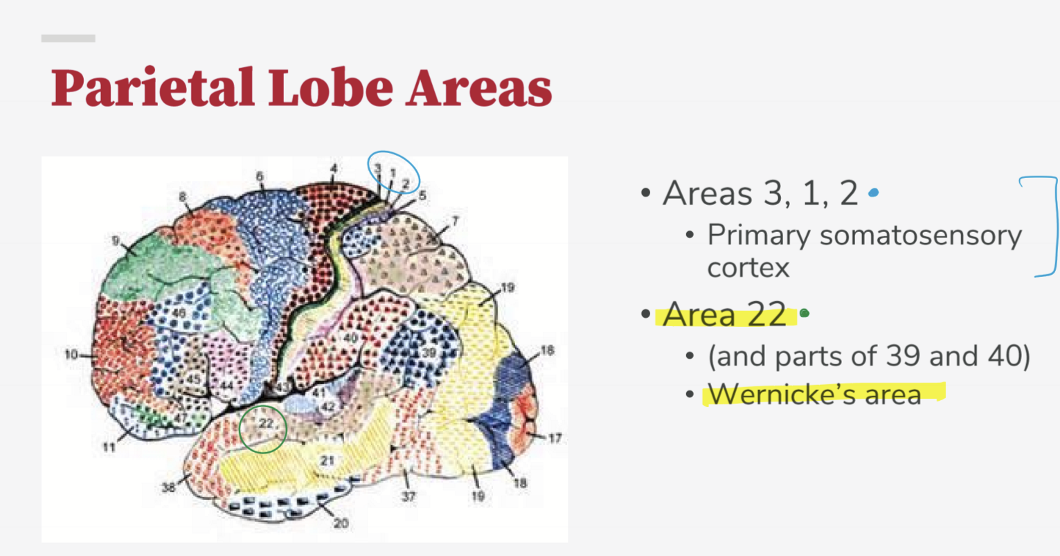
What are BA 3,1,2 responsible for?
Primary somatosensory cortex
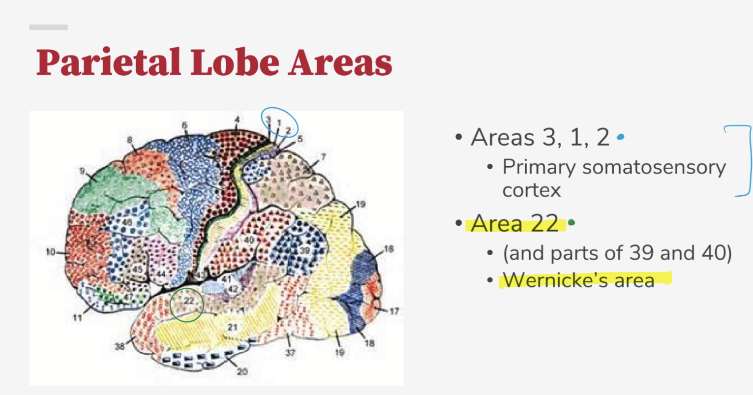
What is BA 22, 39, and 40 responsible for?
Wernicke’s area for language comprehension
What is BA 41 responsible for?
Primary auditory, receiving input from the medial geniculate nucleus and sends to BA 42
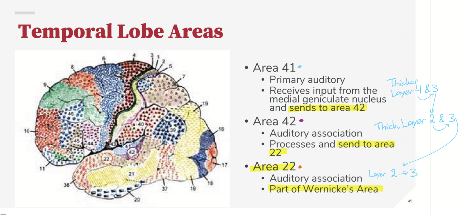
What is BA 42 responsible for?
Auditory association, and sends processed information to BA 22.
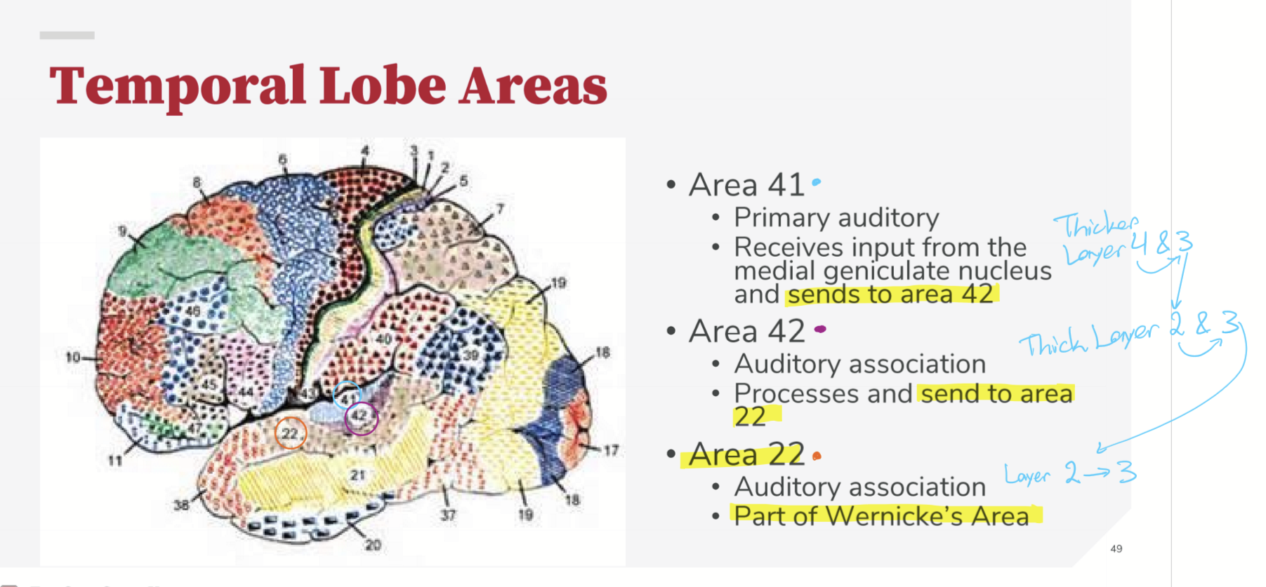
What is BA 22 responsible for?
Part of Wernicke’s Area for auditory association
What is BA 17 (V1) responsible for?
Primary vision cortex, receiving input from LGN. Primarly processes lines of various orientations and circles of color
What is BA 18 (V2) responsible for?
Association vision cortex, receives input from BA 17 and processes further
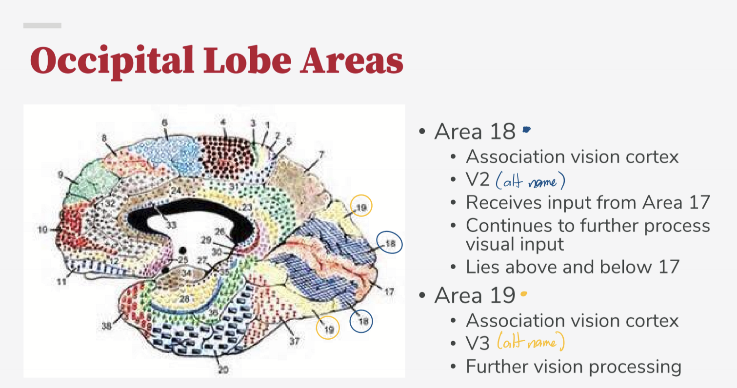
What is BA 19 (V3) responsible for?
Further vision processing
Where does the visual information go to after it is processed?
To the occipito-parietal or occipito temporal regions.
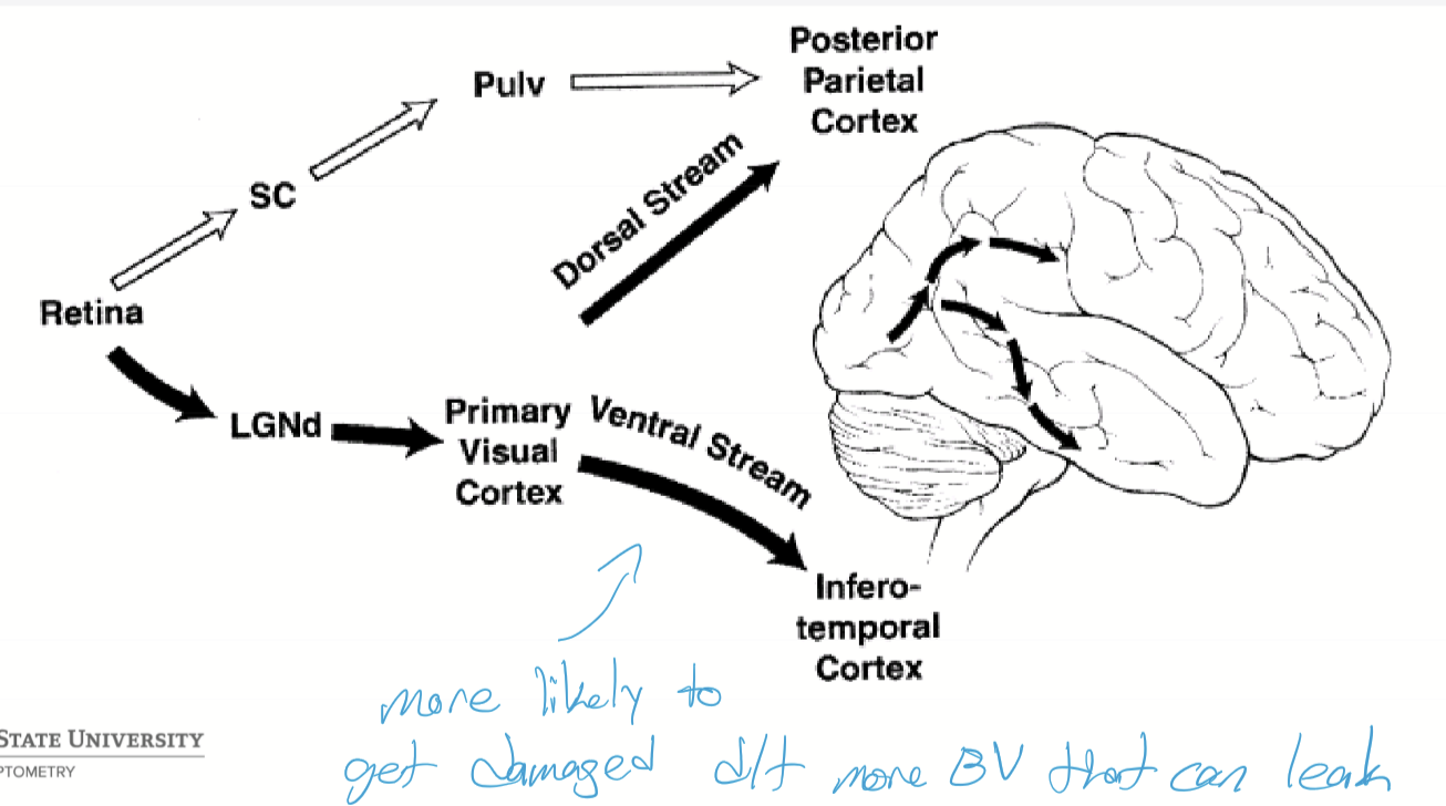
How is information transmitted to the occipito-parietal cortex and what does it do?
It receives information via the dorsal stream and is concerned with spatial awareness, depth perception, and visually guided movements.
How is information transmitted to the occipito-temporal cortex and what does it do?
It receives information via the ventral stream and is concerned with object recognition
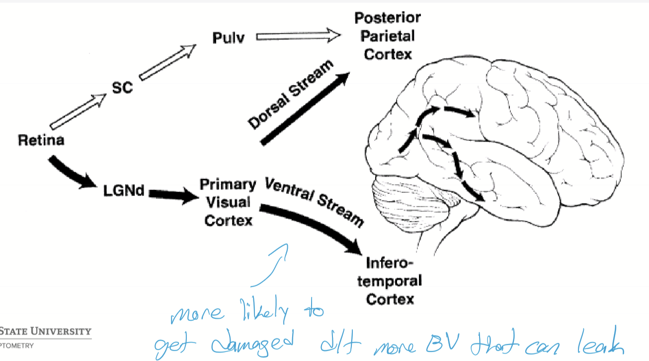
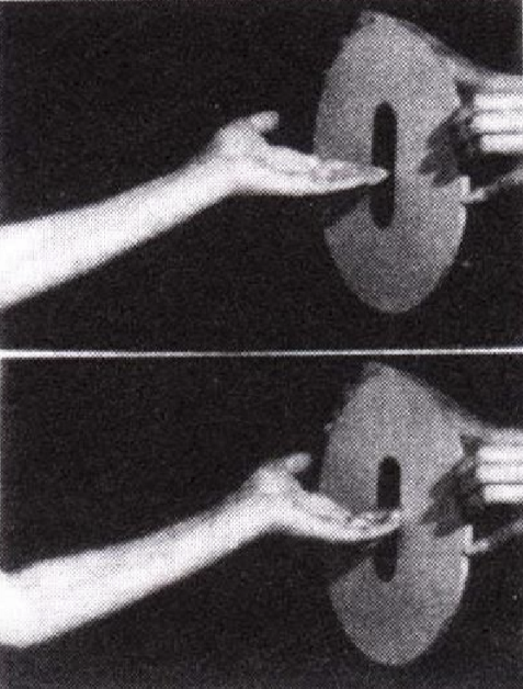
What is optic ataxia?
When the eyes are not communicating with the brain where things are, so can visually identify orientation, but motor action is not accurate.
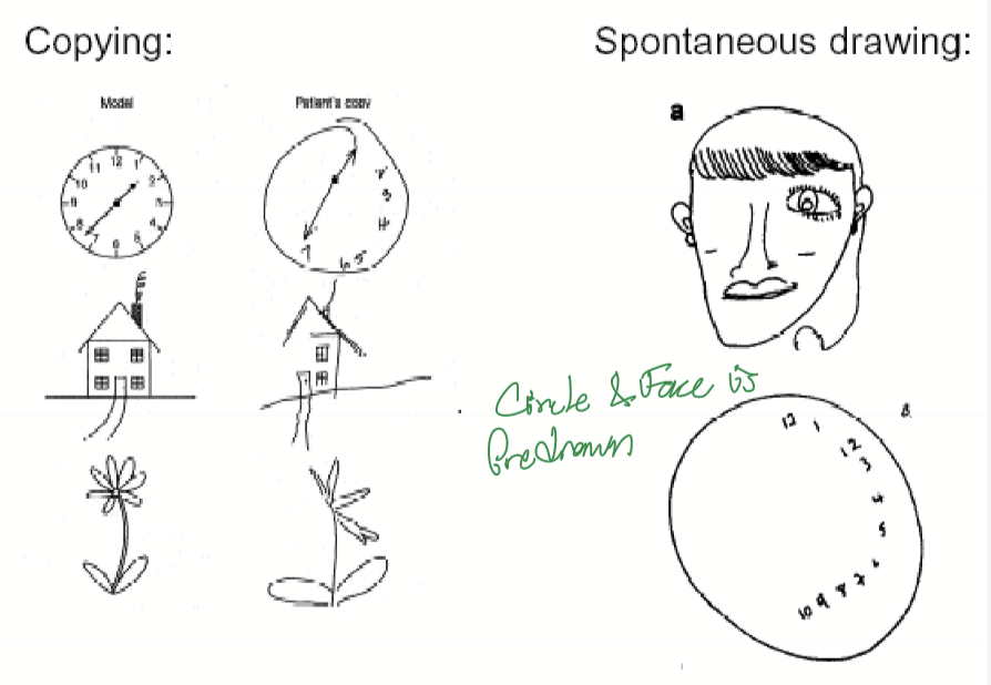
What is hemifield neglect and what causes it?
It is where individuals neglect the left visual field due to damage of the right parietal lobe responsible for spacial awareness and attention.
What is hemianopsia?
It is the loss of a visual field due to damage to the visual pathway (optic tract, LGN, occipital cortex, etc.)
What are some complications if the ventral pathway is disconnected?
Visual-visual: agnosias: the inability to recognize objects
Visual-verbal: Anomias and Alexia: difficulty naming objects even though the object is recognized
Visual-limbic: impair emotional responses to visual stimuli or the ability to associate visual input with personal memories
Types of Visual Agnosias
Achromatopsia: an inability to distinguish different colors
Prosopagnosia: an inability to recognize human faces
Simultagnosia: an inability to recognize multiple objects in a scene/ can’t put the cause & effect together
Object agnosia: inability to recognize objects
Pure alexia: an inability to read
What is Object Form Topology Hypothesis?
Hypothesis suggests that object form representation in the ventral temporal cortex is organized in a continuous, distributed, and overlapping manner.
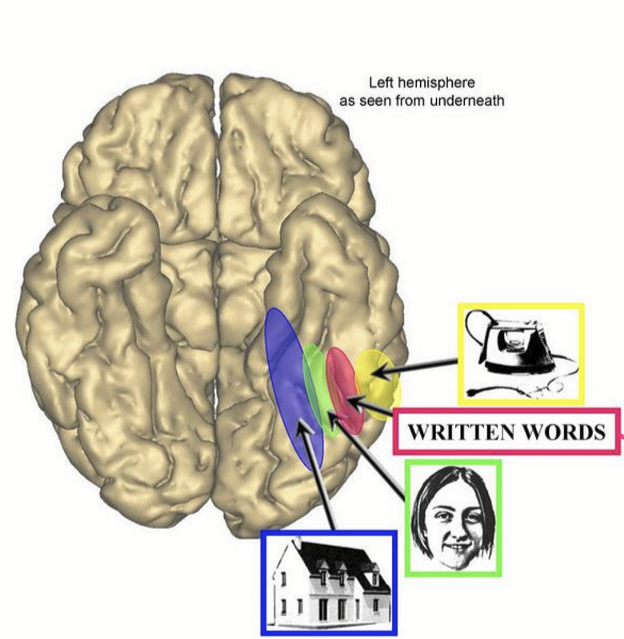
What causes Alexia?
Defect in occipitotemporal corticofugal bundle
Defect in LEFT inferior temporal gyrus
Rostal vs caudal portions of the brain
Rostal is more “ventral” while caudal is more towards the spine.
