LAB: Axial Skeleton
 Call Kai
 Learn
Learn Practice Test
 Spaced Repetition
 Match
Match Flashcards
 Knowt Play
1/96

There's no tags or description
Looks like no tags are added yet.
Name | Mastery | Learn | Test | Matching | Spaced | Call with Kai | Chat |
|---|
No analytics yet
Send a link to your students to track their progress
97 Terms
Frontal bone

Parietal bone

temporal bone

occipital bone

Sphenoid bone (lateral view)

sphenoid bone (inferior view)

ethmoid bone

Zygomatic bone

mandibular fossa
a smooth, oval depression on the temporal bone of the skull where the lower jawbone meets the head

stylod process

mastoid process
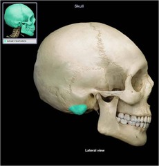
external acoustic meatus
The ear canal leading to eardrum and middle ear, directs sound waves and protects the inner ear from external factors with earwax.

internal acoustic meatus
A short, narrow bony canal inside the skull that acts as a passageway for nerves and blood vessels.

jugular foramen
Located at the base of the skull, positioned between the temporal bone and the occipital bone. Allows for nerve passageway and blood drainage.

carotid canal
Located at the base of the skull within the temporal bone. Passageway for internal carotid artery

foramen magnum
A large opening at the base of the skull through which the brain connects to the spinal cord.

occipital condyles
two smooth, oval bumps on the bottom of the skull that connect your head to your neck. It also helps in rotating the skull back and forth and provides stability and balance.

greater wing of sphenoid bone

lesser wing of sphenoid bone
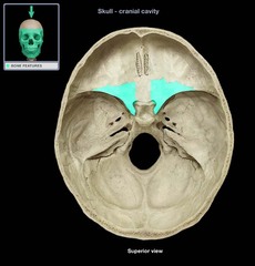
superior orbital fissure
is a narrow cleft-like opening at the back of the eye socket that allows vital nerves and blood vessels to pass between the brain and the eye

sella turcica of sphenoid bone
Located right in the middle of the sphenoid bone at the base of the skull and houses and protects the pituitary gland

optic canals of sphenoid bone
openings in the base of the lesser wings for cranial nerve II (optic nerve) and let the ophthalmic artery (main blood source for eye and eye socket) through

crsita galli

cribriform plate of ethmoid bone
The horizontal plate of the ethmoid bone separating the cranial cavity from the nasal cavity.
Aids in smelling and separating the nasal and cranial cavity.

perpendicular plate of ethmoid bone
superior portion of the bony nasal septum.
Divides the nose into left and right parts and support the nose bridge.

Middle nasal conchae
Located on the side wall inside each of your nasal passages.
Its function is to warm and moisten air, filter dust, and help in drainage for the sinuses

superior nasal concha of ethmoid bone
Found in the uppermost part of the inside of the nose above the middle nasal conchae.
Function is to aid in smelling, airflow, and acts as a drainage passageway for the ethmoid bone.
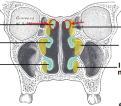
sagittal suture
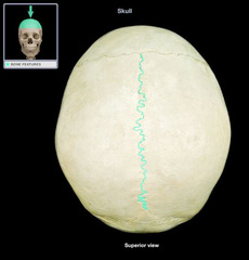
coronal suture

squamous suture

lambdoidal suture

coronoid process of mandible
Located on the mandible right below your cheekbone area.
Its function is to help in chewing.

ramus of mandible

body of mandible

alveolar process of mandible

alveolar process of maxilla
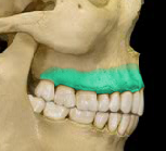
palatine process of maxilla
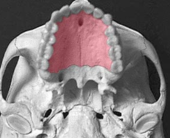
palatine bone
Located deep inside the skull behind the upper jaw
separates the oral cavity (mouth) from the nasal cavity (nose)

zygomatic process of maxilla
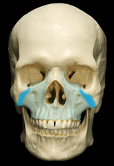
lacrimal bone

lacrimal fossa
Located in the inner corner of the eye.
Houses the lacrimal sac, which helps to drain tears from the nasal cavity

nasal bone

Vomer

inferior nasal conchae

hyoid bone
a U-shaped bone in the neck that supports the tongue.

frontal sinus

ethmoid sinus

maxillary sinus

sphenoid sinus
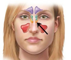
function of sinuses
Reduce weight of head
Warm and humidify air
Trap dirt, dust, and germs in mucus
Absorb impact from head injuries
Modify voice during phonation
Manubrium of sternum

body of sternum

Xiphoid process of sternum

xiphisternal joint of sternum

true ribs
first 7 pairs of ribs; attach directly to sternum

false ribs
Ribs 8-12; attach indirectly to sternum

floating ribs
Ribs 11-12; do not attach to sternum

anterior fontanel
Located at the top of the head
where the sagittal suture and coronal suture meet. Ossifies around 9-18 months of age.

posterior fontanel
back soft spot on fetal skull where the parietal and occipital bone meet. Closes by 1-2 months of age.

sphenoid fontanel
soft spot between frontal, parietal, temporal bones. Ossifies around 3-6 months.

mastoid fontanel
Located where the temporal, parietal, and occipital bones meet. Ossifies between 6-18 months of age.

What is the purpose of fontanelles?
To allow the skull to fold/flex during labor and provide room for rapid brain growth.
How many cervical vertebrae are there?
7

How many thoracic vertebrae are there?
12

How many lumbar vertebrae are there?
five (fused)

How many sacral vertebrae are there?
5 fused

How many coccyx vertebrae are there?
3-5 fused

body of vertebrae

vertebral arch of vertebrae

pedicle of vertebrae

lamina of vertebrae

spinous process of vertebrae

vertebral foramen
canal through which spinal cord passes

transverse process of vertebrae
The portion of the vertebrae that points out laterally, serving as a crucial anchor point for muscles and ligaments.

superior articular process of vertebrae
An upward-projecting bony bump on the posterior arch that connects with the vertebra directly above it. It also provides stability for the spinal column and determines the movement of the spine.

inferior articular process of vertebrae
articular process that arises at the junction between the pedicles and laminae; projects downward and connects with the vertebra directly below it. It also provides stability for the spinal column and determines the movement of the spine.

Transverse foreman of cervical vertebrae
The two holes lateral to the body of a vertebrae that allows vital blood vessels and nerves to safely travel up to the brain.

facets where ribs articulate (thoracic vertebra)

What is this vertebrae?
Atlas (C1)
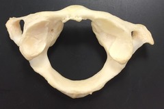
posterior arch of C1?

anterior arch of C1?

superior articular facet of C1
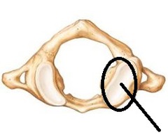
inferior articular facet of C1
the underside of the superior articular facet of C1
What is this vertebrae?
Axis (C2)

Dens of C2

superior articular facets of C2

Inferior articular facets of C2
the underside of the superior articular facet of C2
spinous process of C2

Promontory of sacrum
anterior view

foramina of sacrum
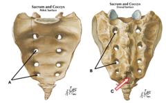
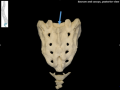
median sacral crest of sacrum
posterior veiw

Body of sacrum posterior view
concave
curves inward
convex
curves outward
Lordosis
abnormal increase in the forward curvature of the lumbar spine

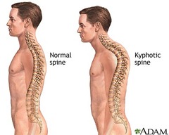
Kyphosis
excessive outward curvature of the upper thoracic spine, causing hunching of the back.
Scoliosis
an abnormal lateral curvature of the spine
