Special Senses Objectives
1/40
There's no tags or description
Looks like no tags are added yet.
Name | Mastery | Learn | Test | Matching | Spaced |
|---|
No study sessions yet.
41 Terms
What are the four defects that affect the organogenesis of the entire eye?
1) Anophthalmia: rare condition in which there is no globe development; seen with other developmental
anomalies
2) Microphthalmia: inadequate growth resulting in a small, disorganized globe; caused by a genetic defect or in utero infection (bovine viral diarrhea virus, merle coat)
3) Cyclopia: single globe (1 optic structure present) vs Synophthalmia: incomplete separation of globes (2 pairs of
optic structures present)
4) Coloboma: something not forming completely (i.e., Failure of the optic fissure to fuse if at 6:00)
What plant causes cyclopia/synophthalmia in lambs? What day of gestation do gravid ewes have to ingest this plant for this to occur?
- Ewes ingesting Veratrum californicum (Corn Lily/False Hellebore)
- Ingestion at day 14 of gestation
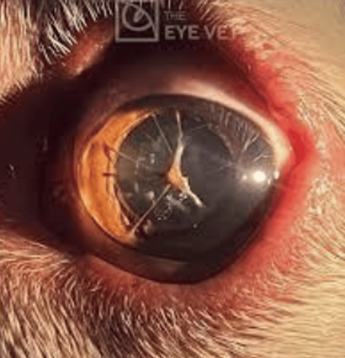
What ocular defect arises from incomplete atrophy of the fibrovascular sheet?
- Persistent pupillary membrane which covers developing iris and lens
What are the characteristics of collie eye anomaly?
- Patchy to diffuse choroidal hypoplasia (choroid is very thin) - may be related to defective RPE
- Optic disc coloboma (optic disc doesn't fully form) with staphyloma (out patching of sclera) - Retinal detachment with retinal rosettes (histological finding)
Compare and contrast Infectious Bovine Rhinotracheitis and Infectious Bovine Keratoconjunctivitis.
- IBR is NOT pink eye, is caused by Bovine Herpesvirus type-1, and leads to purulent conjunctivitis and respiratory disease.
- Bovine keratoconjunctivities IS pink eye, is caused by Moraxella bovis transmitted by the face fly, and leads to lacrimation, blepharospasm, and corneal ulceration.
What parasite is a "true" eye worm (one that inhabits the conjunctival sac)?
- Thelazia – lives in conjunctival sac and lacrimal duct
What layer of the cornea is dehydrate? Why does this matter in corneal ulcers?
- Corneal stroma is dehydrated
- • Ulceration can expose the corneal stroma to fluid and infectious pathogens, which can ultimately lead to a
descemetocele if Descemet's membrane is involved; The stroma uptakes fluorescein stain
Describe what each "color" of the cornea indicates.
- Red - blood vessels
- Blue - corneal edema
- Yellow - cellular infiltrates (ex. Ulcer, hypopyon - accumulation of leukocytes and fibrin within the anterior chamber)
- White - fibrosis (granulation tissue)
- Brown - pigment
- Black -sequestrum, foreign body
What infectious agent causes "blue eye"? What layer of the cornea does this target?
- Canine adenovirus-1 (causes infectious canine hepatitis)
- Targets endothelial cells (destroys the endothelium allowing for edema/fluid to infiltrate)
Compare and contrast the disease presentation of Chlamydophila felis and Feline herpesvirus-1.
- Chlamydophila felis causes bacterial conjunctivitis (usually unilateral), is seen in cats and koalas, and leads to the formation of reticulate bodies in epithelial cells (sign of early disease)
- Feline Herpesvirus-1 causes viral conjunctivitis (usually bilateral) and rhinotracheitis in young cats. In adult cats, it causes keratitis and dendritic ulceration.
Compare and contrast Keratoconjunctivitis sicca and Pannus keratitis.
- Keratoconjunctivitis sicca is a disorder of the tear film which most commonly affects dogs and leads to corneal damage. This is commonly due to a chronic, progressive, immune-mediated disease of the lacrimal gland (can also be neurogenic, drug toxicity, iatrogenic (cherry eye removal). It leads to erosion and ulceration w/o watery eyes; increased mucoid discharge, and cloudy/pigmented cornea.
- Pannus keratitis is a non-infectious progressive, superficial keratitis seen with altitude and UV exposure (immune reaction to corneal epithelial antigens). German Shepherds are predisposed and it leads to vascularization, pigmentation, and scar tissue formation from the limbus to the cornea
Why would removing a "cherry eye" result in Keratoconjunctivitis sicca?
- The third eyelid holds the gland of the third eyelid which contributes to the tear film. Removal therefore affects tear film production, leading to keratoconjunctivitis sicca.
- What layer is affected in Indolent ulcers AKA Spontaneous Chronic Corneal Epithelial Defects?
- Corneal epithelium (re-ulcerating lesion due to poor adhesion of the epithelium to the underlying stroma; persistence of normal superficial stromal degeneration)
What is a feline corneal sequestrum?
- Non-inflammatory necrosis of keratocytes with epithelial healing (stromal pigment due to aggregation of porphyrins and keratocytes); Sequestrum may eventually slough off; Seen in brachycephalic cats; FHV-1 related
What layers of the cornea are present in a descemetocele?
- Descemet's membrane: Very thin and susceptible to rupture
Why might a swab of the superficial cornea in equine mycotic keratitis not yield an infectious agent? What is the fungus typically cultured from equine mycotic keratitis?
- The fungal infectious agent lives in the DEEP portion of the cornea
- Fungus is typically Aspergilllus spp or Penicillum spp (Fungus targets Descemet’s membrane)
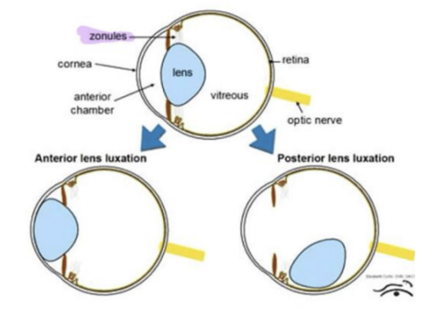
Terrier breeds have an inherited predisposition to anterior lens luxation. What is the mutation that causes this and what is the normal function of this gene?
- ADAMST17 gene mutation - responsible for formation and maintenance of lens zonules; Results in anterior lens luxation
Why do diseases like uveitis and glaucoma result in cataracts?
- Because it results in the disruption of normal composition and flow of aqueous humor
- Causes denatured lens proteins and cellular degeneration
- Seen in diseases like trauma, uveitis, glaucoma, nutritional, radiation, toxins (post inflammatory cataracts)
What species gets diabetic cataracts?
- 70% of diabetic dogs: Due to high glucose in aqueous overwhelms anaerobic glycolic pathways in the lens; Glucose in converted into sorbitol that accumulates in the lens and osmotically attracts water
What part of the immune privilege of the eye mediates the day-to-day leakage of autoantigens? How does this help protect the eye? (
- Anterior chamber-associated immune deviation
- Immunomodulating cytokines alter ocular antigens; APCs can only call limited function lymphocytes (only have antigen-specific cytotoxicity)
What is the difference between phacolytic uveitis and phacoclastic uveitis? For both diseases, which part of immune privilege of the eye gets overwhelmed?
- Phacolytic uveitis: Hyper mature cataract leaks denatured lens protein through intact lens; Antigenic lens proteins result in uveitis; Lymphoplasmacytic uveitis (immune mediated response causes this)
- Phacoclastic Uveitis; Traumatic corneal perforation with lens injury; Lens material ends up in the anterior chamber; This overwhelms normal immune deviation and T-cell tolerance - lens sensitized lymphocytes come to
attach; Result: severe granulomatous uveitis after the cornea heals
What components of the uvea are involved in anterior uveitis? Posterior uveitis?
- Anterior Uveitis: iris and ciliary body
- Posterior Uveitis: choroid +/- retina
Is iris bombe associated with anterior or posterior synechia?
- Posterior synechia: adherence of iris to lens
Name two routes of entry for bacterial uveitis. Which is bilateral? Which is unilateral?
- Hematogenous: Bilateral (Ciliary body and choroid)
- Corneal perforation: Unilateral (Anterior chamber)
What fungal agent typically affects dogs with fungal uveitis? Cats?
- Dogs: Blastomyces dermatitidis
- Cats: Cryptococcus neoformans or Cryptococcus gatii
Bovine malignant catarrhal fever is associated with which herpesviruses in wildebeest and sheep? What clinical signs do cattle affected by this disease present with?
- Alcelaphine herpesvirus (AHV-1) and Ovine herpesvirus-2 (OHV-2)
- CS: Severe lymphocytic uveitis with necrosis, ciliary flush (brush border of vessels migrating in), corneal edema
What is one of the most important causes of blindness in horses? What breed is affected? What infectious agent is often connected to this disease?
- Equine Recurrent Ophthalmitis (aka Moon Blindness)
- Appaloosas
- Leptospiral antigen in eye connected to this disease
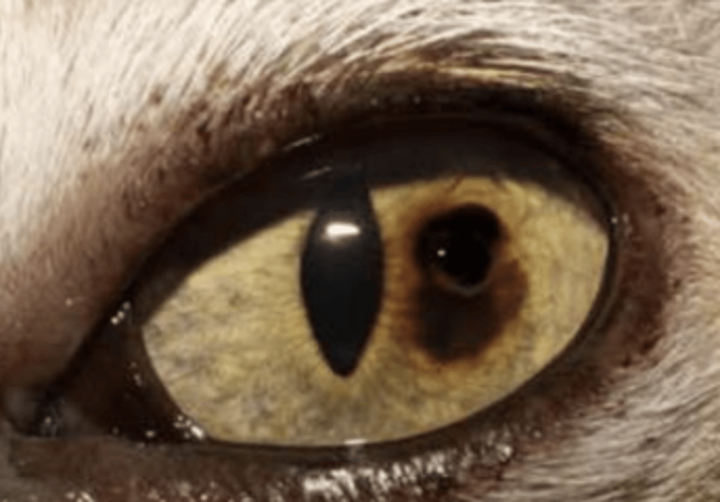
What are two important causes of glaucoma in cats?
1) Idiopathic Lymphonodular Uveitis of Cats: commonly causes glaucoma o Immune-mediated (ID of antigens unknown)
2) Feline Diffuse Iris Melanoma (intraocular neoplasia): Patchy iris pigmentation that progresses over time
What are three diseases/causative agents that cause photoreceptor degeneration?
1) Progressive retinal atrophy of dogs (genetic predispositions): 2-3-month-olds have night blindness (total blindness); also seen in Abyssinian cats
2) Taurine deficiency in cats: Focal lesion of tapetal hyper-reflectivity dorsolateral to the optic disk
3) Enrofloxacin: Can cause retinal degeneration in cats
What are two potential causes of hypertensive retinopathy in cats? What does hypertensive retinopathy result in?
- CKD or hyperthyroidism
- Results in: hypertensive-induced choroid vascular damage, retinal hemorrhage and detachment, acute onset blindness (cats present as looking around at nothing)
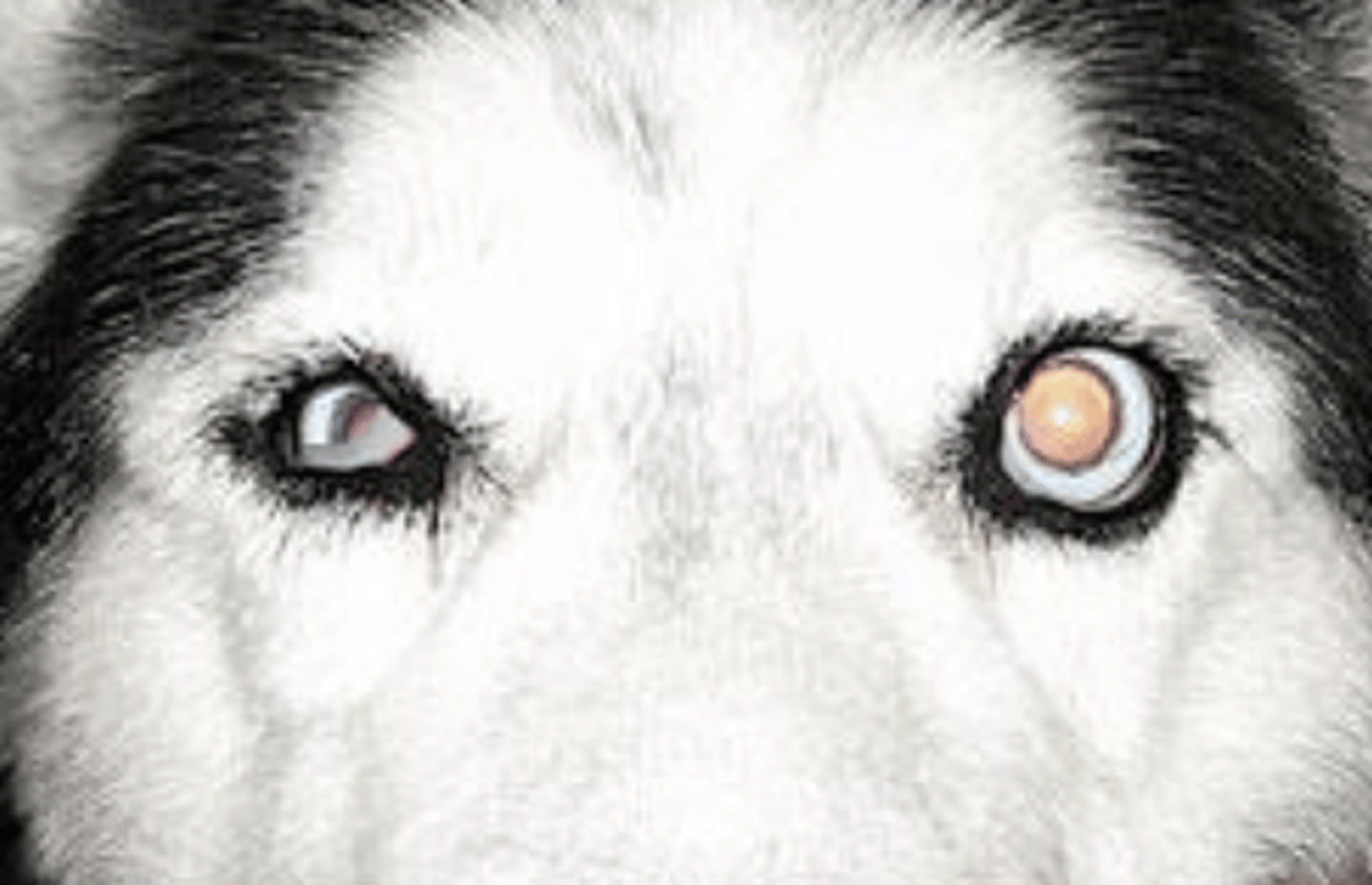
What is glaucoma? What substance builds up that causes glaucoma? Where does this substance normally drain?
- Glaucoma: increase in intraocular pressure resulting from impairment of aqueous outflow
- Aqueous humor build-up - normally drains into scleral venous plexus
What two structures are atrophied in glaucoma? What layer of the retina is atrophied specifically?
- Retinal atrophy (particularly ganglion cell layer)
- Optic nerve atrophy
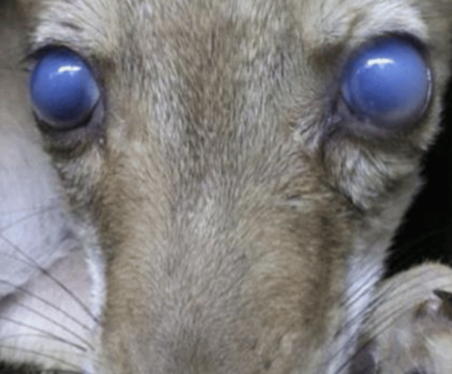
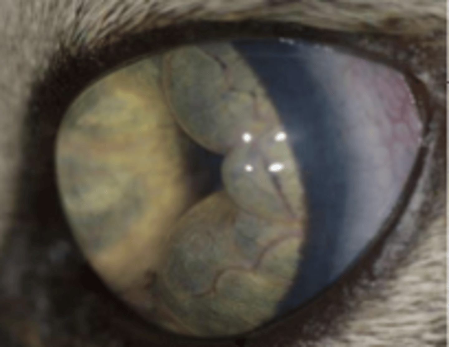
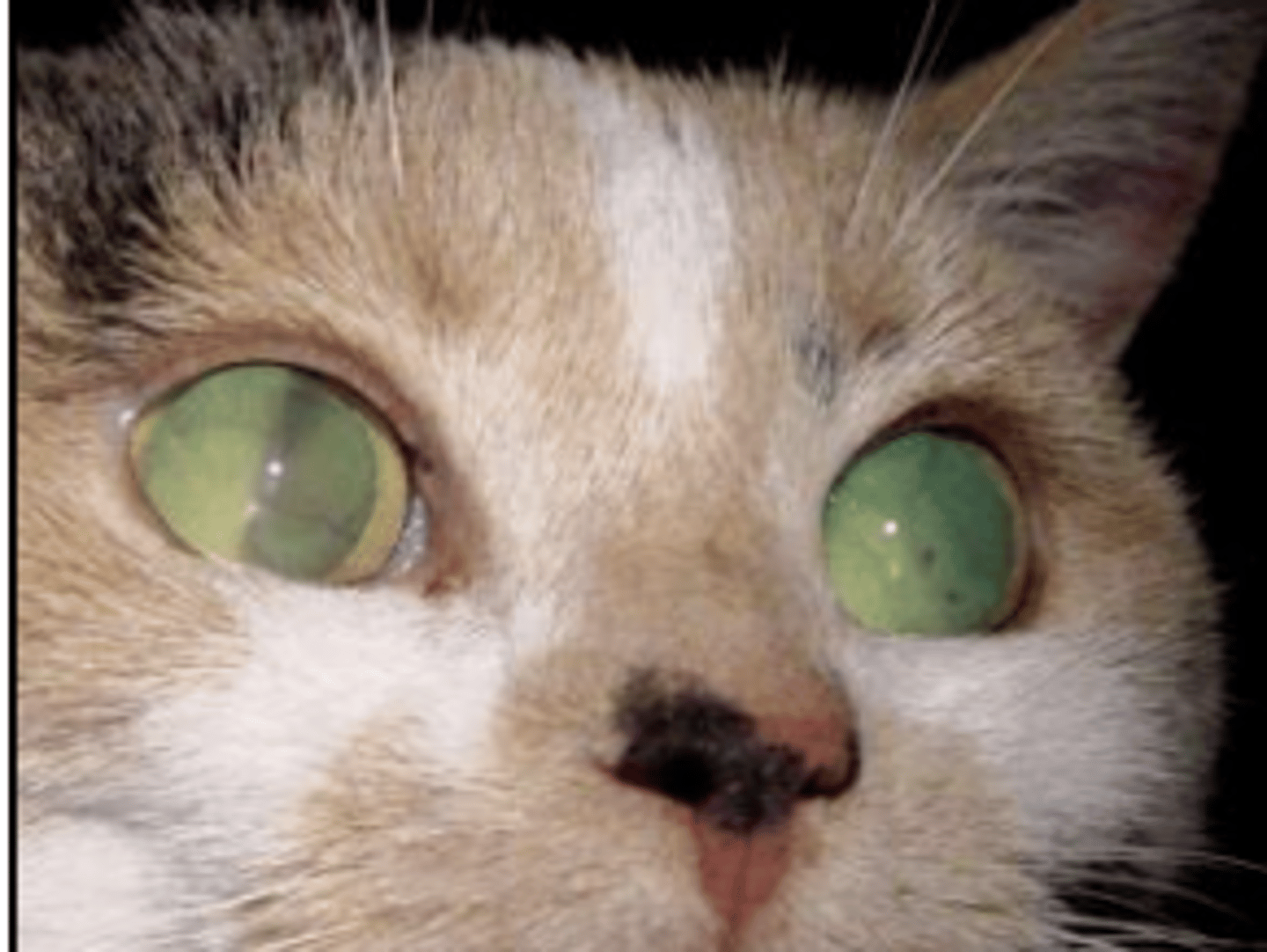
What type of "eye bulge" is seen in glaucoma? How is this different than exophthalmos and proptosis?
- Buphthalmos: Common in glaucomatous eyes; Enlargement of the globe without misplacement in the orbit itself (Left images)
- Exophthalmos: Normal sized globe being pushed out of the orbit
- Proptosis: Pretty much the same as exophthalmos but commonly used to refer to post-traumatic events in which the globe is pushed entirely out of the orbit and requires surgical correction
How are primary glaucoma and secondary glaucoma different? (Think of what causes primary glaucoma vs what causes secondary glaucoma)?
- Primary glaucoma: No prior ocular disease; Malformation of filtration angle (goniodysgenesis)
- Secondary glaucoma: Anterior uveitis, ehmorrhage/fibrin plugging the angle, lens luxation, intraocular neoplasia (Feline Diffuse Iris melanoma)
What are the functions of hair cells? What part of the ear are they located in (outer, middle, inner)?
- Auditory and vestibular function
- Located in the membranous labyrinth of the internal ear
Hereditary deafness is linked to phenotypes of hair coat and eye color in poorly pigmented collies and dalmatians. What cells are defective and why are they important?
- Defective intermediate cells due to diminished pigment (Neural crest derived melanocytes) -> Failure to recycle potassium to maintain endolymph -> Cannot transduce fluid waves into sound recognition
What disease and bacteria results in peripheral vestibular disease in calves?
- Mycoplasma bovis (tympanic membrane fills with pus) - otitis media/interna
What are the clinical signs of Horner's syndrome (loss of sympathetic innervation to the eye)?
- Ptosis (drooping eye)
- Elevated nicitans
- Enophthalmos (sunken eye)
- Miosis (contracted pupil)
- Cutaneous vasodilation
The guttural pouch is a diverticula of what structure?
- Diverticula of the auditory tube (connection of middle ear to pharynx)
Name two diseases of the guttural pouch and what infectious agent they are caused by.
1) Guttural Pouch Empyema (aka Strangles): Streptococcus equi spp qui
2) Guttural Pouch Mycosis: Aspergillus spp; Epistaxis -> fungal erosion of the wall of internal carotid artery (can lead to death)
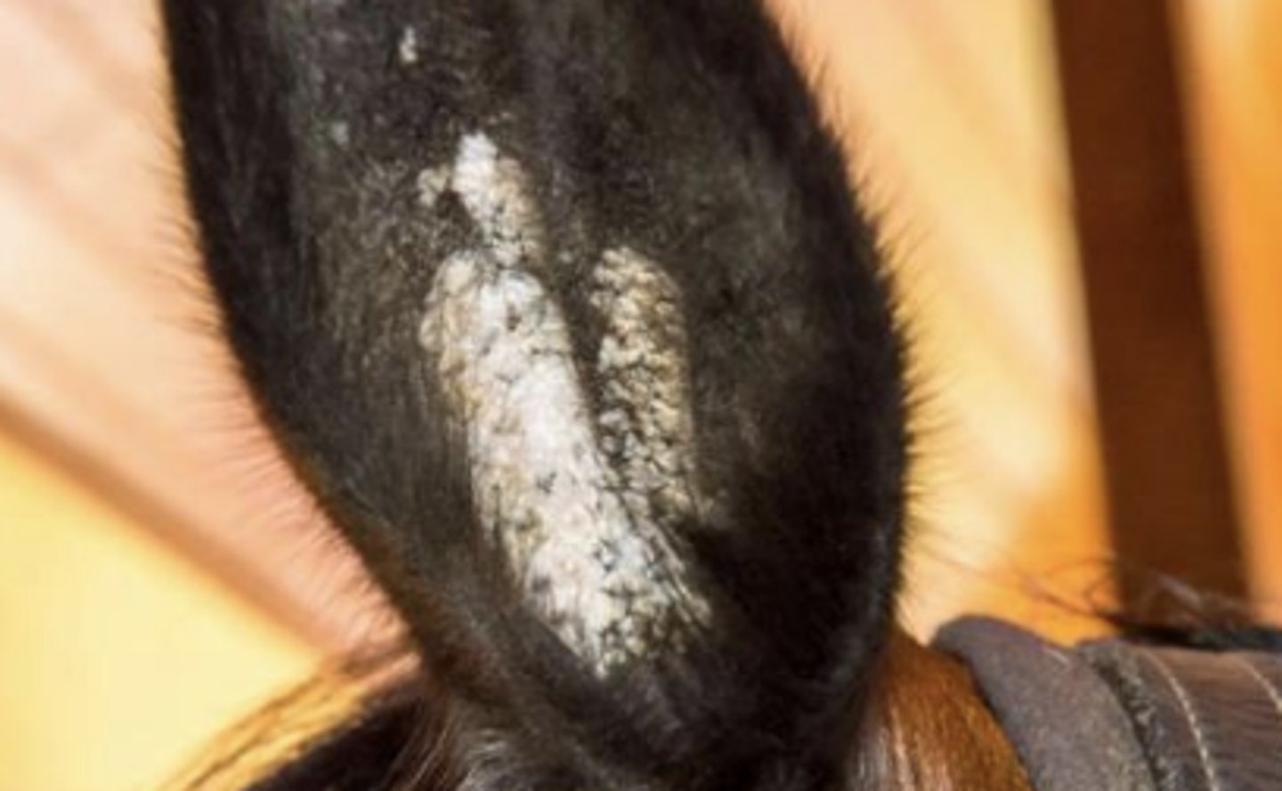
What causes equine aural plaques?
- Equine papillomaviruses (EcPV-3, 4, 5, 6)
- Equine Aural Plaques: depigmented hyperkeratotic plaques along the concave surface of the ear which so not resolve or regress