WBC Lecture 3: Myeloproliferative Disorders, FCM
 Call Kai
 Learn
Learn Practice Test
 Spaced Repetition
 Match
Match Flashcards
 Knowt Play
1/44

There's no tags or description
Looks like no tags are added yet.
Name | Mastery | Learn | Test | Matching | Spaced | Call with Kai | Chat |
|---|
No analytics yet
Send a link to your students to track their progress
45 Terms
Myeloproliferative neoplasms
Clonal hematopoietic disorders caused by gene mutations in HSCs; increase in proliferation of and accumulation of mature erythrocytes, granulocytes, and platelets
10:1
M:E ratio in CML
300×109/L
Approximately 15% of CML patients exhibit hyperleukocytosis (total WBC counts greater than (——)), risk of leukostasis
Decreased
LAP score in CML
Gaucher-like (sea blue)
Green-gray crystals
Describe macrophage characteristics that can be observed in CML (2)
Granulopoiesis: ↑
Erythropoiesis: ↓
Megakaryopoiesis: ↑/N
Reticulin: ↑
BM characteristics in CML (granulopoiesis, erythropoiesis, megakaryopoiesis, reticulin)
Small
Describe megakaryocyte characteristics that may be observed in CML
Hepatomegaly
Splenomegaly
Describe extramedullary tissue characteristics that can be observed in CML (2)
Hyperuricemia
Uricosuria
Secondary gout
Uric acid stones
CML symptoms (4)
Cytogenetic analysis: t(9;22)
qRT-PCR: BCR-ABL 1 transcript
FISH: BCR-ABL 1 fusion gene
Genetic analysis for CML (3)
PBS incubated with ALP substrate → precipitate on bands and seg neuts
Napthol-phosphate substrate and diazo dye at alkaline pH
100 neuts and bands counted → score (color intensity) multiplied by # cells
Normal range: 15-170
How to get a LAP score (3)
Poor response to treatment
More: chromosomal changes, anemia, basophils, abnormal PLT, micromegakaryocytes, megakaryocytic fragments, circulating blasts (10-19%)
Less: Mature neuts, platelets
Transformation to AML/ALL
CML blast crisis characteristics (treatment, increased values (7), decreased values (2), transformation)
Signal transducers and activators of transcription (STAT)
EPO
Polycythemia vera
Normal JAK2 is related to (——) proteins which are activated following the binding of cytokines (——), leading to cell proliferation; in (——) mutated JAK2 activates the JAK/STAT signaling pathway independent of cytokines
Major
Elevated Hb and HCT
BM hypercellularity with trilineage growth (panmyelosis)
JAK2 mutation
Minor:
Low serum EPO
WHO criteria for PV diagnosis (4)
Normal or increased
Polycythemia vera LAP score
Splenomegaly
Hepatomegaly
Generalized vascular engorgement
Inc risk: hemorrhage, tissue infarction, thrombosis
Polycythemia vera symptoms (6)
Toxic vacuolization: 2-4+
Toxic granulation: 2-4+
Dohle bodies; Frequent
Eosinophilia: 0
Basophilia: 0
LAP: High
Ph xm: (-)
Leukemoid reaction characteristics (toxic vacuolization, toxic granulation, dohle bodies, eosinophilia, basophilia, LAP, Ph xm)
Toxic vacuolization: 0-1+
Toxic granulation: 0-1+
Dohle bodies; Rare
Eosinophilia: 1-3+
Basophilia: 1-3+
LAP: Low/0
Ph xm: (+) >90%
CML characteristics (toxic vacuolization, toxic granulation, dohle bodies, eosinophilia, basophilia, LAP, Ph xm)
Red cell mass always increased
EPO decreased
O2 sat normal
Leukocytosis and thrombocytosis
BM myelopoiesis
Splenomegaly
Characteristics of PV that differentiates it from hypoxia (RBC mass, EPO, O2 sat, elevated cells, BM, EM tissue)
Red cell mass can be increased
Only erythroid hyperplasia in BM
EPO increased
O2 sat decreased
Hypoxia characteritics that differentiates it from PV (RBC mass, BM, EPO, O2 sat)
600-2000×109/L
Large masses of PLT aggregates
Abnormal morphology (giant/bizarre/MK fragments)
(generally normal, but can vary)
PLT characteristics in essential thrombocythemia (3)
Elevated WBCs
Neutrophilia with slight left shift
WBC characteristics in essential thrombocythemia (2)
RBCs normal or slightly decreased
nRBCs rare
RBC characteristics in Essential thrombocythemia (2)
Hemorrhage (bleeding tendencies from abnormal PLTs)
Thrombosis (PLTs > 2000×109/L)
Complications associated with polycythemia vera (2)
Primary myelofibrosis
Abnormal clone of megakaryocytes which stimulate the bone marrow fibroblasts to produce collagen
PDGF-β
Fibrosis
Extramedullary hematopoiesis
In primary myelofibrosis, fibroblastic growth factors such as (——) are released; normal bone marrow tissues are eventually replaced by (——) and lead to dry tap in biopsies; over time, this leads to progressive bone marrow failure, eventually leading to (——)
PV
ET
PMF
MPNs associated with JAK 2 mutation (3)
Immature granulocytes
nRBCs
Dacrocytes (+ other bizarre shapes)
Giant PLTs
Primary myelofibrosis characteristics in a PBS (4)
WBC: ↑
RBC: N/↓
PLT: N/↑
Molecular abnormalities: Ph t(9;22), BCR-ABL 1
CML characteristics (WBC, RBC, PLT, molecular abnormalities)
WBC: N/↑
RBC: ↑
PLT: N/↑
Molecular abnormalities: JAK2 V617F (or other JAK2)
Polycythemia vera characteristics (WBC, RBC, PLT, molecular abnormalities)
WBC: N/↓
RBC: N/↓
PLT: ↑
Molecular abnormalities: JAK2 or other mutation
Essential thrombocythemia characteristics (WBC, RBC, PLT, molecular abnormalities)
WBC: N/↑/↓
RBC: N/↓
PLT: N/↑/↓
Molecular abnormalities: JAK 2 or other mutation
Primary myelofibrosis (WBC, RBC, PLT, Molecular abnormalities)
BM aspirate → heparin
BM → fine needle biopsy
PB → EDTA
Lymphoid tissue
Body cavity fluid/solid tissue w/ hematologic malignancy
Specimens most commonly analyzed for flow cytometry in hematology (5)
A: Erythroid and PLT
B: Blast
C: Neutrophils
D: Monocytes
E: Lymphocytes
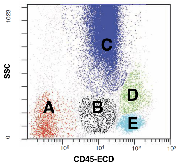
Identify the flow cytometry populations of a BM sample
SSC = complexity
FSC = size
In flow cytometry, side scatter is proportional to (——), while forward scatter is proportional to (——)
SCID
HIV
What can flow cytometry test for with immune deficiencies
CD34
What can flow cytometry test for with stem cell enumeration
CD 55
CD 59
What can flow cytometry test for with PNH
RNA binding dyes (toluene orange)
What can flow cytometry test for with reticulocytes
Ab against HbF, Rh D, or both, or CA
What can flow cytometry test for with FMH (4)
DNA content
What can flow cytometry test for with cell cycle analysis
HLA
Flow cross match
(Donor lymph + pt serum → incubate AHG F(ab)/FITC)
What can flow cytometry test for with transplantation (2)
FMH volume = (% fetal cells) (1800) (1.22)
How to calculate fetal maternal hemorrhage volume from FC analysis
WB/BM stored long with anti-coagulants
WB/BM aspirate delivered not fresh or inappropriate anticoagulant
Tissue sample not in appropriate media
Harsh vortex on cell pellets
Aggressive mechanical disruption of tissue
Cell pellets kept dry for a long time
High concentration cell suspension
Reasons for unacceptable specimens due to compromised cell viability (WB/BM 2, tissue 2, cell pellets 2, cell suspension)