TEST 3 Neurobiology- Motor Systems
1/75
There's no tags or description
Looks like no tags are added yet.
Name | Mastery | Learn | Test | Matching | Spaced |
|---|
No study sessions yet.
76 Terms
What are properties of a skeletal muscle cell?
- has multiple nuclei
- made of long myofibrils (which are fibers of actin and myosin)
- also known as a muscle fiber
What do Myofibrils contain and what do they do? How do they show up in electron micrographs?
- fibers of actin and myosin
- allow cells to have contractile properties (ie shorten and generate force)
-electron micrograph shows the thick and thin filaments of a myofibril:
-----thin filaments show up lighter in color and are made of F actin
-----thick filaments are darker and myosin
-----where the dark bands are show overlap btw f actin and myosin (the myosin filaments have golf club like heads that interact with the F actin heads to bind to actin and pull on it)
What is a sarcomere?
contractile unit of a muscle fiber; they go from z-line to z-line
Describe the cross-bridge cycle
1. ATP binds to the Myosin head, releasing the Myosin head from the actin filament
2. ATP --> HYDROLIZES to ADP + PI... Both stay attached and result in the conformation change of the head to a 90 degree angle (high energy position) and storing energy
3. The cocked head with ADP binds a new actin site , releasing the Pi. causing a conformational change that PULLS the actin to the center of the sarcomere
4. A power stroke motion occurs
5. ADP is released from myosin head, new ATP binds to the myosin and the head detaches--> restarting the cycle
How is muscle contraction calcium dependent?
- tropmyosin is a long thin protein found in muscle fibers that wraps around actin fillaments
- it covers binding sites and blocks myosin binding at rest (aka stops myosin from attaching, pulling, and causing contraction)
- when calcium ions are released, they bind to the
- troponin protein... which changes shape and moves tropomyosin out of the way-> uncovering binding sites
-allows myosin to attach to actin and muscle contraction
How is this calcium released?
1) Motor neuron sends an AP to muscle fiber, that reaches the NMJ and releases ACh into synaptic cleft
2) ACH binds to a nicotinic ACh receptor on the muscle fiber sarcolemma--> NEW AP travels along surface of muscle fiber. The muscle cell membrane depolarizes
3) AP spread down T-tubules (tube like invaginations of the sarcolemma that run deep and carry electrical signal deep into cell)
4) T-tubules depolarize-> open calcium channels in SR-> which releases Calcium ions into cytoplasm around myofibrils
5) the electrical signal reaches EVERY myofibril at nearly the same time-> calcium floods every sarcomere at once-> all sarcomeres contract tat-> entire fiber contracts
- T TUBULES allows for a coord contraction of muscle fiber and rapid depol from one synapse
Where are motor neurons located? What about sensory input? How many muscle fibers can a motor neuron innervate?
- Cell bodies of motor neurons are in ventral (anterior) horn of SC--> has MN cell bodies that leave SC to control muscles
- Dorsal horn (posterior)- receives sensory input coming into spinal chord
Sensory info ENTERS dorsally and motor commands EXIT ventrally
- alpha motor neurons in the ventral horn send AXONS out through peripheral nerves to the muscle fibers. NMJ= spot between muscle fiber and peripheral nerves
- one MN can innervate MANY muscle fibers; one muscle fiber receives input from only one MN (one NMJ)
Describe graded control. How do muscles w/ fine control work as opposed to muscles with gross control?
Graded control states that if a MN only activates one fiber, there will only be a small amount of force generated
muscles w/ precise fine control use small motor units (ie 1 neuron innervates one to a few fibers- like the muscles in eyes/fingers)
Muscles w/ gross control or large mvmts like quadriceps have one neuron innervating HUNDREDS of fibers
what is a motor pool? which motor units activate first? what experiment showed this?
a motor pool is a collection of ALL MNs that innervate one muscle... diff motor pools can be located close in SC region--> allowing for coord mvmt of muscles
the smallest motor units (innervate the fewest fibers)-> activate first bc you start w the least force and work your way up. if more force is needed then activate LARGER motor neurons
-> bigger units join as demand increases, allowing for a smooth increase in muscle force
Henneman Size principle- Henneman applied pressure to muscles-> at low pressure only small units fired and as pressured decreased larger units stopped first
What are Central Pattern Generators?
neural circuits that make rhythmic and patterned outputs (ie breathing, swimming, and walking)- CPGs don't require conscious control OR sensory input. they generate repeating patterns of neural activity AUTOMATICALLY
How is Horse locomotion an example of CPG?
they have distinct movement patterns for each gait... for example walking has a diff timing and coord off the four limbs than galloping and trotting.
the diff gaits of a horse correspond to different CPGs-> each makes a unique rhythm of neural activity.
each gait also requires reciprocal regulation of antagonistic muscles (next slide will describe an example)
Describe antagonistic muscle contraction and biceps example
muscles that perform OPPOSITE actions must ALTERNATE activity to produce rhythmic motion
- for example flexor muscles like the biceps bend a joint whereas extensor muscles like triceps straighten it
- these PAIRS of muscles are called antagonistic muscles... if they contract simultaneously the limb won't move at all
how does the nervous system prevent antagonistic muscles from contracting at the same time
by wiring inhibitory INTERNEURONS between the circuits that control the opposing muscles
- when the flexor motor neuron fires... an inhibatory interneuron surpasses the extensor motor neuron (biceps contract and triceps relax)
What was the chain reflex theory and how was it debunked?
Scientists thought that the alternating electrical activity in opposing muscles/ the fact that flexors and extensors are reciprocally active was caused by a chain of reflexes. This meant that sensory feedback from a step in walking triggered the next step.
ppl thought that rhythm was due to sensory feedback from foot, etc
DEBUNKED: when sensory nerves from legs were cut... animals could still produce rhythmic movments
--> showed that rhythm is generated within nervous system... and is NOT dependent on sensory feedback (could walk when they didn't feel anything in feet)
since it didn't require sensory feedback--> NOT a series of reflexes
How are Stomatogastric Ganglions in Crabs and lobsters structured? what are they? circuit?
- ganglion= cluster of neurons acting as a mini nerves system... the STG controls chewing and churning mvmts of crabs stomach
- STG has motor neurons labeled by muscle they control (PD controls pyloric dilator, LP controls lateral pyloric, and PY controls Pyloric constrictor)
- form a circuit that acts as a CPG along with interneuron Anterior Burster (AB)
Describe the coupling in the STG circuit?
- the AB and pyloric dilator MNs are electrically coupled, sharing ions through Gap juncs--> Synchronized activity... depolarize and fire at same time
- LP on the other hand has opposite firing phase, active when PD is quiet- creates rhythmic alternation
this was discovered by recording electrical activity from each neuron simultaneously--> observing rhythmic alternating depolarization bursts
How did scientists find out which neuron drives the STG circuit rhythm? Can this neuron's firing be modulated?
They disconnected neurons from each other and observed what activity remained. When AB was cut from the MNs, AB kept firing rhythmically but MNs lost their rhythm.... showed that AB is the pacemaker neuron-> intrinsic driver of rhythmic activity
- AB neuron's rhythmic firing is intrinsic coming from NO external output but rather the ion channels within that neuron.
- AB firing can be modulated by NTs or other inputs-> changing rhythm strength or speed
-HOWEVER isolation from the rest of the circuit doesn't stop AB from producing its rhythmic bursts
Vertebrate system: flexor vs extensor muscle process
- have upstream interneurons in spinal chord
- extensor motor neuron is activated by interneuron. same interneuron synapses on an inhibatory interneuron that inhibits flexor motor neuron.. (aka whenever the neuron tells triceps to contract it also tells the biceps to relax)
- this is reciprocal inhibition-> allows for smooth and alternating motion
how do inhibatory interneurons allow for motion in horses?
- during gallop: moves it Left and right kind limb tat while front hind limbs are inhibited (allows 2 limbs to work in parallel while inhibiting opposing muscles)
-
Other than motor neurons, what else does ventral horn of spinal chord contain?
INTERNEURONS- they are local neurons that connect motor neurons to each other or across sides of the spinal chord
- they can coord left-right limb mvmts, allow for alternating flexor/extensor muscle activity, regulating antagonistic and bilateral muscle control during rhythmic behaviors like walking/swimming
nervous system NEEDS interneurons to properly pattern which muscles contract and which relax
what experiment on interneurons in mice tested their role? what did it show?
- focused on V0 interneurons located in ventral horn... they project to opposite side of SC and allow for coord between left and right limbs
- several classes: inhibit interneurons and excitatory interneurons
- researchers genetically deleted or ABLATED V0 interneurons in mice... aka removed mice ability to send inhibit signals across SC midline
- then they recorded the electrical activity in the MN that controlled left and right flexor and extensor muscles. this allowed them to measure whether left/right and flexor/extensor muscle activation was still properly alternated
-RL2 was right side flexer, RL5 was right side extensor, LL2 was left side flexor etc
WT MICE: left and right side show alternating activity
right flexor contracts-> left extensor contracts (V0 controlled... allows for alternating gait)
also when right flexor contracts-> RIGHT extensor relaxes... allowing for limb to bend
-------
V0 ablated mice:
- right flexor contracts->left flexor contracts and right extensor relaxes (the latter is same as before... ablation only affects behavior across midline)
HOPPING MOTION
Why is it that the mouse walk is affected with V0
mouse no longer has interneurons it needs to create opposing limb activity that allows for walking behavior
Recap of muscle fibers, motor neuron, and interneuron
-muscle fibers contract
- motor neuron signal to NMJ to contract muscle fiber
- interneuron regulate opposing muscles/limbs
Motor command nuclei
They are located within the BRAINSTEM and are neurons that signal Down to the spinal motor circuits (to both MNs and interneurons)
Describe how the decerebrate cat experiment demonstrates brainstem control of locomotion
- experimenters surgically disconnected the cerebral cortex from the brainstem/SC of the cat... they left lower motor circuits intact but induced loss of higher control
- animal could still walk on the treadmill-> proved the walking rhythms are generated in SC and brainstem
- found a region of the brain later known as Mesencephalic Locomotor Region (MLR)-> when stimulated with an electrode... cat began to walk faster as intensity of stimulation increased
- low current =slow walking and high current =running
More about the MLR (mesencephalic locomotor region). What are its 2 subregions?
has 2 main parts :cuneiform nucleus (CnF) and Pedunculopontine Nucleus (PPN)... which send descending axons to spinal circuits to control locomotion
The electrical stimulation compresses the rhythmic alternations between limbs (left-right-left-right), corresponding to faster gait cycles.
What is the more recent version of the electrode experiments?
Optogenetics- it uses ion channels that are light sensitive.
Experimenters insert these channels into specific neurons and when a SPECIFIC wavelength light hits the channels, they open or close.--> changes electrical activity of the neuron
allows scientists to activate/silence neurons with millisecond precision
Channelrhodopsin experiment (470 nm)
- channelrhodopsin 2 (ChR2) is from green algae and is blue light sensitive, when blue light hits the channel opens and allows SODIUM ions enter the neuron
- the neuron cell depolarizes-> triggering APs
experiment:
mice engineered with ChR2 in neuron
- increasing frequency of blue light pulses made mouse move faster
- when light stimulation ended, the mice slowed back down
PROOF THAT MLR activation is ENOUGH to drive and modulate locomotor speed
Channelrhodopsin is in algae... what does it allow algae to do
allows algae to move in direction of light
Halorhodopsin (NpHR) inhibition experiment
inhibition using light sensitive proteins
- from archaea and are sensitive to yellow/orange light (580nm) opens Cl- channel when illuminated
- causes hyperpol-> neuron becomes LESS excitable->inhibits neural activity
scientists expressed halorhodopsin in mice MLR... mice were moving freely before
- then a fiber optic cable was used to deliver light directly to MLR
- researchers compared group of mice with halo vs group with a harmless fluorescent protein
- when light was turned on, mice movement slowed DRAMATICALLY and almost stopped moving... when light was off movement resumed
proved MLR is not only SUFFICIENT to drive locomotion but also necessary (stops when inhibited)
Archaerhodopsin (Arch)
an inhibatory opsin that pumps H+ out and hyperpolarizes
2 speed system of MLR
Researchers recorded MLR activity in the CnF (Cuneiform nucleus) and Pedunculopontine Nucleus specifically.
They found that CnF tracked speed linearly even in higher speeds while PN responds better at lower speeds (<15 cm/s)- they create a speed control network together and CnF is accelerator and PPN is cruise control for slow motion
Breathing circuit! describe the phrenic nerve and the abdominal nerve
Ph is phrenic nerve abréviation. when the nerve contracts you breathe in... pulls muscle down which expands space in lungs to breathe in
Abd is abdominal abréviation... when these abdominal muscle contract it does the opposite. it pushes diaphragm back up and presses down on lungs to expel the air
- before releasing air there is a pause called the postinspatory cycle
How is breathing a rhythmic motor behavior? where are the neurons that control respiratory cycle?
generated by a CPG. neurons that control respiratory cycle are in medulla in brainstem (UNLIKE THE SPINAL CHORD IN OTHER SYSTEMS)
respiratory CPG produces a repeating pattern of inspiration and expiration without effort or sensory feedback
What are the roles of spinal chord, medulla, and spinal chord in respiratory cycle?
spinal cord is POSTERIOR to medulla and it receives descending respiratory motor commands
the cerebellum is ABOVE medulla and helps coordinate timing but it doesn't generate rhythm
EXPERIMENT: Do locomotor rhythms arise from a sequence of reflexes triggered by sensory feedback (chained reflex) or from CPGs in SC?
recorded electromyograms in leg muscles (ankle extensor, hip flexor, and knee flexor) in animals with a intact dorsal root (sensory input still connected to SC) and a transected dorsal root
- single EMG recordings have traces that show rhythmic bursts of muscle activity corresponding to alternating flexion and extension. the rhythmic pattern continues in both intact and transected.
- combined EMG summaries show timing of muscle bursts over repeated cycles- both conditions show same alternating rhythm.
CONCLUDED: LOCOMOTOR rhythms are NOT produced by chained reflexes but rather the flexor-extensor pattern is generated by the CPG.
Describe the functions of the key motor nuclei: XII (hypoglossal) facial motor nucleus, PreBotC, PiCo
CPG:
prebotzinger complex is the MAIN rhythm generator aka the pacemaker... it fires rythmically in pace with breathing
botzinger complex creates expiratory patterns and inhibits inspiratory neurons
and PiCo is the post inspiratory complex (more recent discovery) and involved with pause... coordinates transition between inhaling and exhaling.
Motor output nuclei that receive commands from CPG:
XII is the hypoglossal nucleus in the medulla. it controls the TONGUE muscles. during inspiration, pressure drops and creates negative pressure. the tongue muscles contract-> preventing tongue from collapsing backward and blocking the airway due to the neg pressure.
facial motor nucleus is located underneath (ventrally), helps control face muscles that modulate airway resistance and also breathing problems
How are brain slices used to experiment?
you can take a brain out quick and preserve its tissue health then section it. can keep tissue alive for hours
- brainstem slices from neonatal rodents maintain spontaneous rhythmic activity when isolated from the rest of the brain
-electrophysiology is used to record rhythmic bursts of activity of different neurons within the tissue (like XII and PrebotC- which correspond to inspiratory cycle confirming that slice prep retains a functioning CPG)
PreBotC and XII experiment
- XII is the 12th cranial nerve and controls tongue muscles. the experimenters recorded from XII motor region (hypoglossal nucleus where the tongue motor neurons are located)
- the tongue motor neurons extend axons from XII motor region through 12th nerve to the TONGUE
inhalation-> neg pressure in upper airway-> if tongue muscles DIDNT contract the pressure would cause airway to collapse and tongue back into throat
however, each time we inspirit, tongue muscles are synchronously activated to stabilize airway-> so tongue motor neurons are integrated into respiratory cycle
MEASURING ELEC ACTIVITY of XII output/hypoglossal nerve researchers can use it as a PROXY SIGNAL to determine breathing cycle timing
Dbx1 gene KO experiment
- Dbx1 is a transcript factor of a gene that is essential for formation of PreBotC. mice lacking Dbx1 -show no rhythmic activity in medullary region
- calcium imaging showed that WT mice have rhythmic calcium oscillations matching respiratory freq (neuron in preBotC depol-> VGCC open and Ca enters cell-> fluoresces-> burst ends (Repol)-> Ca is pumped out)
PROVES:
preBotC generates inspiratory rhythm, rhythmic depolarizations set timing for motor outputs
Dbx1+ genes are NEEDED and ENOUGH for generating rhythm
Ontogenetic experiment (Stimulate rather than suppress by ablating dbx1)
-put ChR2 into PrebotC and shine light on intact animal while measuring breathing
- when applied between breaths: NEXT inspiration was DELAYED... shows prebotc controls rhythm timing
- when applied at start of inspiration-> breath was amplified and deeper inhilation
proves prebotc is sufficient to drive inspiratory activity and its activation affects respiratory cycle phase
Basal ganglia intro and intro to striatum
- in forebrain (higher up in motor hierarchy- after brainstem and midbrain)
-striatum regulates voluntary mvmd initiation and motor intensity
- striatum receives cortical input and dopaminergic modulation. has 2 main types of medium spiny neurons d1 and d2
difference between D1 and D2 neurons
- D1 part of direct pathway (facilitates movement) and excited by dopamine. depolarize when bind to d1 receptor. They project to internal globes pallidus (GPi)
and substantial nigra pars reticula (SNr)
D2 is part of indirect pathway (inhibits/modulates movement) and inhibited by dopamine. hyper polarize when dopamine binds to d2 recepto. project to external globes pallidus (GPe) and then indirectly to the GPi/SNr through intermediate structures.
D1 and D2 neuron GFP experiment
- put GFP in either d1 or d2 neurons of the striatum...fills the neurons and their axons so we can see where axons go in brain
- showed that d1 projects to GPi (internal) and SNr
- d2 projects to GPe (external)
shows that they express diff receptors and form diff pathways
Optogenetic activation experiment of D1 vs D2 neurons while recording SNr activity
- blue light sensitive ion channel (ie channelrhodopsin) is introduced into D`1/D2 neurons and when exposed to blue light they depolarize-> triggering an AP.
- optogenetics gives millisecond level control bc turning off light stops stimulation immediately
1. researchers activated either d1 or d2 neurons in striatum
2. recorded neuraonal activity from substantial nigra (SNr) - downstream output nucleus in basal ganglia
3. when d1 neurons were stimulation (blue bar represents period of light), SNr was strongly inhibited. before and after swim SNr fired regularly.
4. Before D2 stim, SNr fired regularly, but during D2 stimulation SNr increased in firing frequency.
SNr has a GABA NT that's inhibatory and it sends constant inhibatory signals to motor structures like thalamus. SO D1 stimulation disinhibits movement by inhibiting SNr. D2 activation excites SNr activity and surpasses movement
d1/d2 behavioral experiment with mice in an open arena
scientists recorded baseline movement before stimulating d1/d2 neurons with a blue light laser- mouse explored normally/moved freely
1. during D1 stimulation- mouse movement dramatically increased... moved around arena vigorously. When laser OFF-> mvmd returned to baseline.
2. When D2 stimulated, mouse froze/stayed in place-> normal mvmt resumed when laser off
IMPORTANT NOTE: these experiments activated ALL d1/d2 neurons simultaneously-> not realistic unless in seizure so results represent extreme outcomes of activation not fine tuned natural modulation
IRL: d1 neurons are initiating a specific motor program and d2 neurons suppress its competing motor programs to allow for coordinated behavior. for ex brain inhibits other actions to start one mvmt. IT IS NOT TRUE that D2 neurons only stop motor behavior entirely.
Studies examining natural firing activity in awake primates
-used micro electrode arrays (many small electrodes) to detect electrical changes made by APs of single neurons in striatum. each neuron's AP waveform is different-> scientists can distinguish between signals from individual neurons within the same recording site allowing for simultaneous recording from MULTIPLE single neurons in a behaving animal.
1. a primate was trained to do a motor task in response to a visual cue (when cue appeared monkey had to move lever a specific way to receive a reward)
2. when monkey moved lever, researchers recorded striata neurons
3. many neurons activated...but ONE neuron had low firing rate before cue and as soon as cue appeared firing rate increased just before arm movement began....diff neurons showed different timing patterns (some activating before initiation etc) showing that diff striata neurons encode diff phases of mvmt
CONCLUSION: striata neurons have activity timed around different phases of movement. some plan movement, some initiate it, some give feedback during mvmt, some stop it. STRIATUM is directly involved in controlling voluntary movement
LIMITATION: you can distinguish neurons by waveform but can't always ID which type they are (ie d1 vs d2)- single unit recordings give great detailed temporal Dara but lack cell type specificity. AKA IT TELLS YOU WHAT HAPPENED BUT NOT WHICH CIRCUIT CAUSED IT.
genetic approach to selectively target d1 or d2 during NATURAL mvmt: calcium imaging
KNOW: Calcium makes GFP fluorescence brighter and blue light gives energy to fluoresce
- d1 and d2 neurons have diff genetic markers. researchers used a modified GFP known as GCaMP which combines fluorescent protein with a calcium binding domain (when calcium rises during activation of neuron-> GCaMP changes conformation and becomes more fluorescent. GFP doesn't fluoresce on its own tho and needs incoming light to be excited (absorbs blue light and emits green)
- used Green GCaMP for one neuron population (D1) and red version for D2 allowing for simultaneous imaging
- neurons appeared bright and lit when active... cells sparkle and flash as calcium rises and fall-> translate to neuronal activity bursts
the data allowed for visualization of coordinated activation betweenD1 and D2 neurons during spontaneous natural behavior
-----
side note: since they have diff genetic markers how did they ensure GCaMP is only in D1?? they used a Drd1 promotor:GCaMP complex which is only active in the D1 neuron--> ensuring that GCaMP is only in D1. same for the red variant... they engineered Drd2:RCaMP to express in ONLY D2 neurons.
D1 D2 single behavior selectivity experiment
Researchers recorded an animals natural behavior on video while practicing activity of many neurons simultaneously. They used machine learning to segment their natural behavior into syllables like moving forward, turning left, rearing up, rearing down etc/
- analyzing showed that individual D1/D2 neurons were selective for ONE behavioral syllable.
this means a neuron may ONLY activate when mouse TURNS left and another activates ONLY when moving forward. MOST neurons respond to one syllable only-> single behavior selectivity
Do D1 and D2 neurons oppose or cooperate? Explain.
While ontogenetic results and mass activation may suggest antagonism, They do not simply oppose each other or cancel out actions.
natural activity shows COOP-> D1 helps initiate the selected behavior while D2 supresses competing behaviors---BOTH POP are active at the same time ...D1 and D2 work tgt in parallel to achieve coordinated action select
NOT antagonistic (D1 GO D2 says STOP- one is active and one is silent)
COOPERATIVE (D1 says go this way D2 says don't go those ways)
IF you stimulated the dopamine neurons that signaled to both D1 and D2 striata neurons
- D1 neurons would be excited (Gs coupled, so greater cAMP, depolarized) and D2 neurons would be inhibited (Gi coupled so less cAMP so hyperpolarized)... less excitation of GPi and SNr so thalamus is disinhibited and movement would increase
dual color Fiber Photometry Results Across Behavioral Syllables experiment
- fiber photometry: uses an OPIC FIBER to shine blue light onto a BRAIN REGION and collect BULK fluorescence that returns. thus it collects the population level activity or summed fluorescence from many neurons... shows how the group moves as opposed to one neuron
1. put GCaMP in D1 and RCaMP in D2 using cell type specific promotor
2. implanted a fiber optic cannula abobe striatal region
3. send blue light down the fiber to excite GCaMP and appropriate wavelength for red indicator
4. when neuron becomes active-> calcium rises inside cell-> fluorophores get brighter-> green and red emission travel up the fiber
5. A dichroic mirror blocks blue inout light and lets green/red light pass through so only emission light continues to detector... also separates green and red light
6. photodetectors record intensity over time (delta F/F is change in fluorescence with respect to baseline)
7. also track natural behaviors like running and rearing, and time lock signals to the onset of behavior (t=0) to get condition specific traces
EXAMPLE: running shows that D1 is fluorescent ar onset while D2 has a brief dip then later increases. (initiation-> later suppression of alternatives)
EXAMPLE: rearing has both coactivate which shows that selection and suppression can occur in parallel
Cerebellum (Moving up) and the importance of purkinje cells
purkinje cells are the major output neurons of the cerbellar cortex... they project to the rest of the brain and if they cannot signal the cerebellum cannot function
mice w purkinje cell defects move but uncoordinated-> CEREBELLUM IS CRITICAL FOR COORDINATION AND FINE MOTOR REFINEMENT
anatomy of purkinje cells and its inputs
- it has flat and fanned dendritic arbors (cell body with a VAST planar dendritic tree). they project outside the cerebellum and receive 2 main classes of input: mossy fiber and climbing fiber
----
- mossy fiber pathway: mossy fiber carries sensory, vestibular or other signals from brain to the cerebellum. they synapse onto MANY granule cells distributing info WIDELY. each granule cell receives only up to 4 mossy fiber inputs so they are the most numerous neurons in the brain.
- connectivity from mossy to granule is random (IMP for encoding)
- granule cells ascend and bifurcate to form parallel fibers which synapse on purkinje cells across planar dendritic arbor
----
climbing fibers come from inferior olive (structure in the brainstem that sends error correction signals to cerebellum) and synapse directly on purkinje cells
functional roles of parallel fibers vs climbing fibers
- axons of granule cells ascend and bifurcate to make parallel fibers. THESE FEED FORWARD to purkinje cells, signaling what to expect. mossy fiber-> granule-> parallel->purkinje carries what should happen based on motor commands and sensory predictions.
- climbing fibers provide FEEDBACK signal from inferior olive. what actually happened. IO-> climbing->purkinje carries what ACTUALLY happened. it is the climbing fiber that teaches purkinje to adjust its response when expectation doesn't equal outcome.
when there is a mismatch between expectation and outcome, purkinje cells adjust output to REDUCE ERROR. this is a mechanism of practice driven improvement in fine motor skills
more about granule cells
-each granule cell has 4 dendritic claws which connects to one mossy fiber each. each granule cell samples from 4 different fibers-> combining info from multiple sources (sensory, motor, vestibular, etc)
- each mossy fiber branches to 1000s of granule cells-> massive divergence of info.
RESULT dimensionality expansion: so one small set of input can explode into a billion combos across granule cells aka a high dimensional representation of motor and sensory context.
dimensionality expansion... why is high demnsional reencoding good
each granule cell listens to a few mossy fibers (4 out of the nearby thousands). there are MILLIONS of granule cells so each one rep a diff combo of inputs which spreads the info out to more dimensions
Low-dimensional input (few mossy fibers) = "blurry picture" → hard to tell similar situations apart.
High-dimensional re-encoding (many granule cells) = "high-resolution picture" → subtle differences become clear.
dimensionality expansion means better separation of patterns- the cerebellum can differentiate between slightly different movement contexts (ie moving vs catching) which is essential to correct error and timing
after receiving feedforward and feedback info what do purkinje cells do?
they integrate both streams and adjust their output via plasticity to refine motor timing and coordination
an example of error correction: vestibuloocular reflex
goal of VOR is to keep eyes fixed on target while head turns... if eye slips off target during a head turn, there is a mismatch between expected outcome (eyes rem on target) and actual outcome (eyes moved)
- climbing fiber input signals that the slip occurred...-> purkinje fibers detect error and compare feedback with feed forward expectation and adjust the motor program accordingly
- when expected result MATCHES actual outcome-> circuit is strengthened so corrected behavior is reproduced
dimensionality expansion allows for FINE discrimination of contexts/outcomes needed for error driven learning
same stimulus in different contexts result
context A: one mossy fiber sends input to multiple granule cells. stimulus also causes mossy fiber to diverge to diff granule cells. there is one granule cell that receives BOTH inputs -> coincidence detector fires and strengthens its synapse onto a Purkinje cell->purkinje fires
if same stim happens in a diff context-> a diff granule cell is activated and this one doesn't have prior strengthening so cell is not driven and behavior is inhibited
What is the importance of feed forward signaling in cerebellar learning circuit
provides a predictive model of expected outcome, allowing comparison w actual sensory feedback for error correction
Motor cortex monkey experiment
- electrically stimulating a specific motor cortex region in a monkey would cause stimulation to bring hand to move to mouth regardless of initial hand position
stimulating a single cortical site can drive a goal directed sequence not just one stereotyped muscle twitch. MOTOR CORTEX can organize complex mvmts achieving the same goal via diff trajectories
Motor cortex subregions
has subregions that correspond to diff body parts-> a motor map analogous to sensory homunculus
- hand has notably large region. (in primates, individual fingers aren't strictly segregated into neat subregions)
- single unit recordings in the hand region show that a neuron could respond to the mvmt of multiple fingers. it may have a preferred finger that elicits strongest firing but also fires to other fingers.
spatial plots of neurons show mixed and interdigitated preferences-> no map of fingers. size of a point reflects firing strength and color represents preferred finger.
explain slide 22-24 of lecture
monkey trained to move hand in many directions while recording from SINGLE motor cortex neurons experiment
1. researcher recorded ONE specific motor cortex neuron over many many many trials while making the monkey move his finger in different directions
2. the red marker would describe onset of cue, second red marker is onset of movement, and third is end of movement. the blue darts were the neuron firing. more blue dots means increased firing
3. the neuron had a preferred direction- where firing was increased dramatically right before movement and during movement. however it still fired (albeit less) for other directions
what happened when researchers recorded signals from 244 motor cortex neurons during a SPECIFIC movement? were these accurate?
- each motor cortex neuron's firing was recorded while monkey moved in one direction (for example left to right)
- each neuron's preferred direction was recorded as the line's angle and the line's length was its firing rate during the mvmt
- these vectors were combined to find the POPULATION VECTOR which is very accurate at predicting the direction of the movement it was.
so basically the summation of the different neuron's firing rate and identity (recorded in preferred direction) showed the movement that the monkey was actually making
MOTOR ACTION can be predicted from the POPULATION VECTOR derived from many SINGLE NEURON activities
------
yes these population vector predictions were accurate because in another study the yellow predicted pop vector traces were similar to the red traces (even tho only 244 of the motor cortex neurons were sampled)

what's been covered so far?
what is func of motor neurons in SC, local spinal interneurons (V0), brainstem regions (MLR), basal ganglia (striatum), cerebellum, and motor cortex
We discussed motor neurons in the spinal cord that project to muscles and control their activity (i.e., contractions).
We discussed the importance of local spinal interneurons (e.g., V0 interneurons) that are important for coordinating left and right body movements.
We discussed brainstem regions including the MLR (mesencephalic locomotor region), which encodes rate of movement.
We discussed the basal ganglia, including the striatum, which is important for initiating movements and suppressing competing movements.
We discussed the cerebellum, which improves motor behavior (i.e., motor learning and fine coordination).
We discussed the motor cortex.
The last region we are going to discuss is the premotor cortex.
premotor cortex: what is it? what is the puzzle surrounding it?
its a region important to planning movment
plans mvmts needed to do smth (keeping the plan active until the cue occurs, the cue then activates the program)
motor cortex DRIVES complex movements and premotor helps plan them
---------------------------
puzzle: how can the neurons in PM cortex be very active before movement but there's no behavior? Why doesn't premotor activity itself trigger behavior if its active?
describe the dynamical system of premotor activity
your brain doesn't control mvmt by turning neurons ON or OFF. states that patterns of RELATIVE activity across neurons (not absolute levels of individual neurons) determine behavior
Think of it like this:Imagine each neuron’s firing rate as one axis on a graph. If you have 100 neurons, you’d have a 100-dimensional space (hard to picture, but the idea holds).At any moment, the combined firing rates (ie neuron 1 high, 2-4 low) of all neurons make a single point in this space — the brain’s current “state.-
as the firing rates change over time-> the point moves-> forming a trajectory through the space.... each TRAJECTORY corresponds to a different thing your brain does
- changes in the pattern in a dynamical system (the trajectory in the space) drive transitions like from planning movement to actually moving.
how does the dynamical systems view explain prefrontal cortex planning
even if there are neurons active during planning, the pattern (set of firing rates across all neurons at a moment, depicted by a point) might be inside a null subspac/quiet zone that DOESNT affect the muscles
When a go cue arrives, the population activity shifts — the system’s trajectory moves from the null (planning) subspace into an output subspace.This new pattern does connect to downstream motor pathways, generating muscle activity and visible movement.
this explains why you can have premotor activity ongoing yet NO movement. the state may be in a planning subspace until a transition moves it into an output subspace that drives behavior
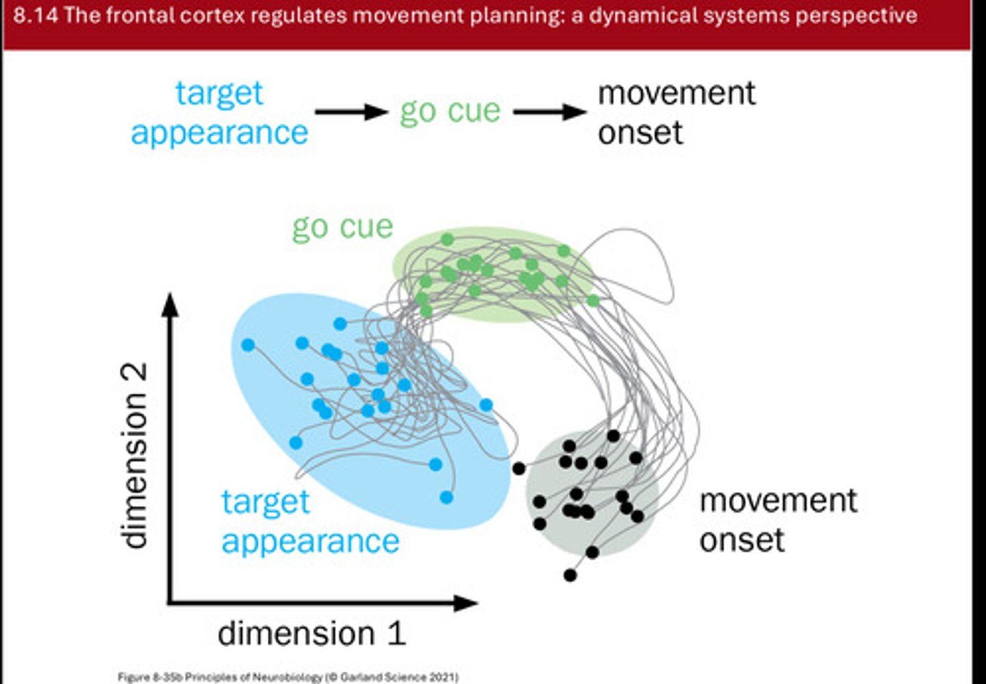
Experiment to show these trajectories in 2D
Scientists recorded from frontal cortex of a primate performing a delayed motor task (the monkey must wait for a cue before performing the action).
Consider the true neural activity as a very high-dimensional space (hundreds of neurons ⇒ hundreds of dimensions).
For visualization, they collapsed that space to two dimensions (e.g., via dimensionality reduction), yielding 2D trajectories.
They observed distinct population patterns for different epochs: A particular pattern when the target appears (the monkey is told to expect the cue), A shifted pattern when the “go” cue appears (the signal to perform), A further shift when the movement begins.
Across many trials (each line = one trial trajectory), similar trajectories appeared: the activity stays in one region until the go cue, moves to another region, and then shifts again as movement begins.
From this dynamical-systems perspective, you can predict movement onset by where/when the population state transitions in this reduced space.

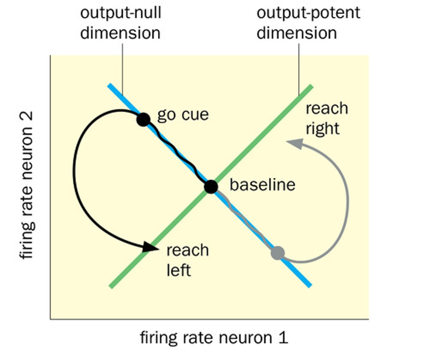
Describe what the following 2 neuron illustration shows:
- where one muscle is influenced by 2 premotor neurons and 2 neuronal firing rates form a 2D state space (x axis is firing rate of neuron 1 and y is neuron 2)
-Within this space, there is an “output null” line (a subspace) along which neurons can change activity but no movement occurs.
- in this specific case, the output null line is when one neuron is very active and the other is silent... as long as the state is on the line there's no movement even though one neuron is active
- breaking out of the line can drive movement, and different regions corr to diff behaviors (both active-> reach right behavior, both inactive->reach left)
- the system then returns to the out null space after the behavior until the next acton... some continuous premotor activity can correspond to no movement
-relative patterns and which subspace the state occupies determine whether and what movement occurs.
describe mouse left ALM inhibition experiment where mice were trained to lick left if anterior whisker was touched and right if posterior was touched.
mouse ALM=prefrontal cortex in mice
-left ALM plans right licking movement and right ALM plans left licking
3 main epochs were being observed: the sample, delay and response period (response was after auditory cue was given) and (sample was when pole touched anterior or posterior whisker)
- experimenters inhibited the mouse left ALM using chanelrhodopsin that they inserted into inhibatory neurons... when blue light flash pyramidal excitatory neurons were silenced and fast spiking inhibatory neurons were activated
-when inhibit ALM occurred during sampling period... little effect occurred on performance but right licking accuracy did decrease. however slight increase in left licking movements (likely due to reduced interference from the contralateral planning side)
- when inhibited ALM during DELAY epoch, licking performance on contralateral side decreased... this is because the mouse loses the plan its supposed to hold bc ALM stores that plan during the delay
SHOWS THAT ALM IS CRITICAL DURING THE DELAY PERIOD and less essential during sensory encoding
describe how even in the same hemisphere of the ALM, cells can demonstrate preparatory activity for licking right or left?
an experiment that recorded single cell activity in ALM as the mouse performs right/left licks. the graph would show how frequently an ALM neuron fired spikes over time.
- in cell one, lots of firing during delay period of right lick trials but not left lick trials. shows it encodes a plan fro right movement. also shows that its not responding to whisker cue but rather planning (bc it fires during delay)--> REPRESENTS PERSISTENT ACTIVITY OF MAINTING RIGHT LICK PLAN UNTIL GO CUE
- cell 2 has high firing during left lick planning... not motor execution but rather motor prep. cell two is left movement direction sensitive.
these cells combined population pattern encodes which movement is planned
about half the cells on each side are contra preferring and other half prefer ipsilateral side.
although there are about half contralateral and half ipsilateral preferring neurons in population, why does each side of ALM prefer contralateral side
Across the entire ALM population:
Roughly half of the neurons in each hemisphere were active for movements on the same side.
The other half were active for movements on the opposite side.
This symmetry suggests a distributed, bilateral coding for movement planning—each hemisphere contributes to preparing both ipsilateral and contralateral movements,
though output dominance is contralateral.
how does the brain hold on to the motor plan generated over time (cue until movement)
motor working memory- brains ability to remember an intended action after receiving a cue but before acting
Our ability to “hold” a planned movement before acting depends on persistent neural activity in a recurrent loop connecting the premotor cortex (ALM in rodents), thalamus, basal ganglia, and cerebellum.
involves the ALM which maintains the motor intention signal and represent the plan
the thalamus provides feedback to sustain cortical firing, the basal ganglia holds back movement until go cue and surpasses competing actions, and cerebellum provides predictive timing and coordinates initiation
form a closed loop network- ALM -thalamus-bg-cerebellum-alm to allow activity persistence after sensory cue disappears
these reverberating circuits allow neural activity to stay elevated for seconds rather than if ALM fired once then went silent-> you'd forget what you're abt to do in a second