Vascular Exam 2: Arterial & Venous Physiologic, Non-Atherosclerotic Pathology
 Call Kai
 Learn
Learn Practice Test
 Spaced Repetition
 Match
Match Flashcards
 Knowt Play
1/179

There's no tags or description
Looks like no tags are added yet.
Name | Mastery | Learn | Test | Matching | Spaced | Call with Kai | Chat |
|---|
No analytics yet
Send a link to your students to track their progress
180 Terms
Atherosclerosis
Plaque buildup
Arteriosclerosis
Hardening of Arteries
Gangrene
Tissue death due to loss of blood supply
Intermittent Claudication
Occurs with exercise, stops with rest
Spinal Stenosis
Occurs with sitting/moving spine
Herniated Disk
Pain radiates down leg
Osteoarthritis
Can be associated with exercise
Elevation Pallor
Pale skin
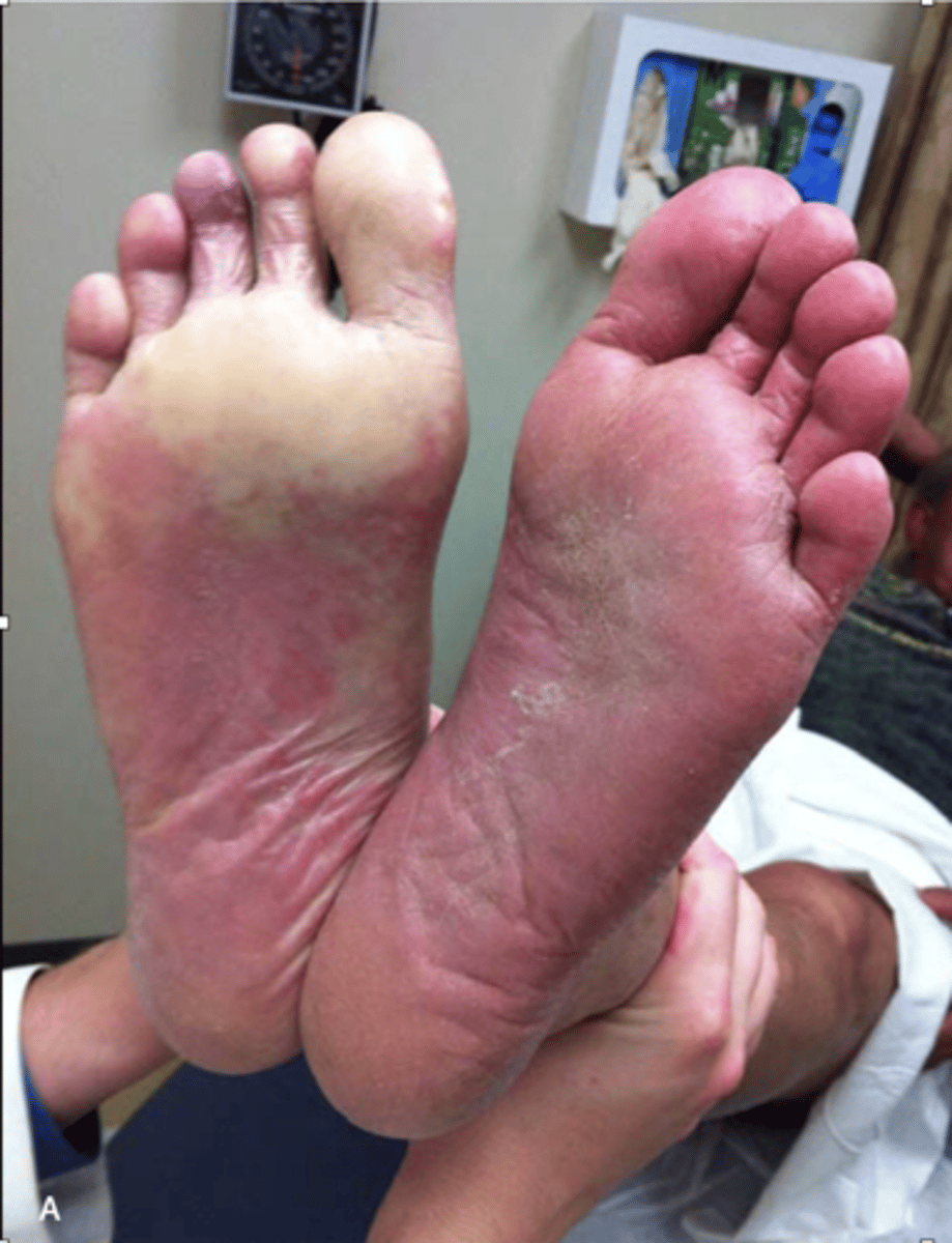
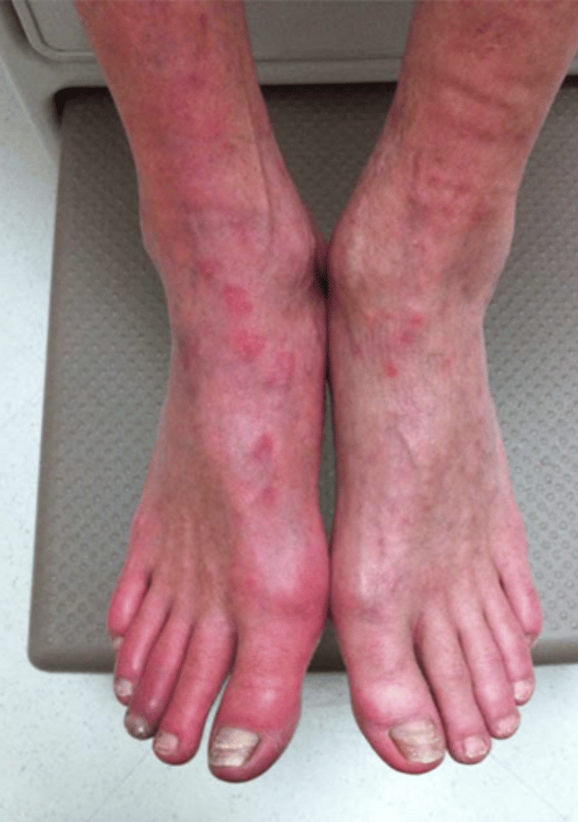
Dependent Rubor
Red skin
Bruit
Low frequency
May not always be heard
Graded on strength and duration
Absence does not rule out disease - 90% disappear
Can strengthen with poor cardiac output
Bruit Grading
1+ = mild
2+ = moderate
3+ = severe -> extends through diastole
Uncontrollable Risk Factors of Arterial Disease
Age
Family history
Gender
Controllable Risk Factors of Arterial Disease
Smoking
Type 2 diabetes
Hypertension
Hyperlipidemia
Obesity
Stress
Symptoms of Acute Arterial Disease
Pain (sudden)
Pallor
Pulselessness
Paresthesias
Paralysis
Symptoms of Chronic Arterial Disease
Claudication
Shiny skin
Loss of hair on legs/toes
Cold to touch
Arterial ulcers
Common Sites of Lower Extremity Arterial Disease
Any bifurcation
Larger vessels
Aorta
Iliacs
Common femoral
Popliteal
Newer in literature:
Anterior tibial/dorsalis pedis
Posterior tibial
Peroneal
Direct Arterial Testing
Arterial duplex
Indirect Arterial Testing
ABI
Segmental pressures
PVR
PPG
CW Doppler
Indications for Lower Extremity Arterial Physiologic Testing
Claudication
Difficulty walking
Atherosclerosis in other locations
Cold limbs
Tingling/numbness
Ulcers
Contraindications for Lower Extremity Arterial Physiologic Testing
Recent graft placement
Suspected venous thrombosis
Arm with shunt or dialysis graft
Information Provided by Physiologic Studies
Presence of arterial disease
Level & severity of blockage
Collateralization
Healing potential
Revascularization improvement
Graft patency
Information Not Provided by Physiologic Studies
Exact location of disease
Patient Prep for Arterial Physiologic Exam
At rest for at least 10-15 min - get history, physical, and place cuffs
Warm room - reduces vasospasm
Restrict nicotine or caffeine - reduces vasoconstriction
Arterial Physiologic Exam Sequence
History
Physical exam
Palpate pulses
CW Doppler tracings
PVR/PPG
Segmental pressures
Stress test (if indicated)
Patient History
Smoking
Hypertension
Hyperlipidemia
TIA or CVA
MI or CAD
Vascular surgery
Diabetes (I or II)
Back pain
Claudication - how long can they walk/exercise
Rest pain
Trauma
Hx. of disease
Previous testing
Family history
Physical Examination
Arterial ulcers
Trophic changes - thickened nails, hair loss (toes, foot), shiny skin
Skin Changes - cool skin temp, color changes (cyanosis/pallor), dependency changes (rubor/pallor), Livedo reticularis (purple patches on foot)
Absent pulses
Dry skin
Bruits
Gangrene
Loss of sub-cutaneous fat
Gangrene
Capillary filling (> 3sec = decreased perfusion)
Where to Palpate Pulses in Lower Extremity
Common femoral
Popliteal
Dorsalis pedis
Post tibial
Pulse Grading System
0 = absent
1+ = weak
2+ = good
3+ = strong
4+ = bounding (aneurysmal)
CW Doppler Signals in Lower Extremity
CFA
SFA (lab dependent)
POP A
DPA
Posterior Tibial
CW Doppler Techniques
Gel
Probe at 45º angle to skin
Hold probe steady
Do not apply too much pressure
Make sure the pass-filter is appropriate
Normal CW Doppler Signal in Lower Extremity
Multiphasic - high resistance, two beats in the antegrade direction
Strong late systolic forward flow
Reversal of flow in early diastole (peripheral resistance)
Forward flow in late diastole (compliance)
Dicrotic Notch
Recoil/contraction of distended arterial walls
Size of notch determined by level of arterial compliance
Incisura
Downward pressure contour on waveform
Start of ventricular diastole
Results in velocity decrease in forward flow
Multiphasic With High Resistance
2 phases
2 forward antegrade beats
Normal
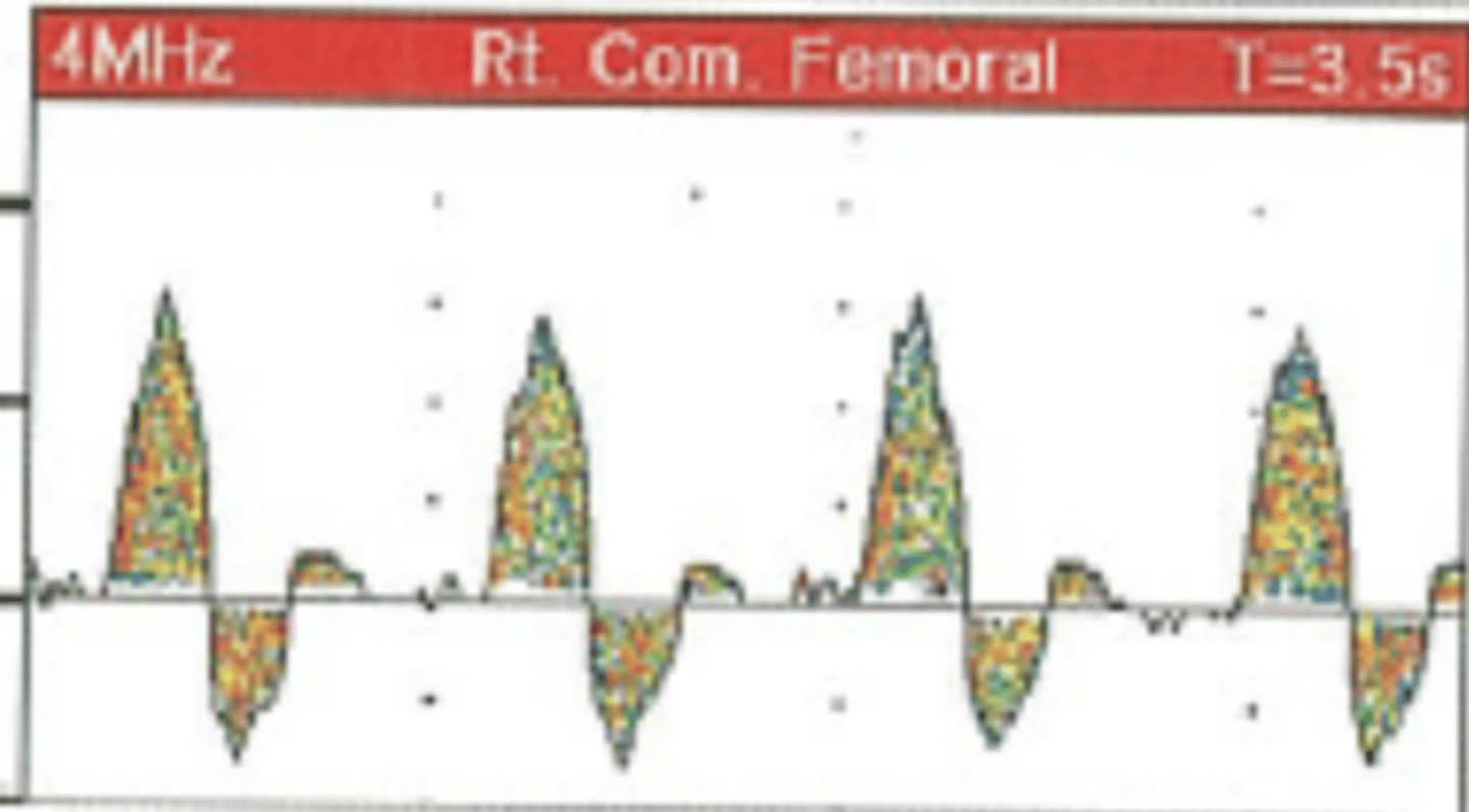
Monophasic with Low Resistance
Mild disease
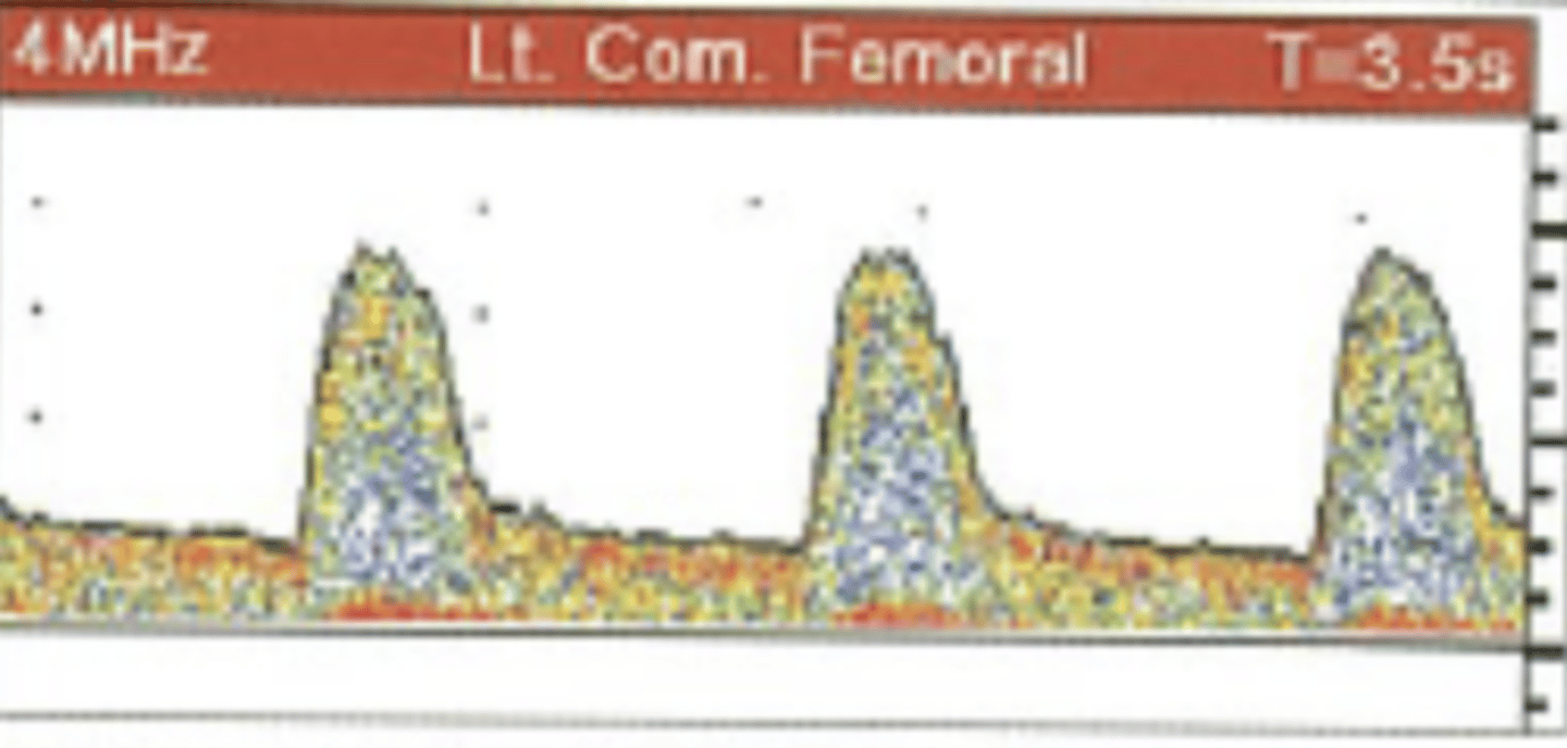
Monophasic without Diastolic Flow
Moderate disease
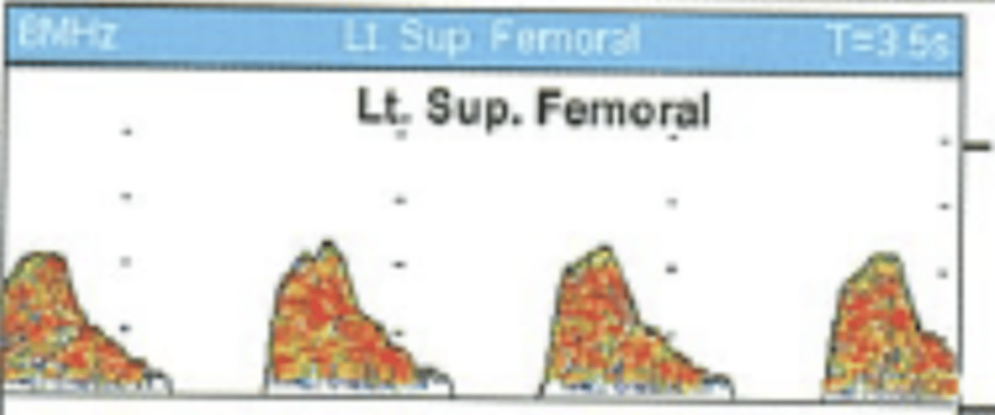
Monophasic with Low Resistance - Tardus Parvus
Severe proximal disease
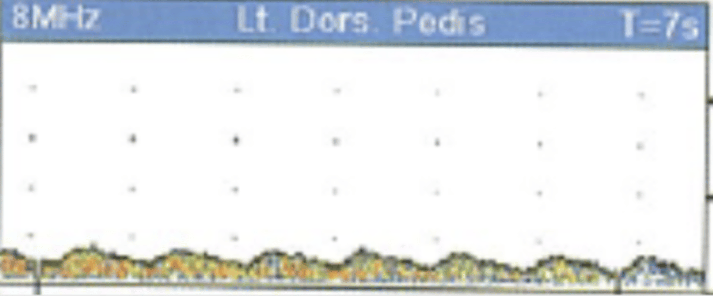
Occluded CW Doppler Signal
Pulse Volume Recordings (PVR)
Assesses overall limb perfusion based on pressure changes
Records change in volume of blood in a limb as blood moves through
Used to measure limb volume related to cardiac cycle-pulsing changes in limb volume - girth of limb increases and air in the cuff is displaced
Ways to Obtain a PVR
Air-filled cuffs - volume changes
Strain gauge - electrical resistance
Impedance - voltage difference
Photoelectric - infrared light
Techniques for PVR Cuff Inflation
Fill cuffs with 55-60 mmHg
Take bilateral brachial PVR first
Begin at ankles and move up
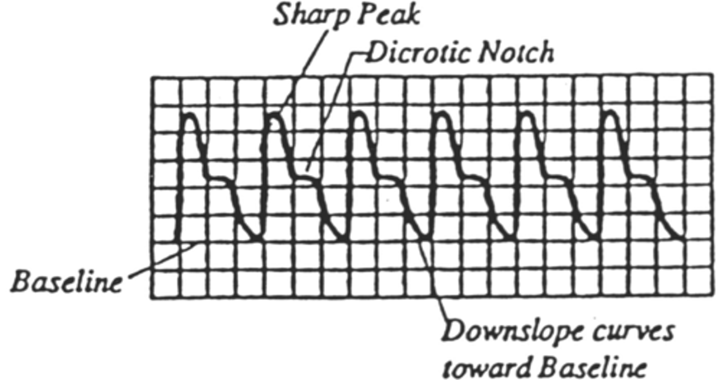
Normal PVR Waveform
Biphasic - always above baseline
Rapid upstroke in systole
Sharp & defined peak
Delayed downstroke
Flat dicrotic notch - on downstroke
PVR Waveform of Mild Disease
Monophasic flow (looks like tardus parvus)
Loss of dicrotic notch
Slightly delayed upstroke
Downstroke bows away from baseline
Sharp peak
May decrease in amplitude
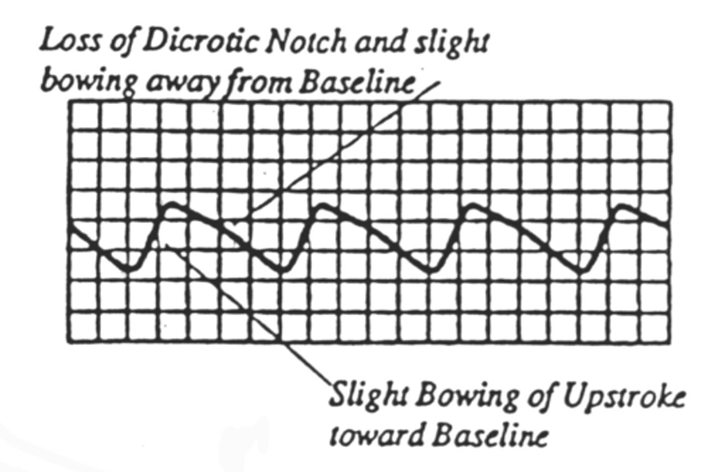
PVR Waveform of Moderate Disease
Delayed upstroke
Rounded
Decreased amplitude
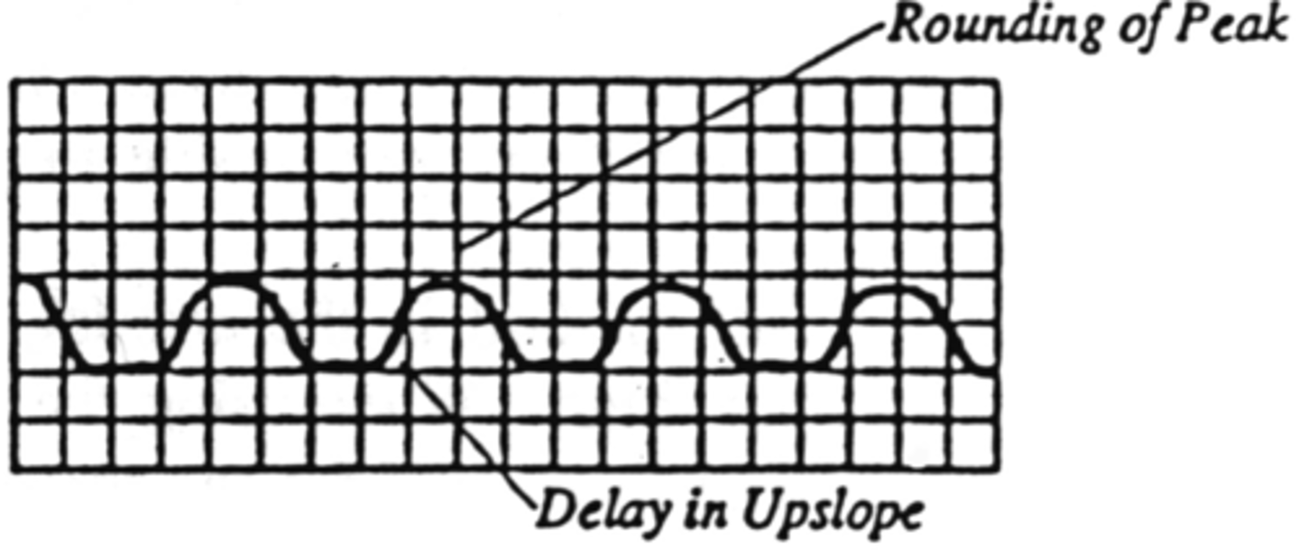
PVR Waveform of Severe Disease
Significant delay in upstroke
Absent or low-amplitude
Equal upslope and down slope time
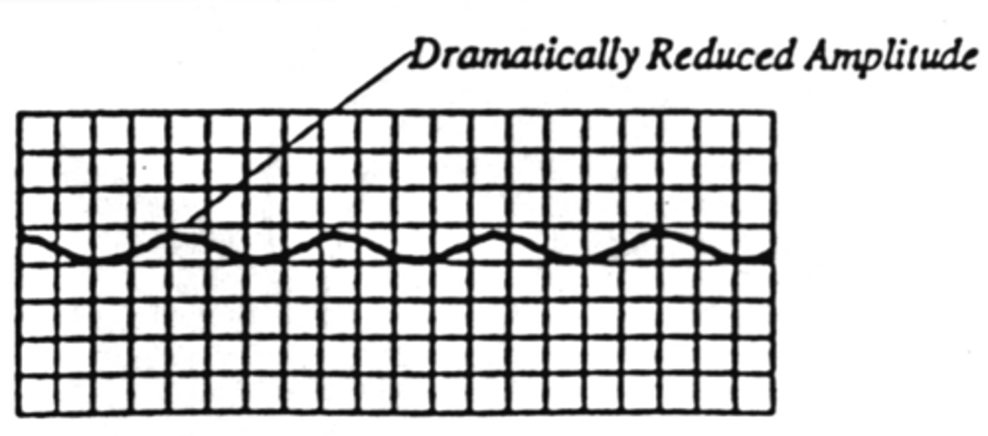
PVR Waveform of Critical Disease
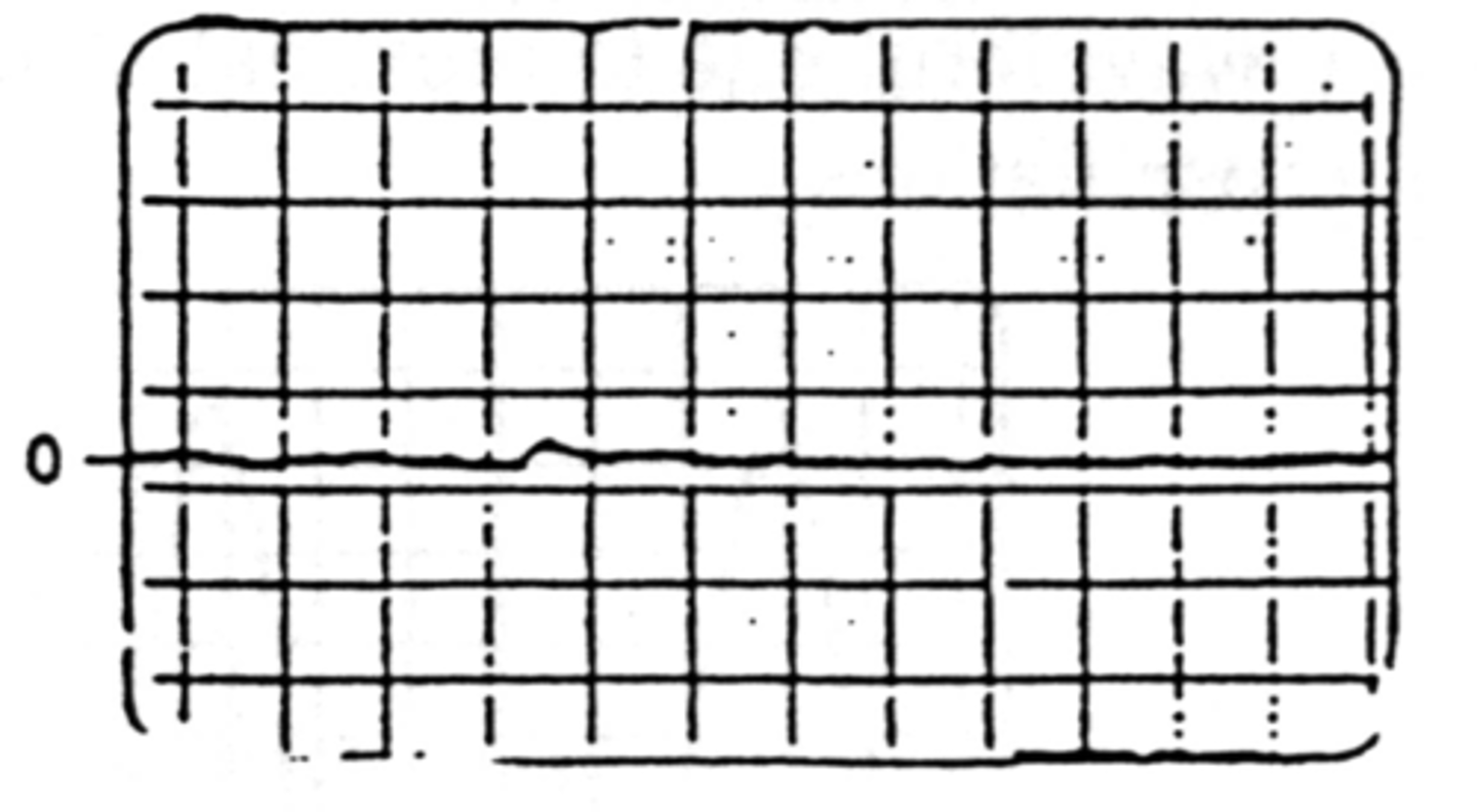
Almost becomes flat line
Seen in severe dz or acute arterial occlusion
Differentiate from increased venous flow
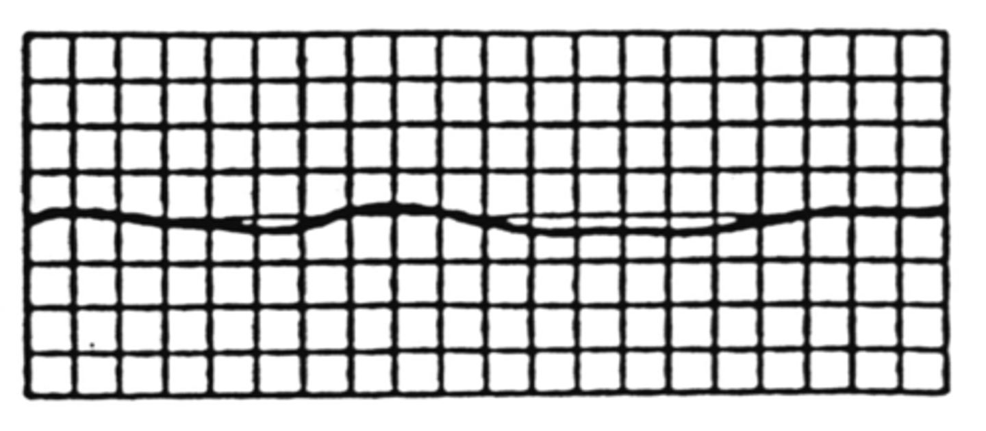
Where is disease on an abnormal PVR waveform?
Above the cuff
Uses of Photoplethysmography (PPG)
Digit waveforms
Penile waveforms
Burn sites
Skin lesions
Healing potentials
PPG Techniques
Attached with tape, clip, or pressure cuff
Use same amount of pressure from site to site
LED surface needs to be flat against skin surface
Reseat probe if abnormal signal is seen
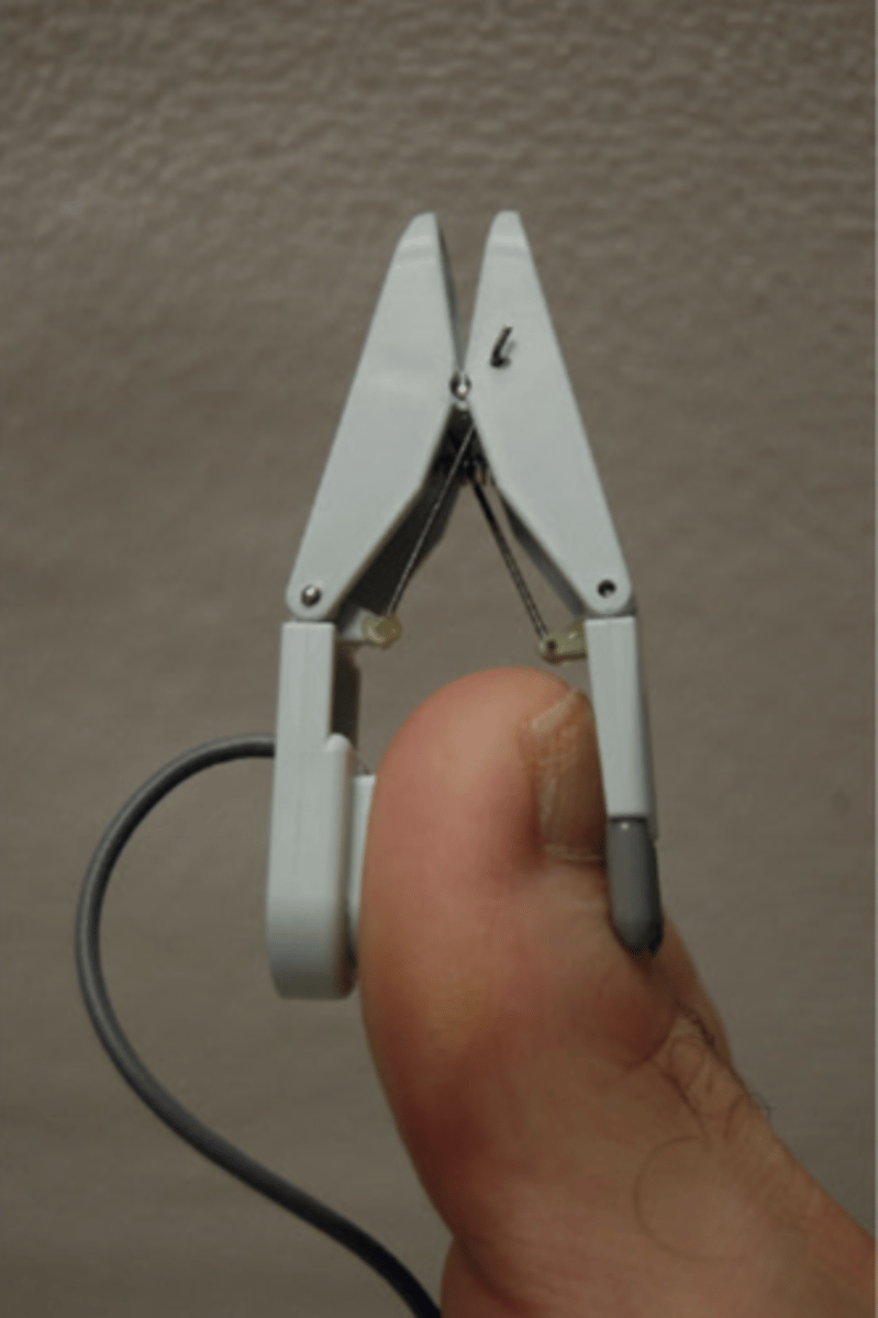
PPG Placement
Lower extremity - great toe
Upper extremity - index finger
May need to obtain signals from all digits - Raynaud's or Buerger's disease
Cuff Bladder Width Sizing
Fit snugly and evenly over artery
20% wider than limb diameter
≥ 50% than limb circumference
Cuff Bladder Width too Wide
Underestimates pressures
Cuff Bladder Width too Narrow
Overestimates pressures
Max pressure for segmental pressure cuffs
220 mmHg
3-Cuff Placement
Thigh - 17 cm
Below knee - 12 cm
Ankle - 10 cm
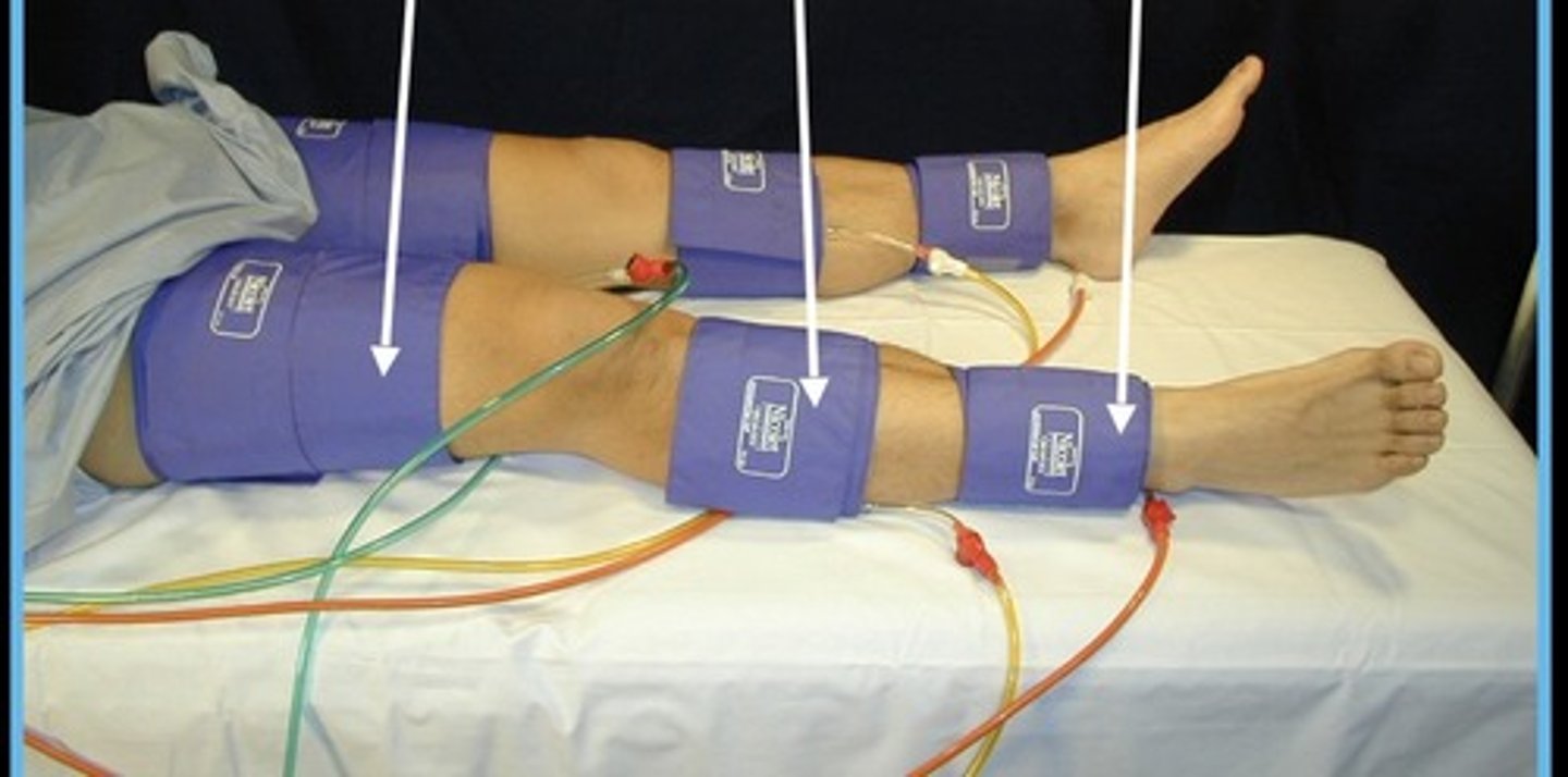
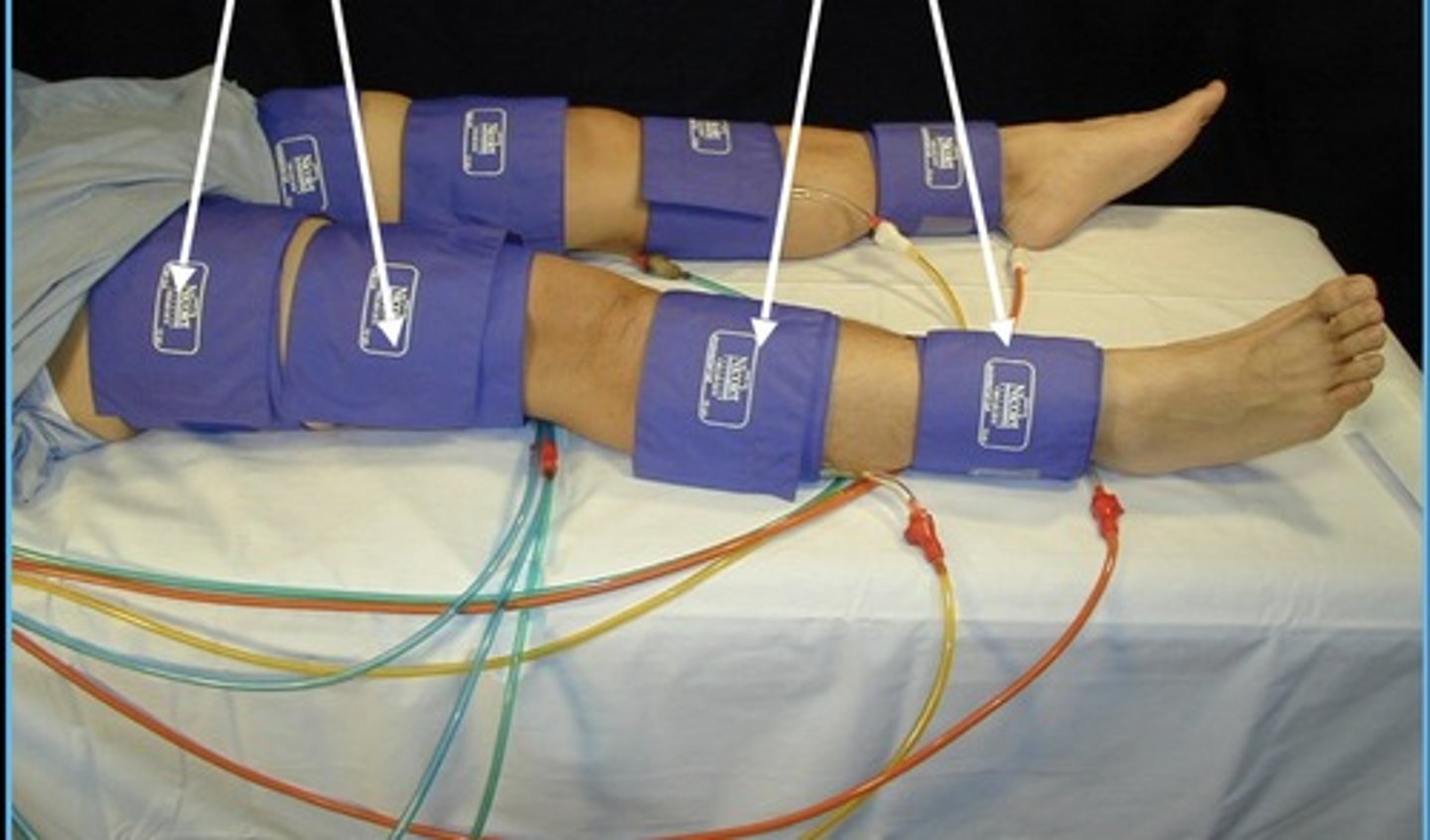
4-Cuff Placement
High thigh - 12 cm
Above knee - 12 cm
Below knee - 10 cm
Ankle - 10 cm
Segmental Pressure Techniques
Start with brachial pressures, then start from ankle and move up the leg
ABI Formula
Highest ankle pressure ÷ highest brachial pressure
Incompressible ABI
> 1.3
Normal ABI
0.9 - 1.3
Mild Disease ABI
0.75 - 0.89
Moderate Disease ABI
0.5 - 0.74
Severe Disease ABI
< 0.5
Ischemia ABI
< 0.35
High Thigh vs. Brachial Pressures in 4-Cuff Method
High thigh should be 30 mHg higher than highest brachial
Thigh vs. Brachial Pressures in 3-Cuff Method
Thigh should be ≥ highest brachial
High Thigh Pressures ≤ Brachial Pressures
Indicates aorta-iliac disease
Pressure Difference Between Adjacent Cuff Sites
< 30 mmHg
Pressure Difference Between Cuff Sites at Same Level on Opposite Legs
< 20 mmHg
Pressure Difference Between Entire Legs
< 40 mmHg
10-15% Pressure Difference Between Brachial Pressures
Positive disease on lower side
When are Toe Pressures More Reliable than Leg Pressures?
Patients with diabetes
Calcified arteries
End stage renal disease
Long-term steroid use
Normal Toe Brachial Index (TBI)
> 0.8
Purpose of Stress Testing
When flow is augmented over the stenosis, distal pressure may drop and reveal the presence of disease
Indications for Stress Testing
Normal study with persistent symptoms
Intermittent claudication
Claudication vs. pseudoclaudication
Presence of collaterals
Contraindications for Stress Testing
Shortness of breath
Hypertension
Cardiac problems
CVA/embolic events
Walking problems
Rest Ischemia
Types of Stress for Stress Testing
Treadmill (slight incline @slow speeds) for 5 min or when symptoms appear
Toe ups (50)
Step ups
Stress Testing Protocol
Take baseline pressures of symptomatic & opposite leg, and one arm
Immediately after exercise
Every 2 minutes for 10-20 minutes or until pressures return to normal
Single Level Stenosis
Pressures return to baseline within 5 minutes
Multi-Level Stenosis
Pressures return to baseline within 10-12 minutes
Ischemic Disease
Pressures remain low for ≥ 15 minutes
Post-Exercise Pressures ≤ 60 mmHg
Confirms vascular etiology of disease
Lower Extremity Venous Anatomy
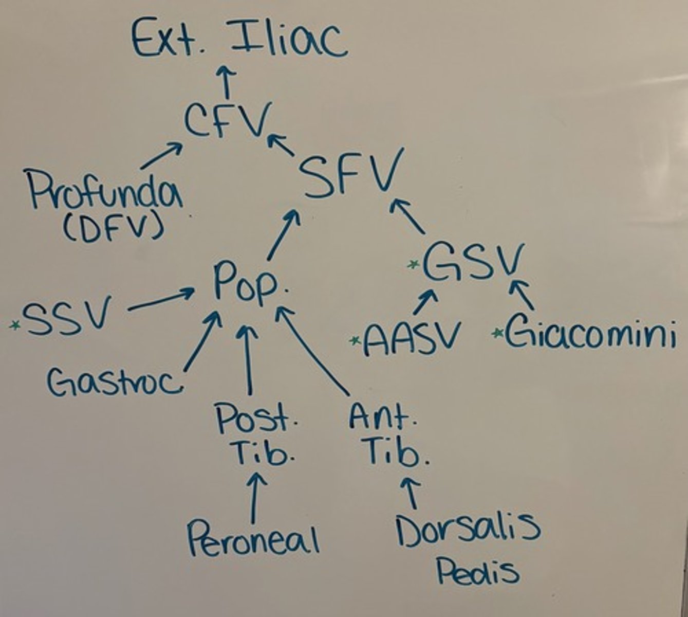
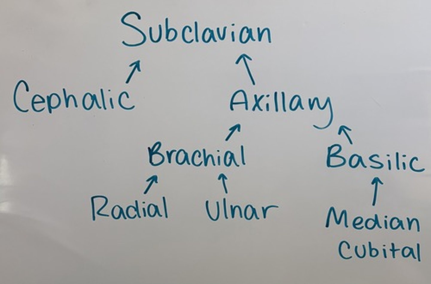
Upper Extremity Venous Anatomy
Anterior Accessory Vein
Runs parallel and anterior to femoral vein
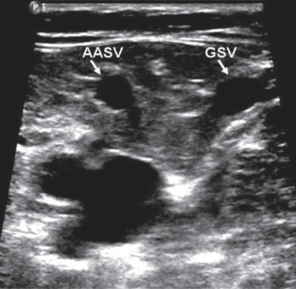
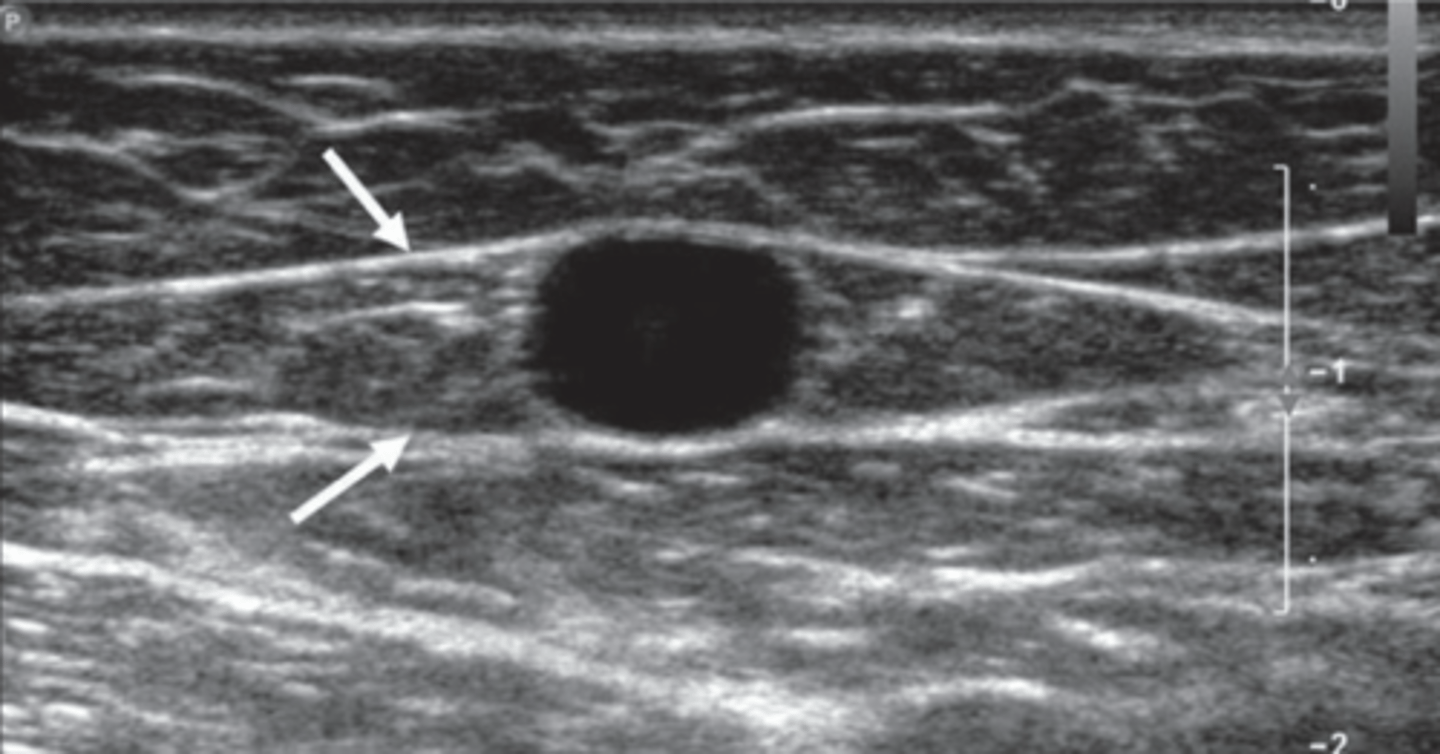
GSV
Saphenous Eye
Between facial layers
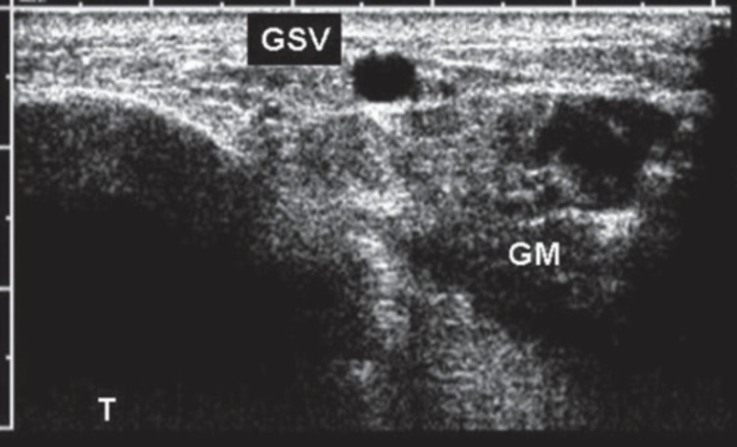
GSV in Calf
Angle Sign
Triangular form between gastrocnemius muscle and tibial bone
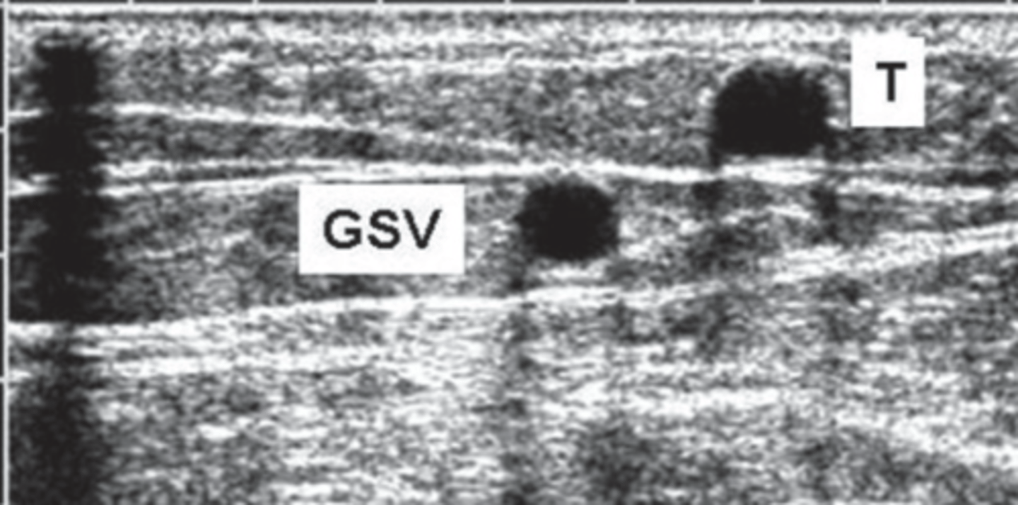
Tributary
Outside fascial plane
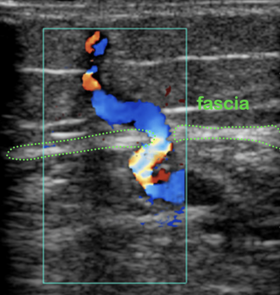
Perforator
Pass through muscular fascia layers - connect deep system to superficial system
Venous Valves in Lower Extremity
Femoral 1-6 valves
Pop 0-4 valves
GSV 10-12 valves
SSV 7-9 valves
Calf veins 8-19 (every 2 cm)
Perforations 1 each
Veins with No Valves
Soleal
Internal iliac
Common iliac
IVC
Subclavian VV
Innominate
SVC
Distribution of Venous Valves in Deep System
2.1 %
Distribution of Venous Valves in Superficial System
16.8 %
Distribution of Venous Valves in Perforator System
8.4 %
Risk Factors for Leg Swelling
Heart dysfunction (CHF)
Kidney disease
Liver disease
Lymphatic system/Lymphedema
Pulmonary HTN
High BMI
Medications (calcium channel blockers)
Multifactorial
Age
Family History
Obesity
Oral Contraceptive use
Smoking/Tobacco
Pregnancy
History of DVT
Prolonged standing or sitting
Sedentary lifestyle
Gender (women)
Primary Valvular Insufficiency
Congenital absence of veins
Secondary Valvular Insufficiency
Damaged valves caused by thrombotic events
Chronic Venous Insufficiency
Obstruction & valve insufficiency
Chronic Venous Valvular Insufficiency
Impaired superficial or deep flow
Causes venous hypertension
Duplex is primary modality for diagnosing
Symptoms of DVT
Swelling
Shortness of breath - emboli
Redness
Warmth
Painful
Throbbing
Phlebitis/Thrombophlebitis