Psychology AOS 2 - Chapter 4
1/58
There's no tags or description
Looks like no tags are added yet.
Name | Mastery | Learn | Test | Matching | Spaced | Call with Kai |
|---|
No analytics yet
Send a link to your students to track their progress
59 Terms
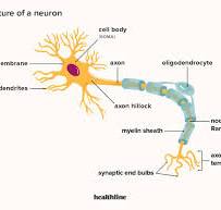
Nerve cell diagram
2 old-timey approaches to understanding the brain
2000 years ago - Greek philosophers contemplated brain function (bring vs heart debate crucial time for heart side)
19th century - scientists dissected animal and donated brains to observe structure, biological perspective
Whether the brain or the heart is the root of human thoughts, feelings and behaviours. ‘Brain hypothesis’ is now widely accepted.
Worked as gladiator doctor 129-216 CE
Observed impact on behaviour of head injuries
Observed nerves from sense organs went to brain
Took brain side of brain vs heart debate
Questions of how brain activity relates to conscious experience, that is, the relationship between what our brain does and our awareness of our own existence and environments
Left hemisphere is language centre
Right hemisphere can see and draw things it sees, but can't verbally articulate it alone.
When asked to articulate something seen in the left visual field, split-brain patients believed they saw nothing at all
Techniques that produce a scan showing brain structure, e.g. CT and standard MRI
Structural X-ray based neuroimaging technique that builds a black and white horizontal cross-section of the brain.
Used for tumours, brain damage, abnormalities, and physical changes due to a disorder
Used for very small brain anatomy and tissue changes, myelin loss, nerve degeneration and blood clots or leaks
(dynamic neuroimaging) Techniques that show some brain function and activity, and can do structure too. E.g. PET and FMRI
Contains outer cerebral cortex and masses of neural tissue. Cerebrum and cortex are responsibility for a lot of what we think, feel and do. Cerebrum is split into 2 hemispheres divided by longitudinal fissure and connected by corpus callosum. Each hemisphere has 4 lobes (in cerebral cortex)
Cerebral cortex (Grey matter)
Layer covering cerebrum. Functions of its areas can be organised into 3 broad categories. Has 4 broad lobes
'Higher order' cognitive functions, e.g. perception, learning, memory, language, thinking and problems. Also processes incoming sensory info and involved with planning and controlling voluntary movements.
Motor Areas - Initiates and executes voluntary movements, made of motor neurons
Association Areas - Surrounds sensory and motor areas, deals with functions requiring integration of inputs from multiple areas.
Verbal functions:
speech production
comprehension
reading
writing
Analytical functions
maths
sequential tasks
evaluation
logical reasoning
Controls voluntary movement and receives and processes sensory info for right side of body
Right cerebral hemisphere specialisations
Non-verbal tasks not dependant on language:
Processing the 'whole' rather than in bits
Creativity and Fantasy
Art & music appreciation
Recognising and expressing emotions
Rhythm and time
Spatial and Visual functions:
Puzzle solving
Recognising patterns and faces
Map reading
Visualising a location
Controls voluntary movement and receives and processes sensory info for left side of body
Has association areas for spatial recognition and attention, plus taste perception (insula cortex)