Retroviruses
 Call Kai
 Learn
Learn Practice Test
 Spaced Repetition
 Match
Match Flashcards
 Knowt Play
 Podcast
1/45

There's no tags or description
Looks like no tags are added yet.
Name | Mastery | Learn | Test | Matching | Spaced | Call with Kai | Chat |
|---|
No analytics yet
Send a link to your students to track their progress
46 Terms
What are the different groups in the Retroviridae family?
Alpha- Rous Sarcoma Virus (RSV)
Beta- Mouse Mammary Tumor Viruses (MMTV)
Gamma- Murine leukemia virus (MLV)
Delta- Human T cell leukemia viruses type 1 (HTLV-1)
Epsilon- Walleye dermal sarcoma virus (WDSV)
Lentiviruses- Human immunodeficiency virus (HIV)
Spumavirus- Simian foamy viruses (SFV)
Which groups in the Retroviridae family are linked with human disease and give rise to cancers?
Delta- Human T cell leukemia viruses type 1(HTLV-1)
Lentiviruses- Human immunodeficiency virus (HIV)
*Except HIV doesn’t give rise to cancer and rather causes severe immunosuppression.
Why is studying retroviruses important?
As retroviruses like HIV give rise to the possibility of severe immunosuppression which requires lifelong treatment and monitoring and can lead to opportunistic infections and various malignancies.
HTLV-1 is linked with the aggressive T-cell cancer: Adult T-cell leukemia (ATL), making understanding retroviruses crucial for public health.
Reverse transcriptase enzyme produced by retroviruses has applications in gene expression studies and is necessary for molecular diagnosis of RNA viruses i.e. RT-PCR
How can reverse transcriptase be used in gene expression studies?
Lets say you want to study the effects of a section of RNA in an organism, you must first convert it into complementary DNA (cDNA) using reverse transcriptase, allowing for further analysis such as quantifying gene expression.
What is the morphology like for HIV?
Spherical morphology, characterized by a lipid envelope surrounding a conical core
Contains two single strands of RNA and essential enzymes such as RT
It’s characteristic capsid is composed of the p24 protein and is crucial for protecting the viral RNA during the infection process.
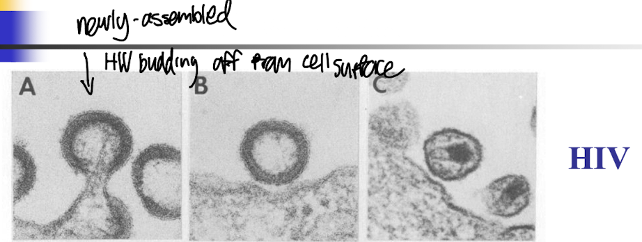
What is the morphology like for HTLV-1?
Morphology is similar to HIV, featuring a spherical shape with a lipid envelope.
Contains two single strands of RNA and essential proteins, including the enzyme reverse transcriptase.
Characterized by a distinct immunodominant protein that helps in immune evasion and pathogenesis.

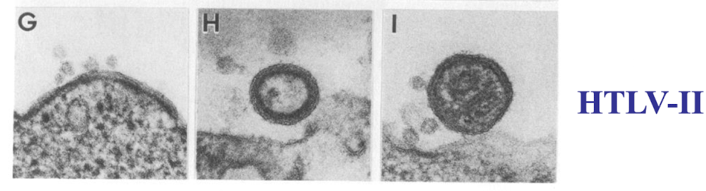
What is the morphology like for HTLV-2?
Similar to HTLV-1, with a spherical shape and lipid envelope.
Contains two single strands of RNA and essential proteins, including reverse transcriptase.
Displays a varied immunodominant protein that may influence its pathogenesis.
What are two essential features of retroviruses?
Has RNA as genetic material in virus (cell free virus) which is converted to DNA in infected cells by reverse transcription
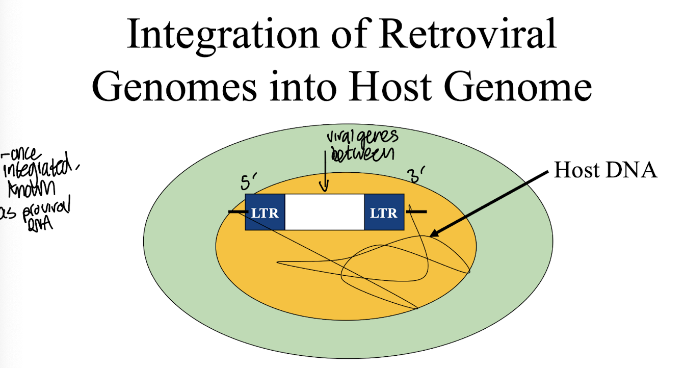
Integration of the viral DNA into the host cell genome (proviral DNA) is a critical part of the lifecycle - not accidental like in some cases of Papilloma virus infection
How does the retroviral genome get integrated into the host genome?
Gets integrated between 5’ and 3’ LTR regions (long terminal repeats)
Involved in integration of viral DNA into host cell genome
Contains the promoter for viral gene expression; contains binding sites for cellular transcription factors → essential for viral gene expression in host
LTR - more detail
LTR is present on either side of the viral genome.
It harbors cis-acting elements, which are required for RNA synthesis
Is the initiation site for transcription of the viral genome.
LTR consists of three regions:
U3 (unique, 3′ end)
R (repeated)
U5 (unique, 5′ end).
Various elements present in U3 help in direct binding of RNA polymerase II (pol II) to DNA templates.
What are the 3 types of oncogenic retroviruses?
◼ Transducing retroviruses eg RSV
◼ Cis-activating retroviruses eg MMTV
◼ Transactivating retroviruses eg HTLV-1
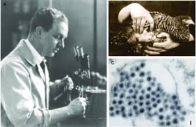
How was RSV shown to cause cell-free transformation of cancer from one chicken to another?
Isolation of the viral from one chicken’s tumour tissues were then transferred to healthy chickens.
The infected chickens then developed similar tumours, indicating that RSV can promote oncogenesis in host cells.
Investigated by Peyton Rous
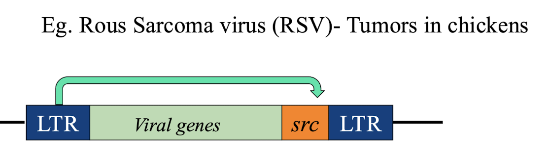
How is RSV an example of a transducing retrovirus?
The viral v-src oncogene was found to be originated from cellular counterpart, c-src giving rise to the idea that cancers might arise from abnormal expression Or alterations in normal human cellular gene lead to uncontrolled cell division:
This demonstrates how RSV captures and utilises cellular oncogenes to promote cancer.
What is an example of a cis-activating retrovirus?
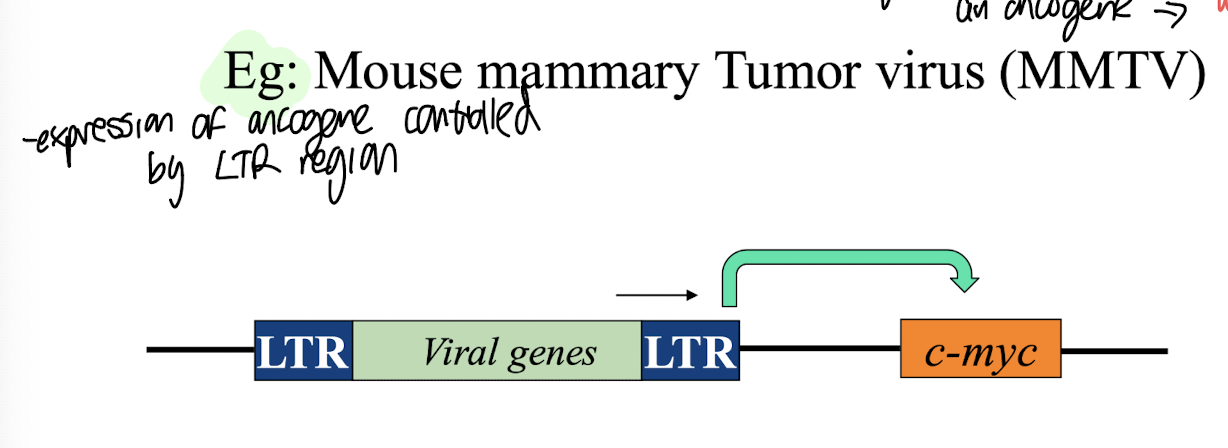
An example of a cis-activating retrovirus is MMTV (Mouse mammary tumour virus), which promotes oncogenesis through the activation of cellular genes adjacent to its integration site.
Oncogenesis is highly dependent on where the viral genes integrate, thus if they integrate near an oncogene, it can enhance its expression and lead to tumour development → cancer
Integration away from oncogenes, will typically result in no transformation or minimal effects on cellular function.
The LTR region in cis-activating retroviruses acts as a promoter, driving the transcription of nearby oncogenes.
What is an example of a trans-activating oncogenic retrovirus?
HTLV-1 which contributes to Adult T cell leukemia (ATL)
No cellular oncogenes compared to the transducing retroviruses
Integration predominantly in transcriptionally active chromatin
Oncogenic proteins Tax 1/HBZ
What is the relationship between HTLVs and STLV counterparts?
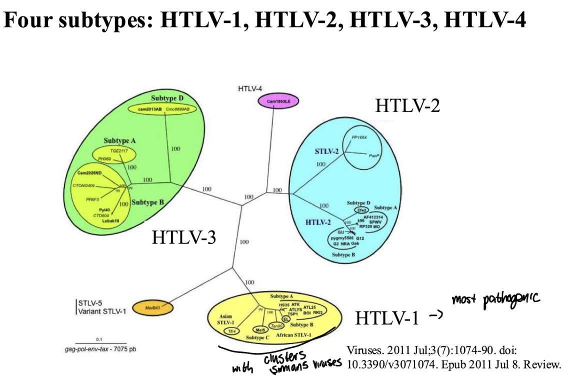
HTLVs share a close evolutionary relationship with STLVs (Simian T-cell Leukemia Viruses) often clustering close together in phylogentic trees, with HTLVs likely originating from STLVs through cross-species transmission.
Both groups of retroviruses are known to infect T-cells and are implicated in similar oncogenic processes.
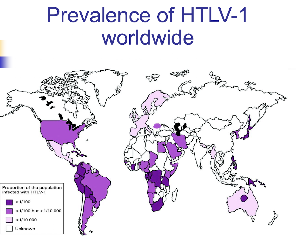
What is the worldwide prevalence of HTLV-1 like worldwide?
Primarily is distributed in Japan, The Caribbean, areas of Subsaharan Africa and parts of South America, with higher prevalence rates in specific populations i.e. central Australia with indigenous communities having high percentages of adults with the virus
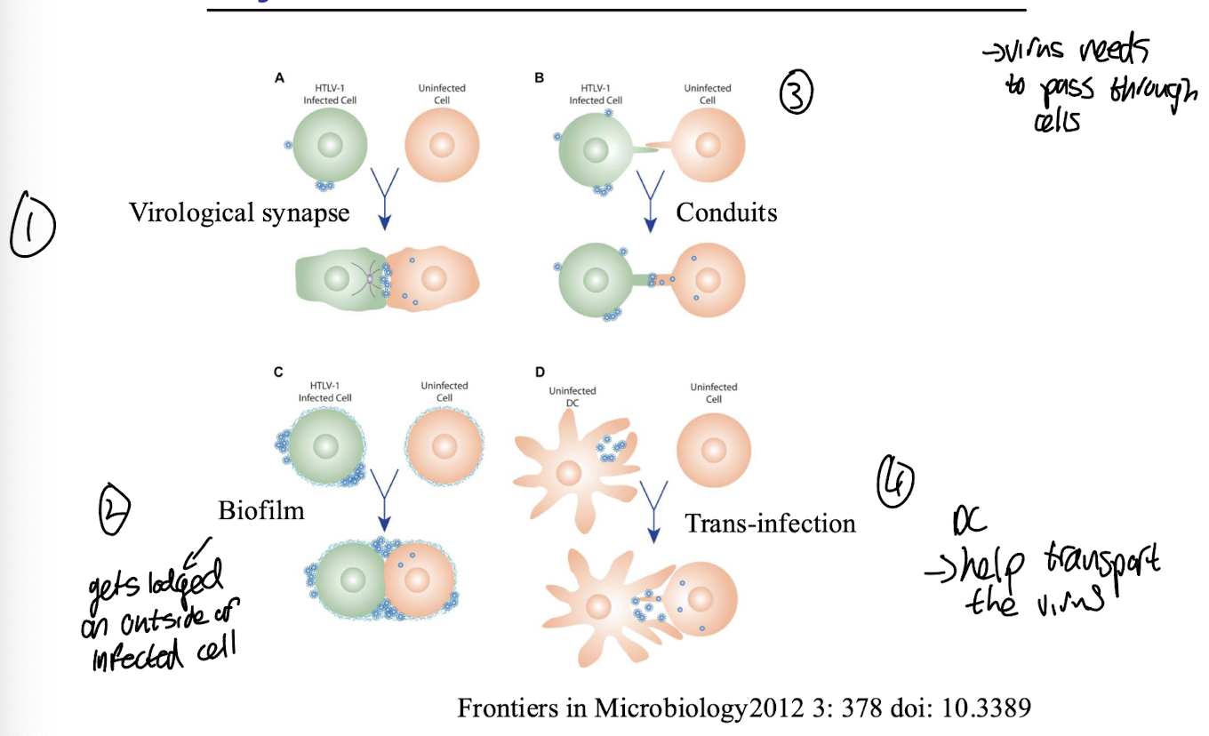
What is the predominant way HTLVs are spread and what are the 4 main types of how it is spread?
Spread via cell-to-cell transmission:
Virological synapse
Biofilm between an infected/uninfected HTLV-1 cell, where the virus gets lodged on the outside of the cell
Conduits between infected and uninfected cells.
Trans-infection via uninfected dendritic cells that present viral antigens to T cells, causing them to become infected.
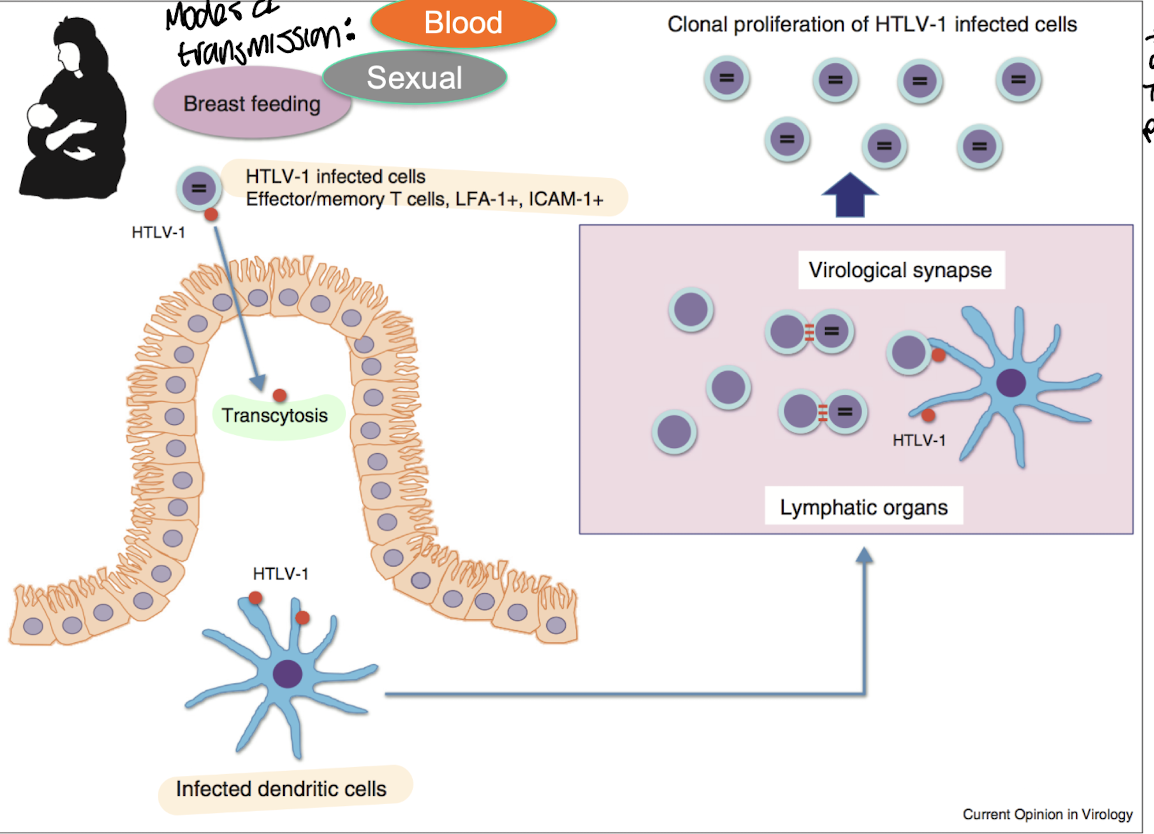
What are the main transmission routes of HTLV-1?
HTLV-1 is primarily transmitted through unprotected sexual contact i.e. spread of sexual fluids i.e. semen, breast feeding, contaminated blood products spreading infected lymphocytes, and sharing of needles or syringes.
These routes facilitate the infection and spread of the virus among hosts.
Pathogenesis of HTLV-1
HTVL-1 infected cells i.e. effector/memory T cells that are positive for ICAM-1 allow binding to the integrin LFA-1 for enhanced adhesion and migration across epithelial cells → Transcytosis
Dendritic cells become infected with HTLV-1, facilitating viral transmission to T cells by presenting the viral antigens to it and allowing spread into the lymphatic organs
Virological synapse occurs when infected T cells form a direct contact with uninfected T cells, allowing for the transfer of viral components and T-cell proliferation
Clonal proliferation of HTLV-1 infected cells
What is ATL (Adult T-cell leukemia) characterised by?
Aggressive leukemia of CD4 +, CD8 - T cells that takes 40-50 years until initial symptoms show after being infected
Creates elevated, morphologically abnormal T cells
Sx incl: Lymphadenopathy, hepatosplenomegaly, skin lesions, hypercalcemia
Associated with lytic bone lesions and poor prognosis, no effective treatment
Different ATL subtypes have varying survival times
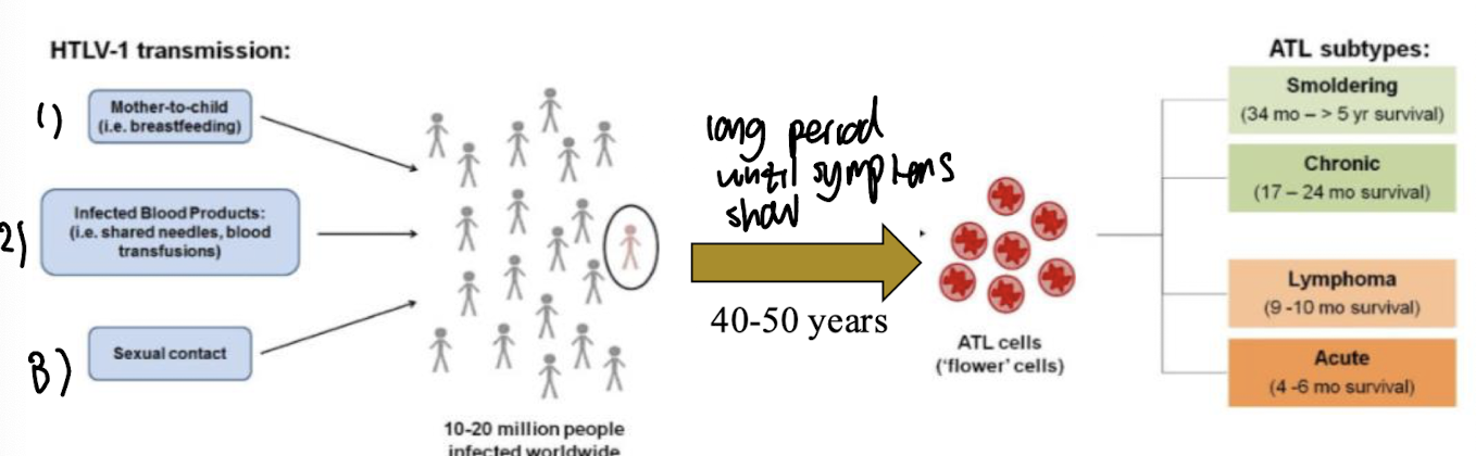
What are the 3 main classifications of ATL?
1. Smouldering ATL: 5% or more abnormal T cells; normal lymphocyte
count; skin lesions; no hypercalcemia, no lymphadenopathy
2. Chronic ATL: 19% or more abnormal T cells; elevated T cell counts;
no hypercalcemia; may have lymphadenopathy
3. Acute ATL: > 60% abnormal T cells, skin lesions, systemic lymphadenopathy, hypercalcemia; metabolic disorders
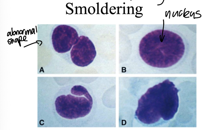
What is the characteristic T cell morphology of Smoldering ATL?
T cells appear with a large nucleus-to-cytoplasm ratio, minimal cytoplasm, and are often irregularly shaped.
These cells exhibit mild atypia but maintain their overall cellular integrity.
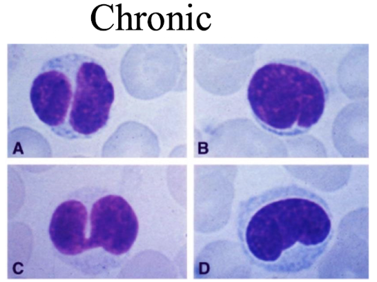
What is the characteristic T cell morphology of Chronic ATL?
T cells show moderate pleomorphism (marked variability in the size, shape, and staining of cells) and/or their nuclei
Irregular nuclei and a higher nucleus-to-cytoplasm ratio compared to normal T cells.
They may present with more pronounced atypia and are associated with a higher percentage of abnormal cells.
What is the characteristic T cell morphology of acute ATL?
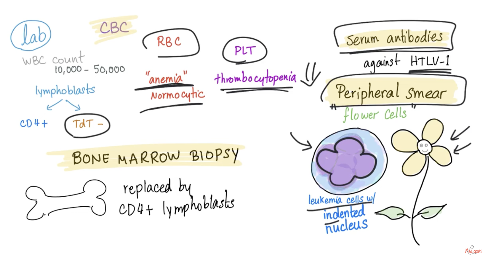
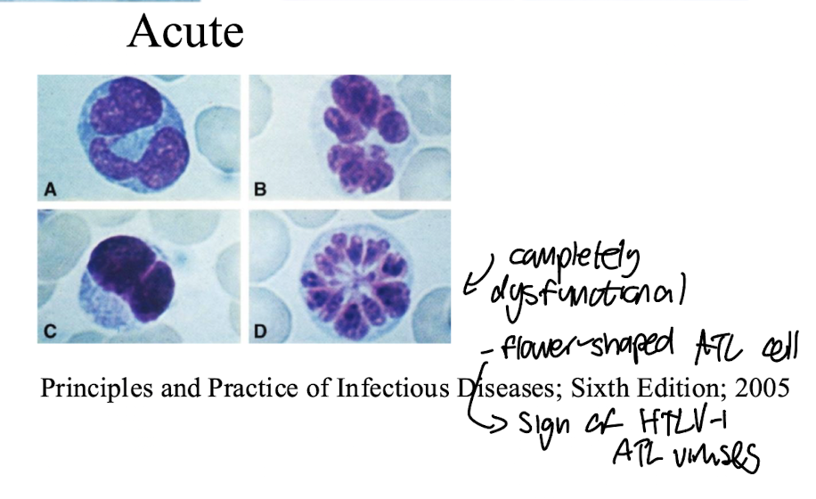
Characteristic ‘flower-like’ cells with a large nucleus-to-cytoplasm ratio, prominent atypia, and irregular shapes.
Exhibit significant pleomorphism with varying sizes and nuclear characteristics i.e. indentations
Cells are completely dysfunctional
Key initial sign of ATL caused by HTLV-1, indicating chance of malignancy and aggressive behavior.
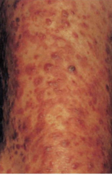
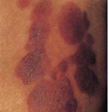
What are the 2 common types of skin lesions associated with ATL?
Prescence of multiple skin papules and nodules which can become widespread, biopsy can be sequenced to indicate active virus
Plaques which are often erythematous and scaly, potentially evolving into tumors, result of viral skin invasion
What is HAM/TSP
HTLV-1-associated myelopathy/ Tropical Spastic Paraparesis:
Chronic inflammatory disease of the CNS caused by infection with HTLV-1
Leads to progressive weakness, spasticity, and sensory disturbances in the lower limbs as well as back pain and urinary incontinence
Commonly seen in females in their forties
Causes non-specific brain lesions
Inflammatory markers elevated in cerebrospinal fluid (CSF) tests.
Characteristics of HTLV-2:
Subtypes include HTLV2A, HTLV-2B, HTLV2C
+sense
Distribution in Central/West Africa, native populations North America, Central and South America and IVDUs in the US and Europe
Infects CD8+ (cytotoxic) T cells
No clear disease associations
Can be used as a comparative tool for understanding pathogenic properties of HTLV-1
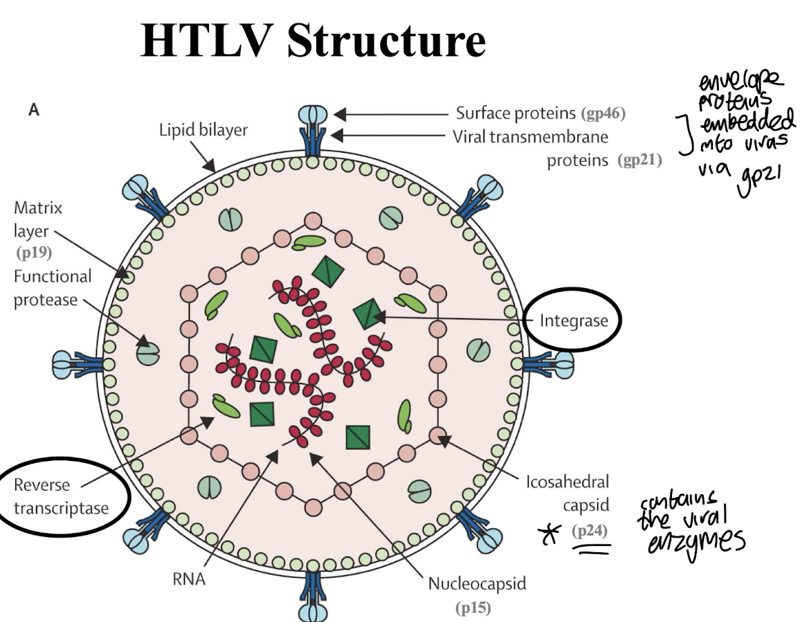
What is the structure of HTLV?
ssRNA genome surrounded by a lipid bilayer, with a protein coat consisting of gag, pol, and env genes.
Surface proteins e.g. gp46 are embedded into virus via viral transmembrane proteins e.g. gp21
Matrix layer made up of p19
Has an iscosahedral capsid (p24) which contains key viral enzymes:
Functional proteases
RTases and integrases which allow the virus to integrate into the host's DNA and replicate.
What is the structure of the HTLV genome?
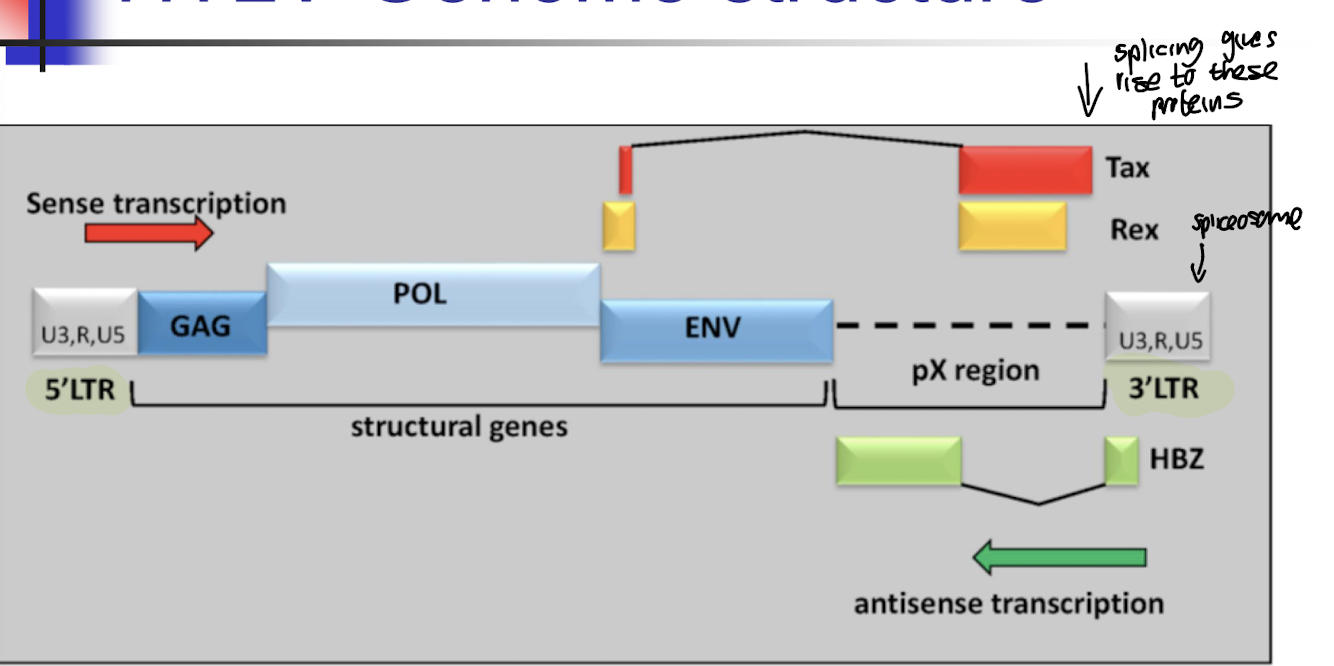
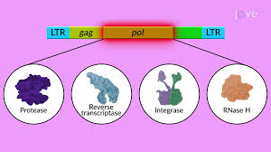
Genes located between the 5’ (sense transcription) and 3’ (antisense transcription) LTR encode key viral products such as gag, pro, pol, env, tax, rex, and hbz which are essential for viral assembly, replication, and regulation.
What does the gag gene encode in HTLV?
Core proteins needed for viral assembly and protection of the RNA genome:
- matrix (p19)
- capsid (p24)
- nucleocapsid (p15)
What does the pro gene encode in HTLV?
Protease - cleaves Gag and Pol polyproteins to produce individual protein components necessary for viral assembly and replication.
What does the pol gene encode in HTLV?
Polymerase - includes reverse transcriptase and integrase
What does the env gene encode in HTLV?
Envelope proteins necessary for entry into host cells, facilitating viral fusion with the host membrane such as surface envelope (gp46)/transmembrane (gp21)
What does the tax gene encode in HTLV?
Encodes the viral oncogenic protein Tax which plays a crucial role in viral replication and the transformation of host cells.
What does the rex gene encode in HTLV?
Viral RNA transport from nucleus to cytoplasm
What does the hbz gene encode in HTLV?
Encodes the HTLV-1 basic leucine zipper protein (HBZ), which is involved in viral anti-sense transcription regulation and allows for the proliferation of infected cells by counteracting the activity of Tax1
What are the main stages of the HTLV lifecycle?
Attachment and entry into host cells,
Reverse transcription of viral RNA to DNA
Integration of proviral DNA into the host genome
Viral transcription and translation
Assembly and release of new virions.
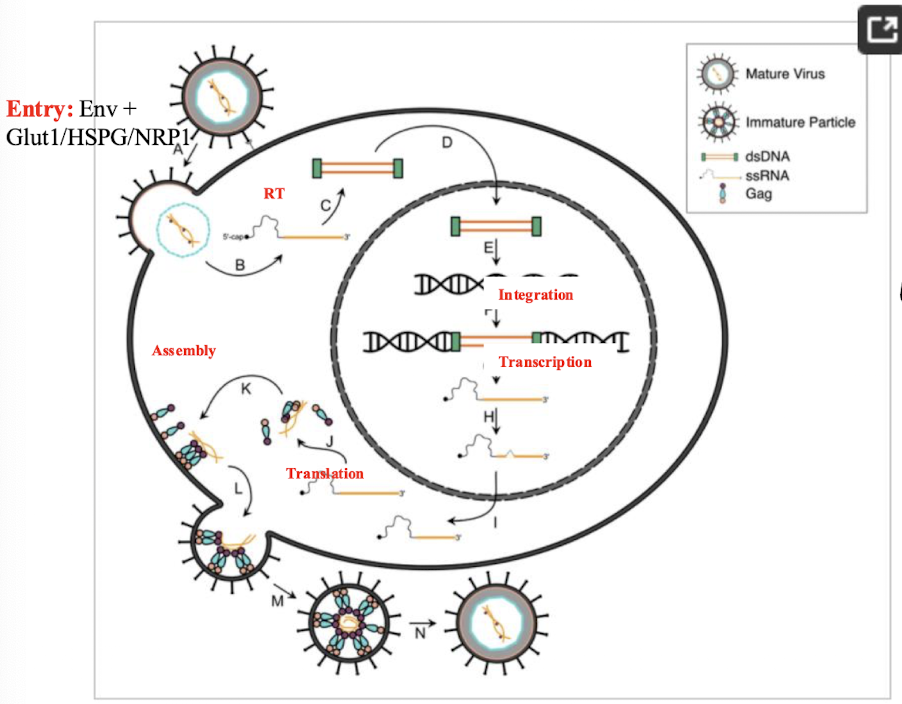
How is permanent infection established after viral RNA is converted into DNA using RTase?
Permanent infection is established through the integration of proviral DNA into the host genome, allowing for the continued replication and expression of viral genes.
What are properties of the HTLV-1 Tax protein?
Powerful transactivator of HTLV-1 transcription eg CREB pathway
Activates numerous cellular pathways eg NFkB, AP-1, AKT pathways
Transforms animal and human cells in vitro
Tumors/ATL like disease in transgenic animals
Why is Tax a powerful transactivator?
Tax1 activates viral gene expression via the CREB pathway which is involved in regulating cell growth and differentiation, thereby enhancing the transcription of HTLV-1 genes.
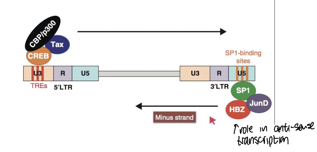
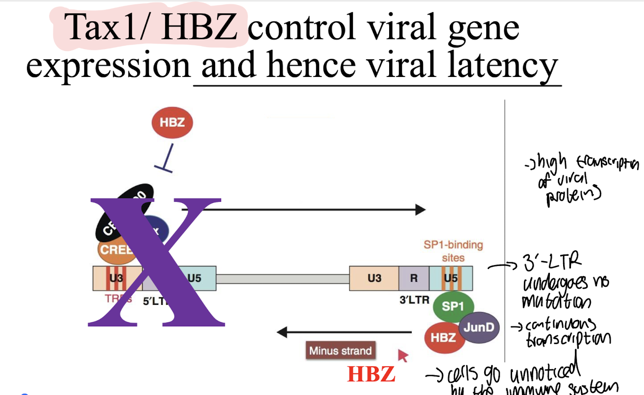
How do Tax1 and HBZ control viral latency?
They regulate the expression of genes involved in maintaining viral latency and activation from the latent state, with Tax1 promoting viral transcription and HBZ maintaining latency.
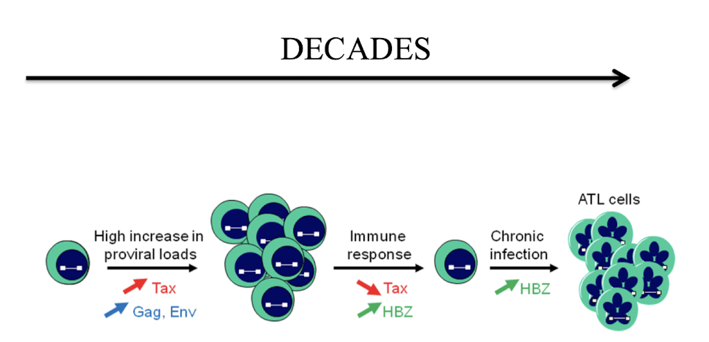
5’ LTR is mutated in most ATL cases meaning that HBZ is the only protein produced in infected cells
Continuous transcription occurs as the 3’ LTR is not mutated.
Immune evasion occurs as HBZ interferes with immune responses i.e. promotes immunosuppressive IL-10 and modulates the host's immune system to prevent elimination of infected cells.
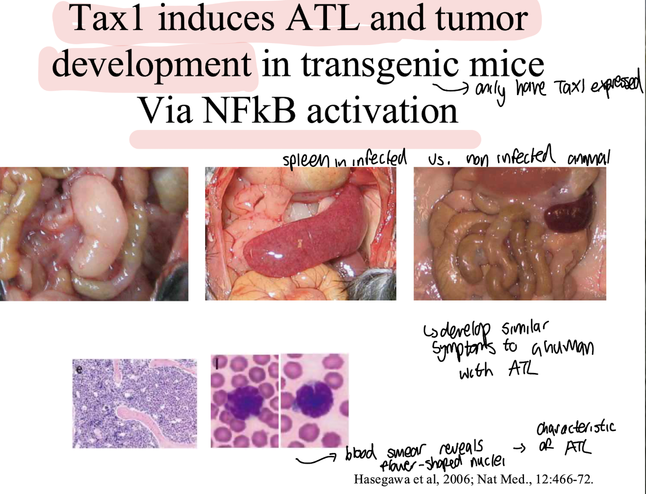
How does the Tax1 protein induce ATL and tumour development in transgenic mice?
Via NFkB activation leading the similar symptoms to those observed in human ATL cases, including increased cell proliferation and survival.
Spleens often are enlarged with abnormal cells i.e. flower-like resembling those of ATL due to effects of Tax1.
HBZ summary
Transforms primary CD4+ T cells
HBZ transgenic animals develop ATL –like disease and a range of inflammatory disorders
Drives proliferation and maintenance of ATL cells
Only viral protein expressed in ATL cells
HTLV-1 pathogenesis is determined by a balance between ____________ and __________
viral gene expression and host immune response
3 main diagnostic strategies of HTLV?
Serology - antibody testing on blood sample by ELISA for Ab against envelope proteins e.g. gp46
Confirmation Test of positive ELISA by Western Blot differentiates HTLV-1 from HTLV-2
PCR for subtype identification and proviral load i.e. level of integrated DNA in blood