Mirco Lab
1/79
There's no tags or description
Looks like no tags are added yet.
Name | Mastery | Learn | Test | Matching | Spaced | Call with Kai | Chat |
|---|
No analytics yet
Send a link to your students to track their progress
80 Terms
What is an appropriate way to streak?
The quadrant streak pattern
- dividing the plate into four sections
T-streak
- divided in three sections
ALL DONE IN A ZIGZAG

Orange
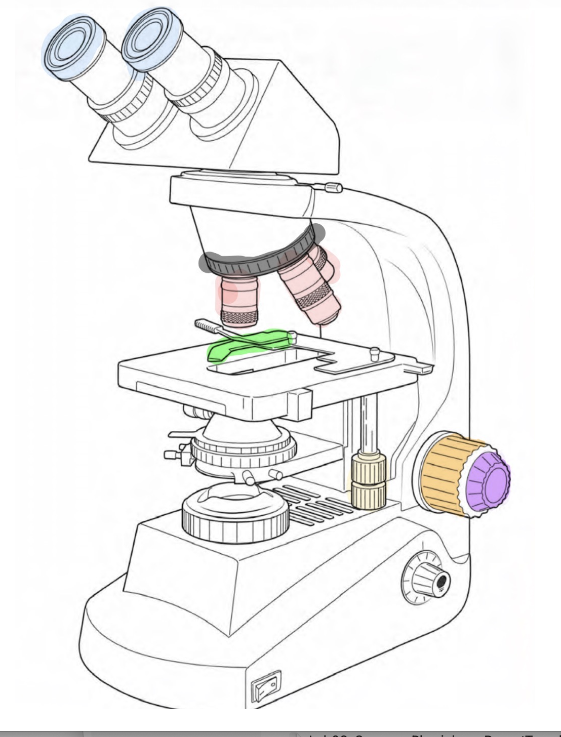
Coarse-focus

purple
Fine-focus

slide- positioning
yellow

red
objective lense

blue
ocular

black
revolving nosepiece
green
slide clip
Appropriate clean-up procedures
lens paper to remove oil
dispose slides in sharps (red box)
wipe down table
Capsule stain protocol
1 loopful sheep serum
1 drop congo red
whatever mix we are working one
smear using second slide
let dry
Maneval’s reagent
- 1 min
Gently rinse with water
Gram Satin protocol
heat fix
crystal violet
- 1 min and then rinse with water
gram’s iodine
- 1 min then rinse
Decolorize with alcohol, then rinse with water
Safranin
- 1 min
Blot dry
Endospore stain protocol
heat fix
Malachite green
- 10 min exposure
rinse
safranin
- 1 min then rinse with water
Acid-Fast stain protocol
heat fix
rinse smear with water
acid-alcohol decolorize
- 1 min, rinse with water
counterstain with methylene blue
1 min, rinse
blot dry
Capsule Stain: Positive result:
Clear halo (capsule) surrounding the bacterial cell.
Capsule Stain: Negative result:
No halo present; capsule absent or not visible
Capsule Stain: Type of stain
Structural stain
Also uses an indirect (negative) staining technique because the background is stained instead of the capsule.
Capsule stain: Why each step matters
Background stain creates contrast.
Air drying preserves the capsule.
Counterstain colors the cell.
Capsule stain:If mistakes are made
Heat fixing: Capsule shrinks or disappears (false negative).
Skipping Congo Red: No dark background.
Skipping Maneval's stain: Cells difficult to see.
Overwashing: Capsule may wash away.
Gram stain: Positive result
Purple cells
Gram-positive
Thick peptidoglycan cell wall
Gram stain: Negative result
Pink/red cells
Gram-negative
Thin peptidoglycan with outer membrane
Gram stain:Type of stain
Differential stain
Gram stain: Why each step matter
Crystal violet stains all cells.
Iodine locks the stain inside cells.
Alcohol differentiates Gram-positive from Gram-negative bacteria.
Safranin makes Gram-negative cells visible.
Gram stain: If mistakes are made
Forget iodine → Crystal violet washes out → Gram-positive cells may appear Gram-negative.
Over-decolorize → Gram-positive cells become pink.
Under-decolorize → Gram-negative cells stay purple.
Skip safranin → Gram-negative cells appear colorless.
Endospore Stain:Positive result
Green endospores inside or outside pink vegetative cells.
Organism is capable of producing endospores.
Endospore Stain: Negative result
Only pink vegetative cells.
No spores present.
Endospore Stain: Type of stain
Structural stain
Endospore Stain: Why each step matters
Heat opens the tough spore coat.
Water removes stain from regular cells.
Safranin provides contrast
Endospore Stain:If mistakes are made
No heat → Spores stay colorless.
Skip water rinse → Everything stays green.
Skip safranin → Vegetative cells are difficult to see.
Too much heat → Distorted cells.
Acid-Fast Stain: Positive result
Bright red or fuchsia cells.
Organism contains mycolic acids in the cell wall.
Acid-Fast Stain:Negative result
Blue cells.
Lacks mycolic acid-rich cell walls.
Acid-Fast Stain: Type of stain
Differential stain
Acid-Fast Stain:Why each step matters
Carbolfuchsin stains all cells.
Heat helps stain penetrate the waxy cell wall (Ziehl-Neelsen method).
Acid-alcohol differentiates acid-fast from non-acid-fast bacteria.
Methylene blue stains decolorized cells
Acid-Fast Stain: If mistakes are made
Too much decolorizer → Acid-fast cells may lose the red stain (false negative).
Too little decolorizer → Non-acid-fast cells remain red (false positive).
Skip methylene blue → Non-acid-fast cells appear colorless.
Insufficient heating (Ziehl-Neelsen) → Poor penetration of carbolfuchsin into acid-fast cells.
Blood Agar (BAP): Type of media
Differentia
Blood Agar (BAP): Tests for the ability to
produce hemolysins, which lyse (break open) red blood cells.
Blood Agar (BAP): Important ingredient
5% Sheep blood
Blood Agar (BAP): Differential agent
Red blood cells (sheep blood)
Blood Agar (BAP): positive
(Beta) hemolysis = complete destruction of red blood cells; clear zone around colonies
(Alpha) hemolysis = greenish or brown discoloration around colonies
Blood Agar (BAP): negative
(Gamma) hemolysis = no hemolysis; agar unchanged
MacConkey Agar (MAC): Type of media
Selective and Differential
MacConkey Agar (MAC): What does it test?
Growth of Gram-negative bacteria
Ability to ferment lactose
MacConkey Agar (MAC): Important ingredients
Bile salts
Crystal Violet
MacConkey Agar (MAC): Differential agents
Lactose
Neutral Red (pH indicator)
MacConkey Agar (MAC): positive
Pink/red colonies = Positive for lactose fermentation
MacConkey Agar (MAC): negative
Colorless and lacks lactose fermentation |
Mannitol Salt Agar (MSA): Type of media
Selective and Differential
Mannitol Salt Agar (MSA):What does it test?
Salt tolerance
Mannitol fermentation
Mannitol Salt Agar (MSA): Important ingredients
7.5% Sodium Chloride (NaCl)
Mannitol Salt Agar (MSA): Differential agents
Mannitol
Phenol Red
Mannitol Salt Agar (MSA): postive
Yellow agar = Positive for mannitol fermentation
Mannitol Salt Agar (MSA): negative
Red/pink agar = Negative for mannitol fermentation
Eosin Methylene Blue (EMB): What does it test?
Growth of Gram-negative bacteria
Lactose fermentation
Eosin Methylene Blue (EMB):Selective agents
Eosin Y
Methylene Blue
Eosin Methylene Blue (EMB): Differential agent
Lactose
Eosin Methylene Blue (EMB): positive
Metallic green sheen =
|
Eosin Methylene Blue (EMB): negative
Purple/dark colonies = Weak lactose fermenter |
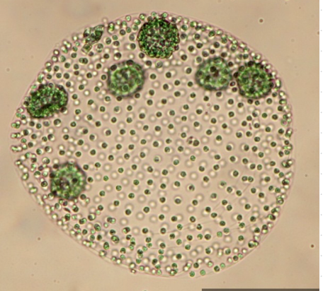
Volvox
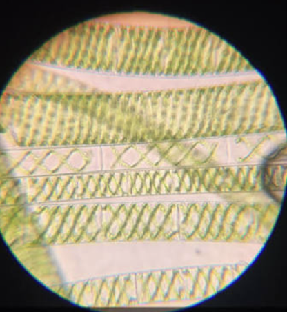
Spirogyra
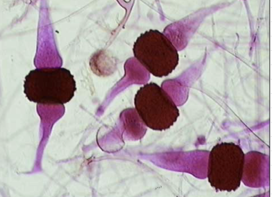
Rhizopous stolonifer
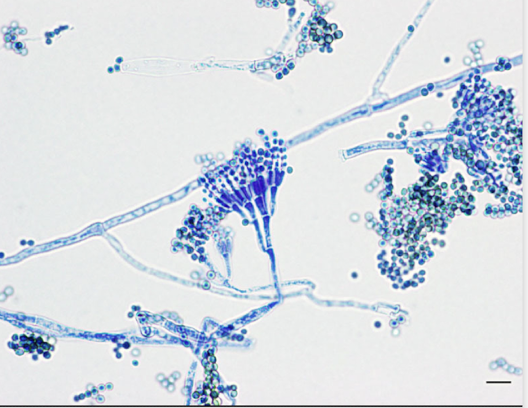
Penicillium roquefortii
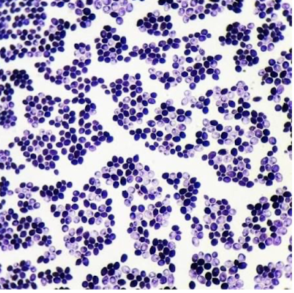
Candida albicans
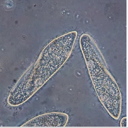
Paramecium caudatum
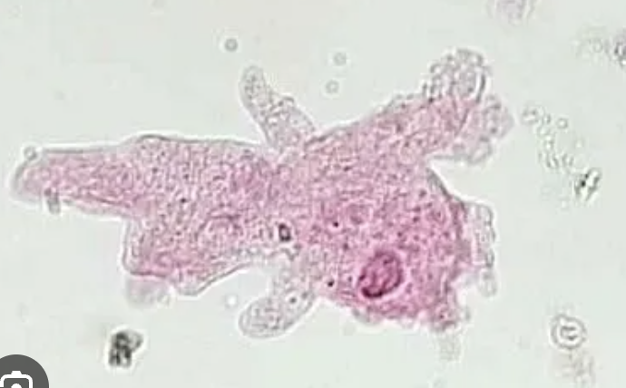
Amoeba proteus
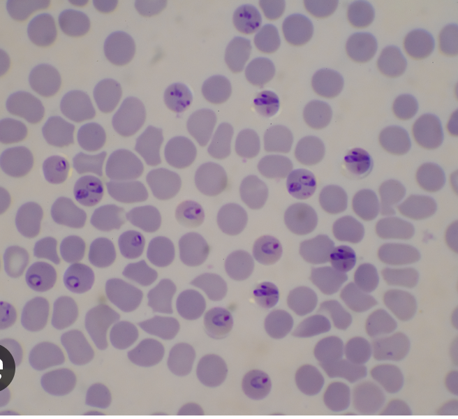
Plasmodium falciparum
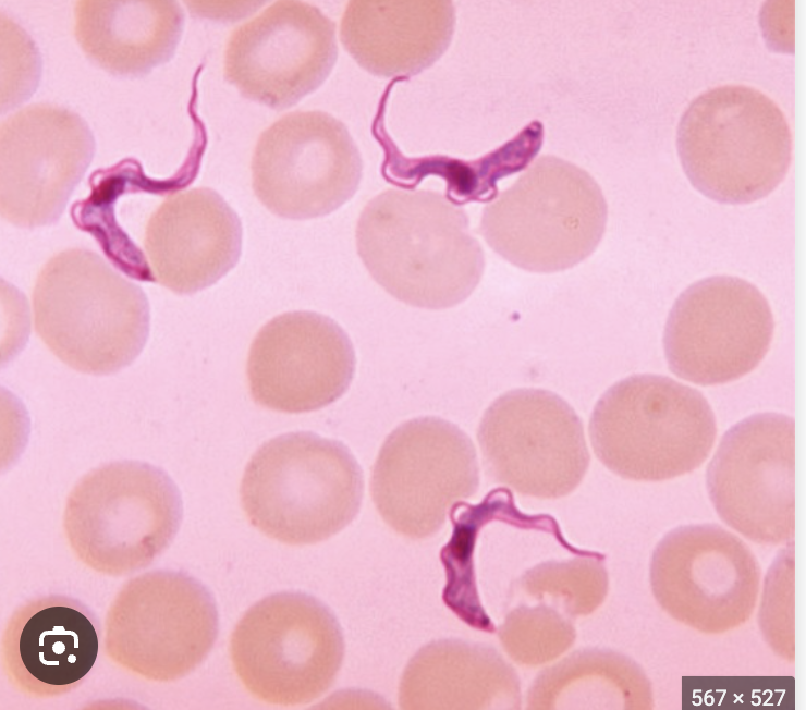
Trypauosoma cruzi

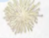
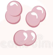
Colony shape
round
Colony shape
irregular
Colony shape
spindle
Colony shape
Filamentous
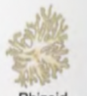
Colony shape
Rhizoid

Arrangement
streptococcus
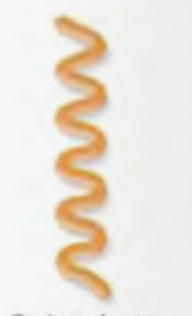
Arrangement
spirochete
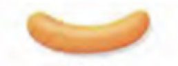
Arrangement
Vibrio

Arrangement
Streptobacillius

Arrangement
Diplobacillius

Arrangement
Palisades
Arrangement
Diplococci

Arrangement
Tetracoccus

Arrangement
Staphylococci:

Arrangement
Sarcinae