Week 6: Upper GIT

0.0(0)
studied byStudied by 0 people
learnLearn
examPractice Test
spaced repetitionSpaced Repetition
heart puzzleMatch
flashcardsFlashcards
Card Sorting

1/21

encourage image

There's no tags or description

Looks like no tags are added yet.

Study Analytics
Name
Mastery
Learn
Test
Matching
Spaced

No study sessions yet.

22 Terms

1
New cards

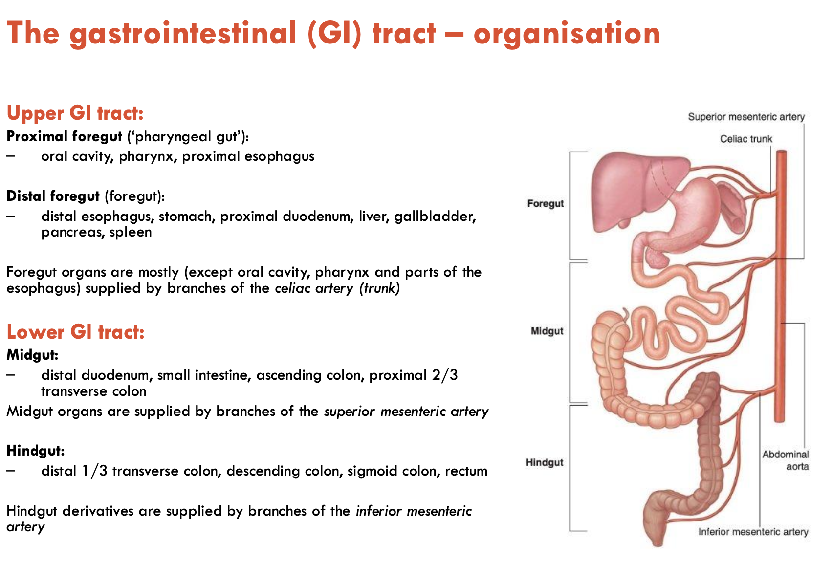
Describe the organisation of the GIT (4)

Upper GIT

Proximal foregut (‘pharyngeal gut’):

  • oral cavity, pharynx, proximal oesophagus

Distal foregut (‘foregut’)

  • distal oesophagus, stomach, proximal duodenum, liver, gallbladder, pancreas, spleen

Foregut organs are mostly supplied by branches of the coeliac artery (except oral cavity, pharynx, and parts of the oesophagus)

Lower GIT

Midgut:

  • distal duodenum, small intestine, ascending colon, proximal 2/3 transverse colon

  • supplied by branches of sup. mesenteric artery

Hindgut:

  • distal 1/3 transverse colon, descending colon, sigmoid colon, rectum

  • supplied by branches of inf. mesenteric artery

<p><u>Upper GIT</u></p><p><strong>Proximal foregut</strong> (‘pharyngeal gut’):</p><ul><li><p>oral cavity, pharynx, proximal oesophagus</p></li></ul><p><strong>Distal foregut</strong> (‘foregut’)</p><ul><li><p>distal oesophagus, stomach, proximal duodenum, liver, gallbladder, pancreas, spleen</p></li></ul><p>Foregut organs are mostly supplied by <strong>branches of the coeliac artery</strong> (except oral cavity, pharynx, and parts of the oesophagus)</p><p></p><p><u>Lower GIT</u></p><p><strong>Midgut:</strong></p><ul><li><p>distal duodenum, small intestine, ascending colon, proximal 2/3 transverse colon</p></li><li><p>supplied by branches of <strong>sup. mesenteric artery</strong></p></li></ul><p><strong>Hindgut:</strong></p><ul><li><p>distal 1/3 transverse colon, descending colon, sigmoid colon, rectum</p></li><li><p>supplied by branches of <strong>inf. mesenteric artery</strong></p></li></ul><p></p>
2
New cards

List the 3 stages of swallowing

  1. Oral stage

  2. Pharyngeal stage

  3. Oesophagal stage

3
New cards

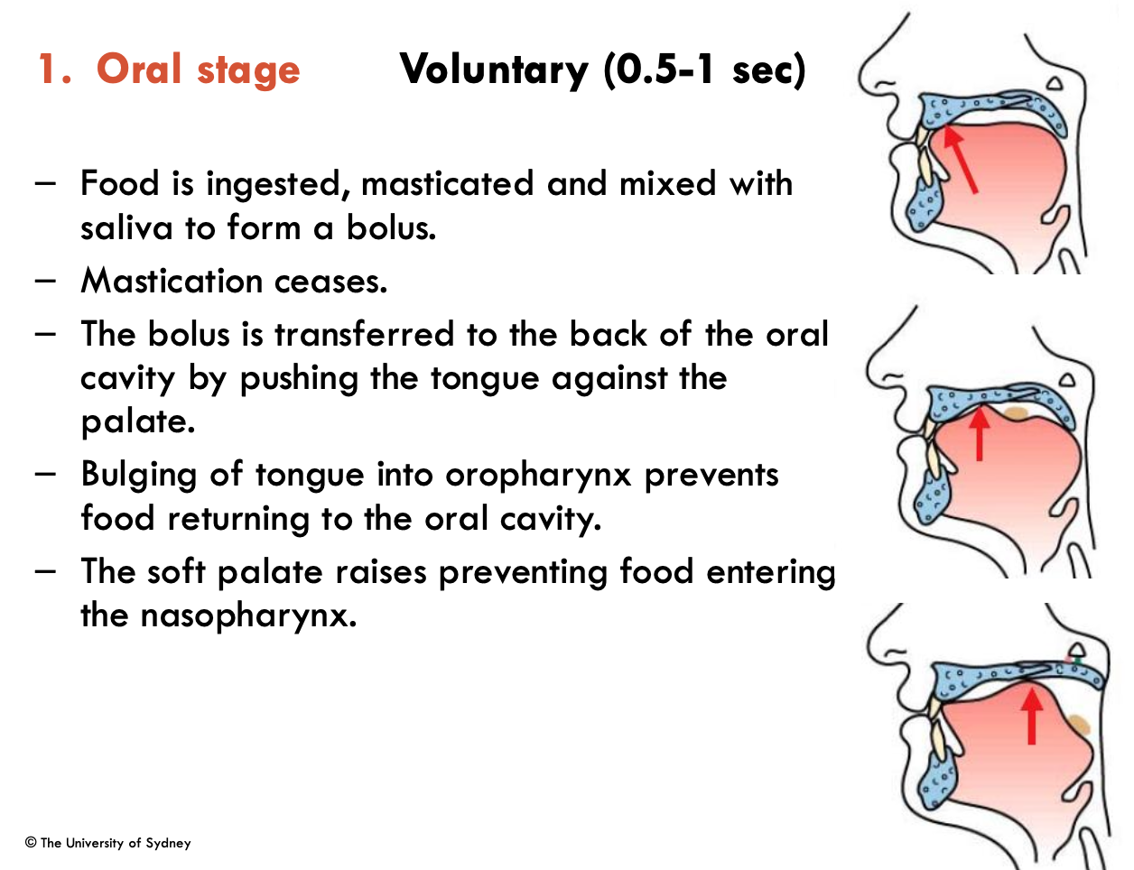
Describe the oral stage of swallowing (5)

Voluntary (0.5-1 sec)

  1. Food is ingested, masticated, and mixed with saliva to form a bolus

  2. Mastication ceases

  3. Bolus is transferred to back of oral cavity by pushing tongue against the palate

  4. Bulging of tongue into oropharynx prevents food returning to oral cavity

  5. Raising of soft palate prevents food from entering nasopharynx

<p>Voluntary (0.5-1 sec)</p><ol><li><p>Food is ingested, masticated, and mixed with saliva to form a bolus</p></li><li><p>Mastication ceases</p></li><li><p>Bolus is transferred to back of oral cavity by pushing tongue against the palate</p></li><li><p>Bulging of tongue into oropharynx prevents food returning to oral cavity</p></li><li><p>Raising of soft palate prevents food from entering nasopharynx </p></li></ol><p></p>
4
New cards

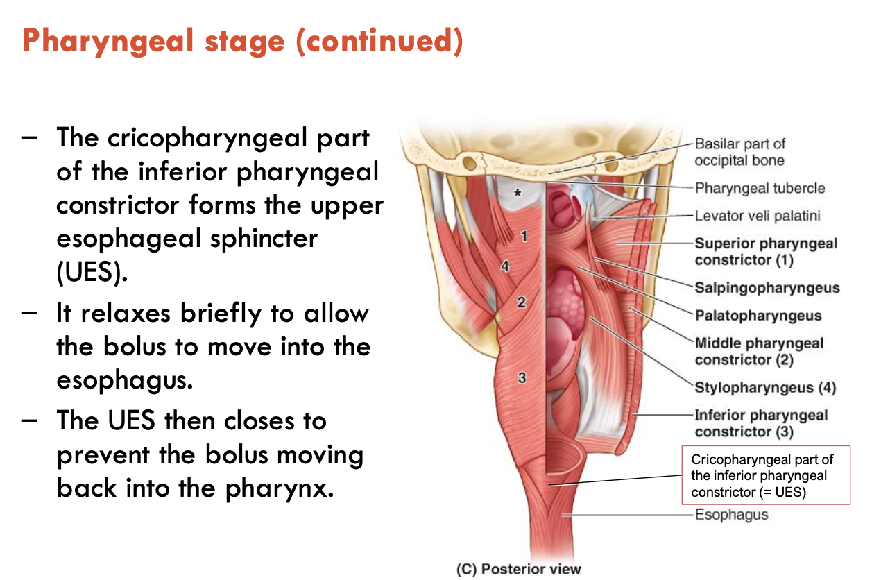
Describe the pharyngeal stage of swallowing (6)

Involuntary (1 sec)

  1. Swallowing reflex is triggered by the bolus, stimulating tactile receptors on the faeces and uvula

  2. Epiglottis is deflected by the food bolus passing over it and by the larynx raising

  3. This closes the larynx, preventing aspiration of food into the airways

  4. The superior, middle, and inferior pharyngeal sphincters contract sequentially

  5. The upper oesophageal sphincter (UES - formed by the cricopharyngeal part of the inf. pharyngeal constrictor) relaxes briefly to allow the bolus to move to the oesophagus

  6. The UES then closes to prevent the bolus from moving back into the pharynx

<p>Involuntary (1 sec)</p><ol><li><p>Swallowing reflex is triggered by the bolus, stimulating tactile receptors on the faeces and uvula</p></li><li><p>Epiglottis is deflected by the food bolus passing over it and by the larynx raising</p></li><li><p>This closes the larynx, preventing aspiration of food into the airways</p></li><li><p>The superior, middle, and inferior pharyngeal sphincters contract sequentially</p></li><li><p>The upper oesophageal sphincter (UES - formed by the cricopharyngeal part of the inf. pharyngeal constrictor) relaxes briefly to allow the bolus to move to the oesophagus</p></li><li><p>The UES then closes to prevent the bolus from moving back into the pharynx</p></li></ol><p></p>
5
New cards

Describe the oesphageal stage of swallowing (5)

Involuntary (8-20 sec)

  1. Bolus is moved down the oesophagus by peristaltic contractions - involuntary waves of sequential contraction and relaxation of smooth muscle

  2. Epiglottis returns to normal position

  3. The lower oesophageal sphincter (LES) is tonically contracted

  4. LES relaxes briefly during swallowing to allow the bolus to enter the stomach

  5. The LES closes to prevent reflux/regurgitation back into the oesophagus

6
New cards

What is dysphagia?

Dysphagia is a subjective awareness of difficulty or obstruction during swallowing

  • caused by functional or structural abnormalities of the oral cavity, pharynx, oesophagus, and/or gastric cardia

  • relatively common (esp. with aging and in women)

  • fluoroscopy barium swallow study is the main imaging assessment

    • endoscopy may be used to examine oesophageal mucosa

    • cross-sectional imaging for further evaluation of masses/humans

7
New cards

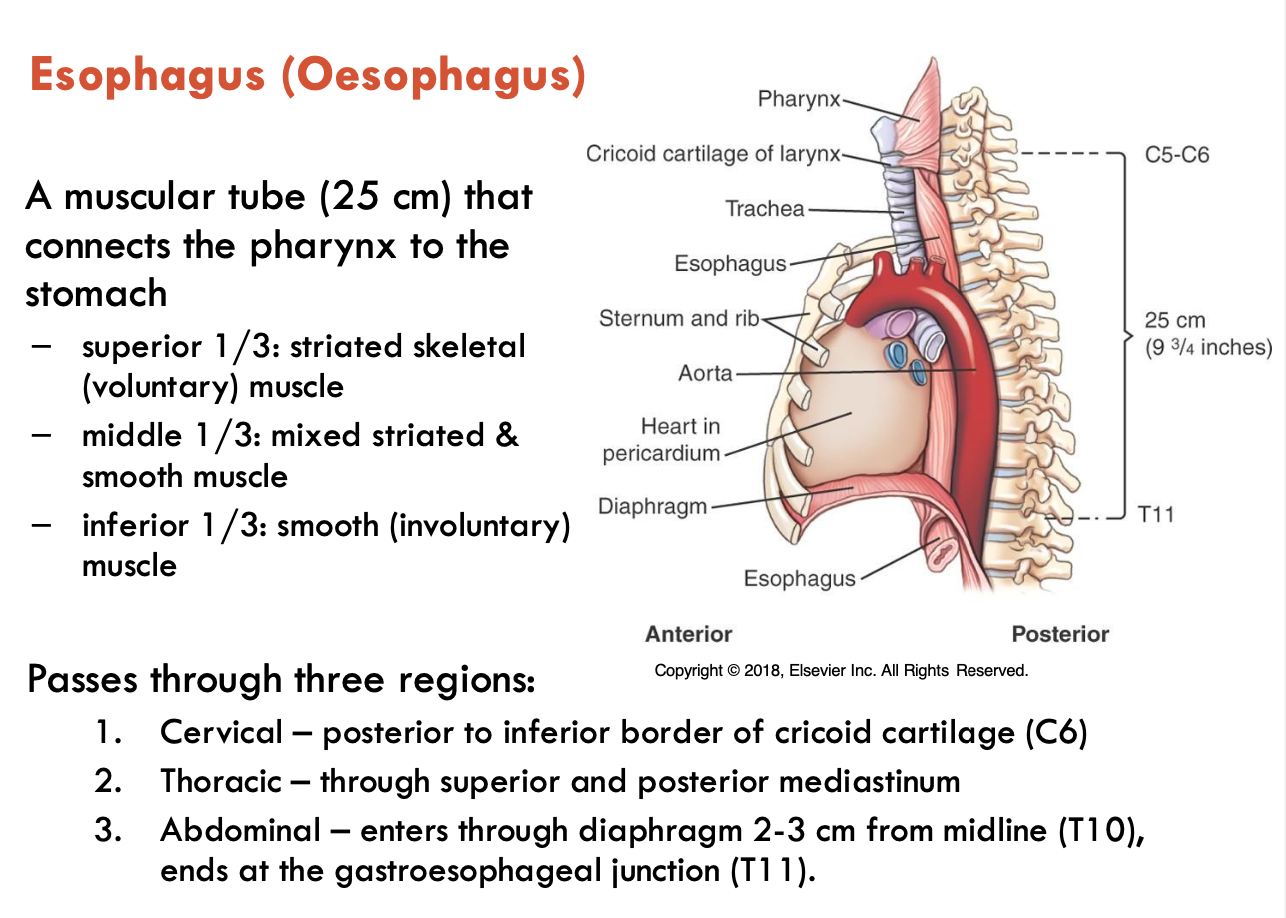
What are the features of the oesophagus?

Oesophagus = muscular tube (25 cm) that connects the pharynx to the stomach

  • superior 1/3: striated skeletal muscle

  • middle 1/3: mixed striated & smooth muscle

  • inferior 1/3: smooth muscle

Passes through 3 regions:

  1. Cervical - posterior to inf. border of cricoid cartilage (C6)

  2. Thoracic - through superior and posterior mediastinum

  3. Abdominal - enters through diaphragm 2-3 cm from the midline (T10), ends at the gastroesophageal junction (T11)

<p>Oesophagus = muscular tube (25 cm) that connects the pharynx to the stomach</p><ul><li><p><strong>superior 1/3:</strong> striated skeletal muscle</p></li><li><p><strong>middle 1/3:</strong> mixed striated &amp; smooth muscle</p></li><li><p><strong>inferior 1/3:</strong> smooth muscle</p></li></ul><p>Passes through 3 regions:</p><ol><li><p><strong>Cervical</strong> - posterior to inf. border of cricoid cartilage (C6)</p></li><li><p><strong>Thoracic</strong> - through superior and posterior mediastinum</p></li><li><p><strong>Abdominal</strong> - enters through diaphragm 2-3 cm from the midline (T10), ends at the gastroesophageal junction (T11)</p></li></ol><p></p>
8
New cards

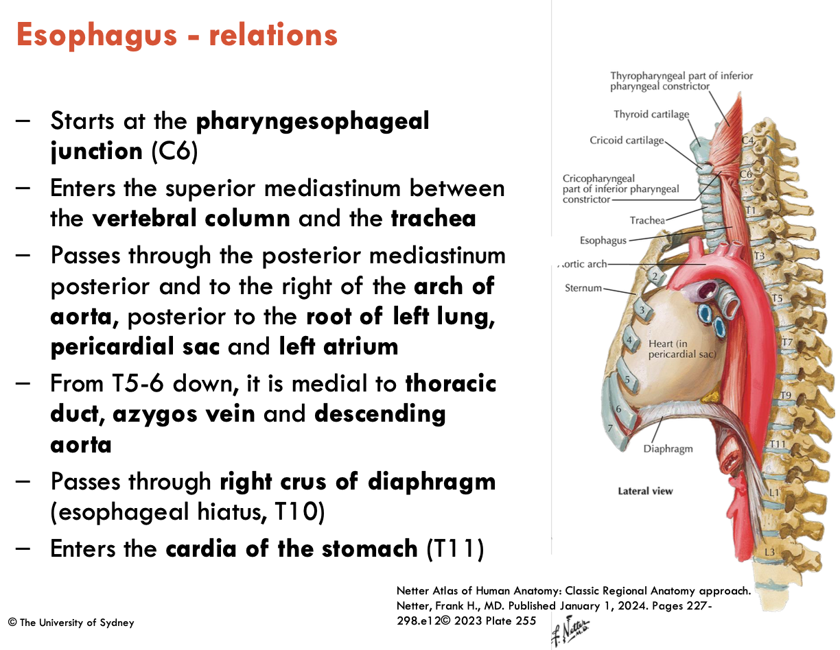
Describe the relations of the oesophagus

  • starts at the pharyngoesophageal junction (C6)

  • enters the sup. mediastinum between the vertebral column and the trachea

  • passes through the post. mediastinum posterior to the right of the aortic arch, posterior to the root of the left lung, pericardial sac, and left atrium

  • From T5-6 down, it is medial to the thoracic duct, azygos vein, and descending aorta

  • passes through the right crus of diaphragm (oesophageal hiatus: T10)

  • enters the cardia of the stomach (T11)

<ul><li><p>starts at the <strong>pharyngoesophageal junction (C6)</strong></p></li><li><p>enters the sup. mediastinum between the <strong>vertebral column</strong> and the <strong>trachea</strong></p></li><li><p>passes through the post. mediastinum posterior to the right of the <strong>aortic arch</strong>, posterior to the<strong> root of the left lung, pericardial sac, </strong>and<strong> left atrium</strong></p></li><li><p>From T5-6 down, it is medial to the <strong>thoracic duct</strong>, <strong>azygos vein,</strong> and <strong>descending aorta</strong></p></li><li><p>passes through the <strong>right crus of diaphragm</strong> (oesophageal hiatus: T10)</p></li><li><p>enters the <strong>cardia of the stomach</strong> (T11)</p></li></ul><p></p>
9
New cards

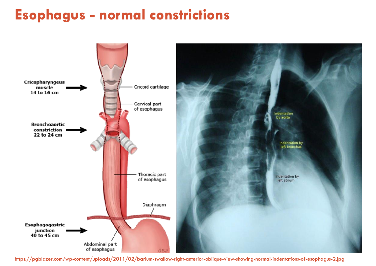
List the normal constrictions of the oesophagus (3)

  1. UES (upper oesophageal constrictor - cricopharyngeal part of the inf. pharyngeal constrictor)

  2. Bronchoaortic constriction

  3. Oesophagogastric junction

Dysphagia can cause abnormality in these constrictions

<ol><li><p><strong>UES</strong> (upper oesophageal constrictor - cricopharyngeal part of the inf. pharyngeal constrictor)</p></li><li><p><strong>Bronchoaortic constriction</strong></p></li><li><p><strong>Oesophagogastric junction</strong></p></li></ol><p></p><p>Dysphagia can cause abnormality in these constrictions</p>
10
New cards

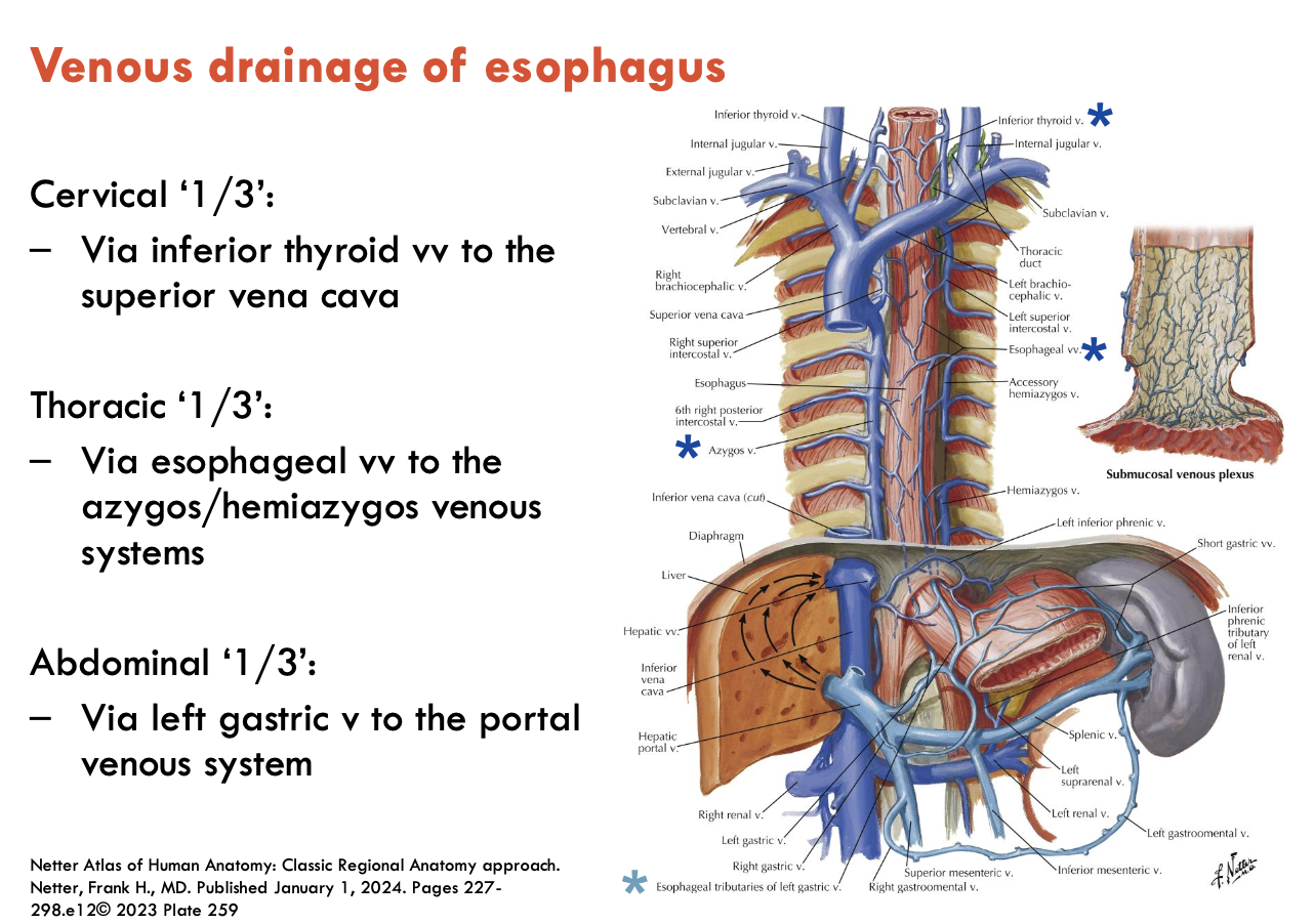
Describe the blood supply to the oesophagus

Cervical 1/3:

  • Arterial: inf. thyroid artery

  • Venous: inf. thyroid veins → SVC

Thoracic 1/3:

  • Arterial: oesophageal branches of the abdominal aorta

  • Venous: oesophageal veins → azygos/hemiazygos

Abdominal 1/3:

  • Arterial: left gastric art. & left phrenic art.

  • Venous: left gastric v. → portal venous system

<p>Cervical 1/3: </p><ul><li><p>Arterial: <strong>inf. thyroid artery</strong></p></li><li><p>Venous: <strong>inf. thyroid veins → SVC</strong></p></li></ul><p>Thoracic 1/3: </p><ul><li><p>Arterial: <strong>oesophageal branches of the abdominal aorta</strong></p></li><li><p>Venous: <strong>oesophageal veins → azygos/hemiazygos </strong></p></li></ul><p>Abdominal 1/3:<strong> </strong></p><ul><li><p>Arterial: <strong>left gastric art. &amp; left phrenic art.</strong></p></li><li><p>Venous: <strong>left gastric v. → portal venous system</strong></p></li></ul><p></p>
11
New cards

What are oesophageal varices?

Varicose (enlarged) veins around the lower part of the oesophagus

  • due to portal hypertension often associated w/ liver disease (e.g. cirrhosis)

  • only lower 1/3 of oesophagus because they contribute to portal vein

12
New cards

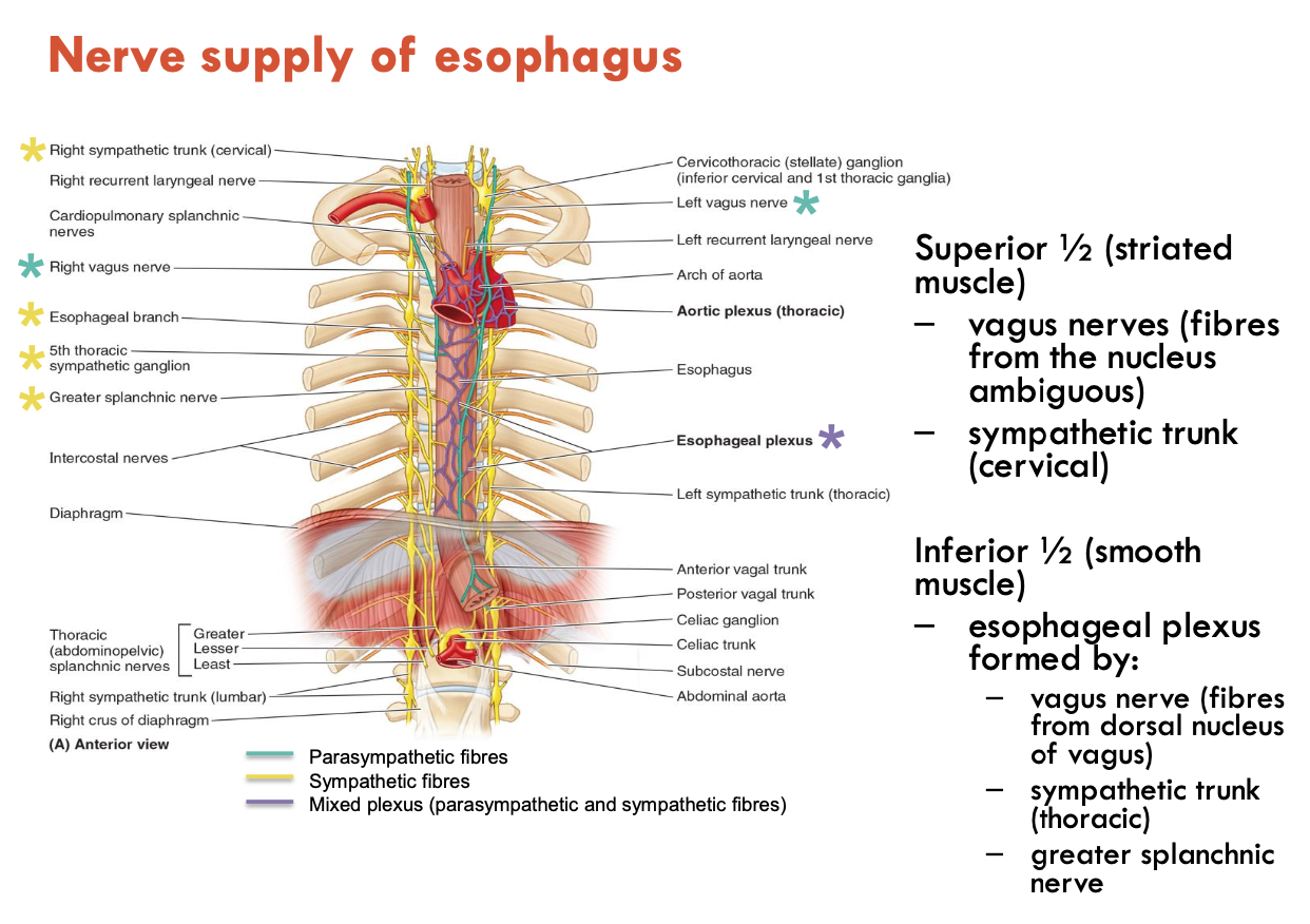
Describe the nerve supply of the oesophagus

Superior ½ (striated muscle):

  • vagus nerves (fibres from nucleus ambiguus)

  • sympathetic trunk (cervical)

Inferior ½ (smooth muscle):

  • oesophageal plexus formed by:

    • vagus nerve (fibres from dorsal nucleus of vagus)

    • sympathetic trunk (thoracic)

    • greater splanchnic n.

<p>Superior ½ (striated muscle):</p><ul><li><p><strong>vagus nerves</strong> (fibres from nucleus ambiguus)</p></li><li><p><strong>sympathetic trunk</strong> (cervical)</p></li></ul><p>Inferior ½ (smooth muscle):</p><ul><li><p><strong>oesophageal plexus</strong> formed by:</p><ul><li><p><strong>vagus nerve</strong> (fibres from dorsal nucleus of vagus)</p></li><li><p><strong>sympathetic trunk</strong> (thoracic)</p></li><li><p><strong>greater splanchnic n.</strong></p></li></ul></li></ul><p></p>
13
New cards

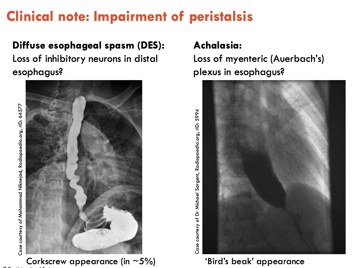
What conditions arise from peristalsis impairment?

Nerve supply to the oesophagus controls peristalsis

Impairment to this can lead to:

  • Diffuse oesophageal spasm (DES): loss of inhibitory neurons in distal oesophagus (corkscrew appearance)

  • Achalasia: loss of myenteric (Auerbach’s) plexus in oesophagus (Bird’s beak appearance)

<p>Nerve supply to the oesophagus controls peristalsis</p><p>Impairment to this can lead to:</p><ul><li><p><strong>Diffuse oesophageal spasm (DES):</strong> loss of inhibitory neurons in distal oesophagus (corkscrew appearance)</p></li><li><p><strong>Achalasia:</strong> loss of myenteric (Auerbach’s) plexus in oesophagus (Bird’s beak appearance)</p></li></ul><p></p>
14
New cards

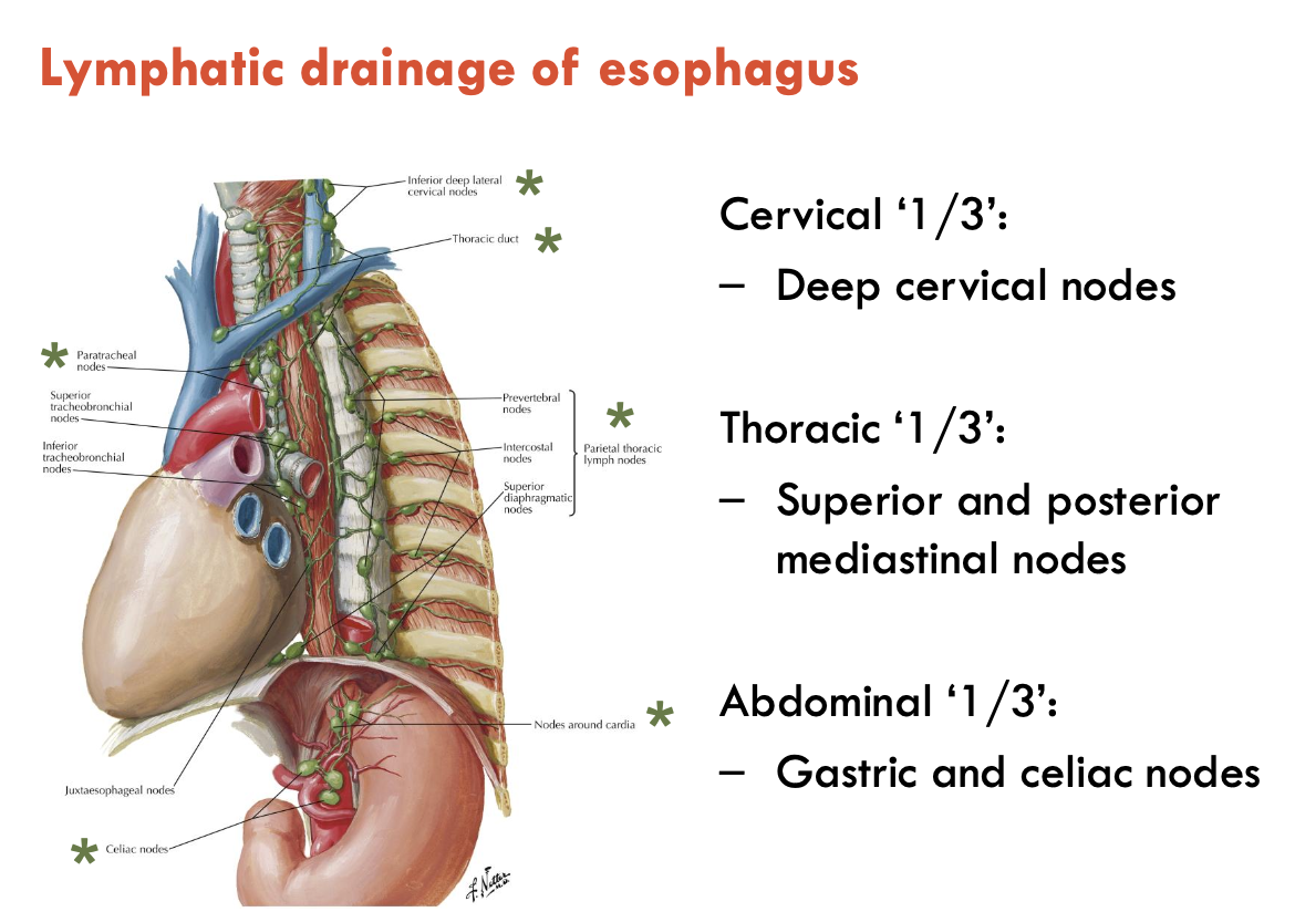
Describe the lymphatic drainage of the oesophagus

Cervical 1/3: deep cervical nodes

Thoracic 1/3: sup. and post. mediastinal nodes

Abdominal 1/3: gastric and coeliac nodes

<p>Cervical 1/3: <strong>deep cervical nodes</strong></p><p>Thoracic 1/3: <strong>sup. and post. mediastinal nodes</strong></p><p>Abdominal 1/3:<strong> gastric and coeliac nodes</strong></p>
15
New cards

List the 4 regions of the stomach

  1. cardia

  2. fundus

  3. body

  4. pyloric part: antrum, canal, sphincter/pylorus

<ol><li><p>cardia</p></li><li><p>fundus</p></li><li><p>body</p></li><li><p>pyloric part: antrum, canal, sphincter/pylorus</p></li></ol><p></p>
16
New cards

List the 2 curvatures and 2 sphincters of the stomach

Curvatures:

  1. Lesser - shorter concave border

  2. Greater - longer convex border

Sphincters:

  1. Lower oesophageal sphincter (LES)

  2. Pyloric sphincter

<p><strong>Curvatures:</strong></p><ol><li><p>Lesser - shorter concave border</p></li><li><p>Greater - longer convex border</p></li></ol><p><strong>Sphincters:</strong></p><ol><li><p>Lower oesophageal sphincter (LES)</p></li><li><p>Pyloric sphincter</p></li></ol><p></p>
17
New cards

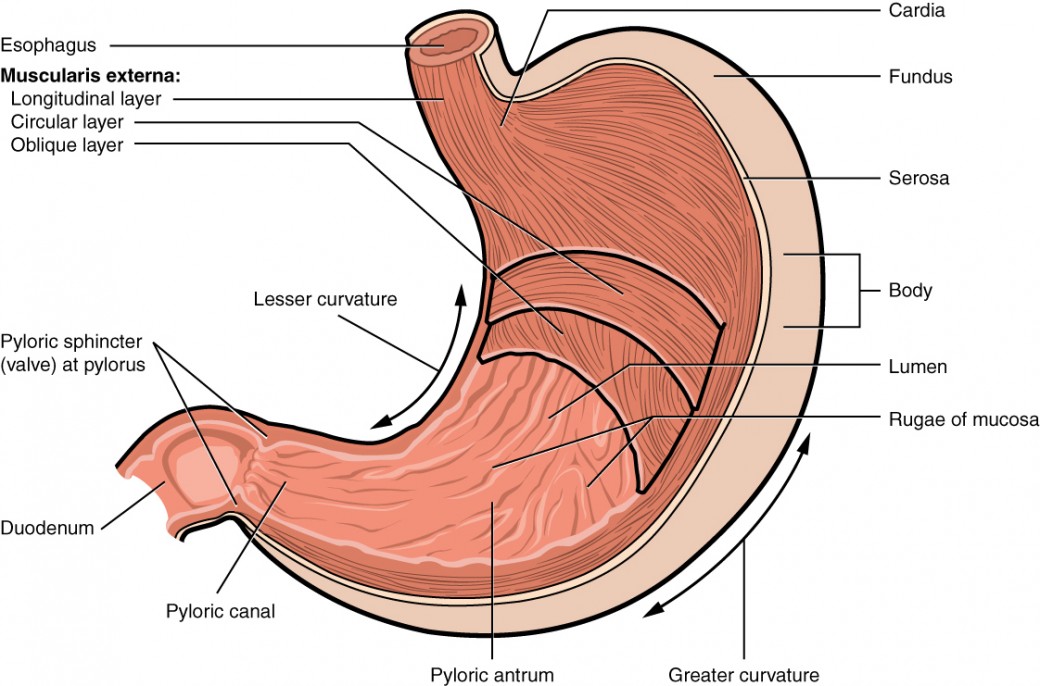
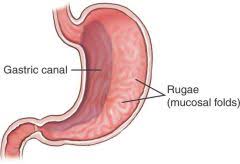
What are the internal features of the stomach (3).?

The stomach is highly distensible (can contain 45 - 1500 mL), and has the following internal features:

  • gastroesophageal junction marked by the ‘z-line’ - the transition between oesophageal (stratified squamous) and gastric (columnar) mucosa (more of a histological feature, hard to see on specimens)

  • longitudinal gastric folds (rugae) - most developed in the pyloric part and along the greater curvature

  • gastric canal - forms along the lesser curvature during swallowing

<p>The stomach is highly distensible (can contain 45 - 1500 mL), and has the following internal features:</p><ul><li><p><strong>gastroesophageal junction</strong> marked by the ‘z-line’ - the transition between oesophageal (stratified squamous) and gastric (columnar) mucosa (more of a histological feature, hard to see on specimens)</p></li><li><p><strong>longitudinal gastric folds </strong>(rugae) - most developed in the pyloric part and along the greater curvature</p></li><li><p><strong>gastric canal</strong> - forms along the lesser curvature during swallowing</p></li></ul><p></p>
18
New cards

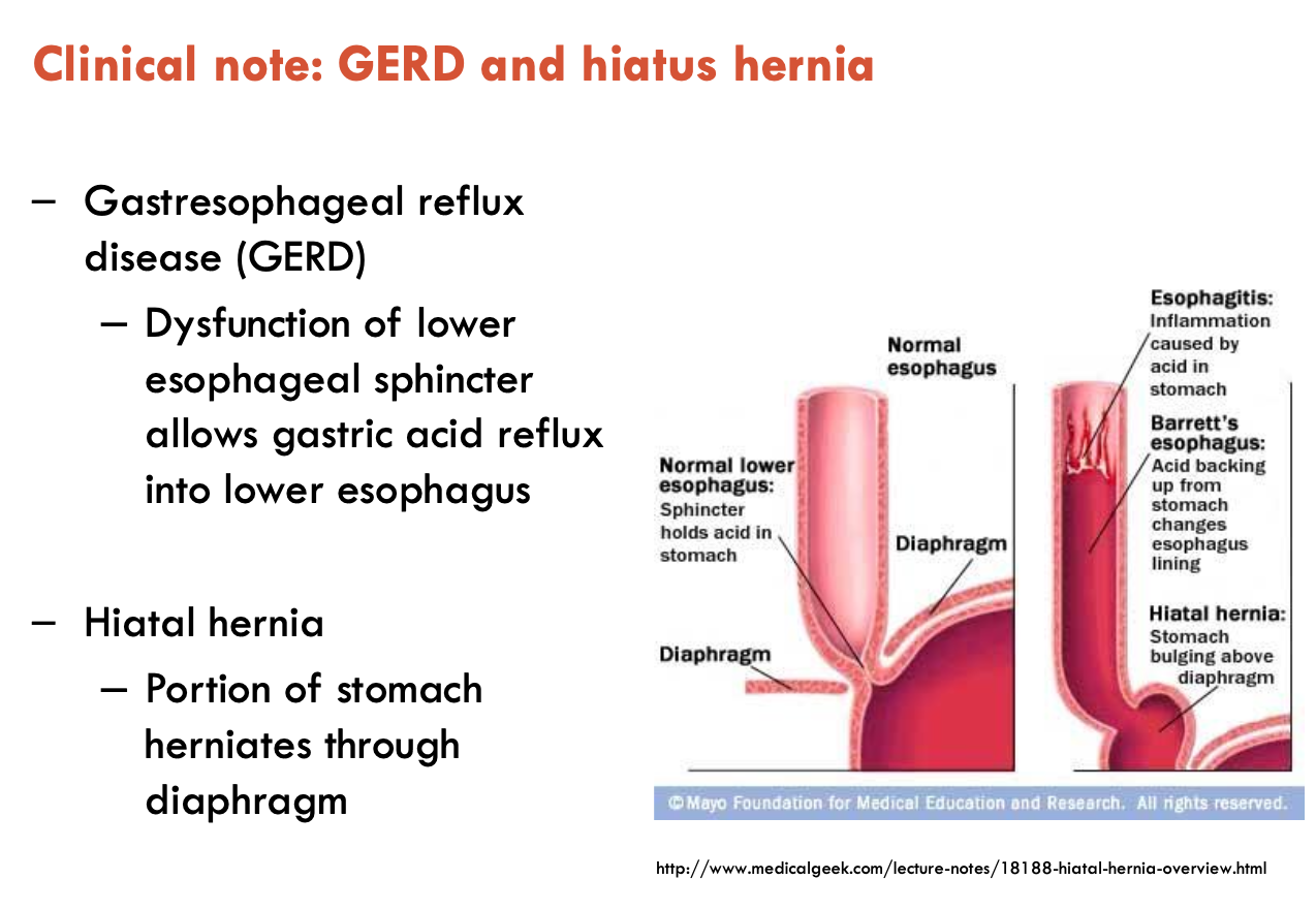
What is gastroesophageal reflux disease?

Gastroesophageal reflux disease (GERD)

  • dysfunction of lower oesophageal sphincter allows gastric acid reflux into lower oesophagus, can lead to hiatal hernia

Hiatal hernia: portion of stomach herniates through the diaphragm

<p><strong>Gastroesophageal reflux disease (GERD)</strong></p><ul><li><p>dysfunction of lower oesophageal sphincter allows gastric acid reflux into lower oesophagus, can lead to hiatal hernia</p></li></ul><p><strong>Hiatal hernia: </strong>portion of stomach herniates through the diaphragm</p><p></p>
19
New cards

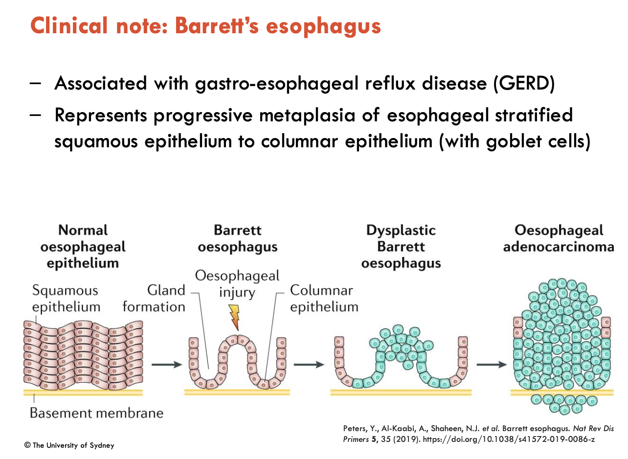
What is Barrett’s oesophagus?

Associated with GERD, Barrett’s oesophagus represents progressive metaplasia of oesophageal stratified squamous epithelium to columnar epithelium (with goblet cells)

  • difficult to detect using radiographic (barium swallow) imahing

  • often detected incidentally when using upper endoscopy for assessment for GERD - confirmed on biopsy

  • if metaplasia continues, can lead to cancer

<p>Associated with GERD, Barrett’s oesophagus represents progressive metaplasia of oesophageal stratified squamous epithelium to columnar epithelium (with goblet cells)</p><ul><li><p>difficult to detect using radiographic (barium swallow) imahing</p></li><li><p>often detected incidentally when using upper endoscopy for assessment for GERD - confirmed on biopsy</p></li><li><p>if metaplasia continues, can lead to cancer</p></li></ul><p></p>
20
New cards

What are gastric ulcers?

Gastric ulcers are characterised by a decrease in mucosal barrier

  • one of the most common chronic GIT diseases

  • risk of perforation (hole)

  • major risk factors:

    • Helicobacter pylori infection

    • frequent, long-term use of NSAIDs

21
New cards

Image of Blood Supply of Stomach

Arteries

  • coeliac trunk

  • L & R gastric arteries

  • short gastric arteries

  • L & R gastro-omental arteries

Veins

  • portal vein

  • L & R gastric veins

  • short gastric vein

  • L & R gastro-omental veins

<p><u>Arteries</u></p><ul><li><p>coeliac trunk</p></li><li><p>L &amp; R gastric arteries</p></li><li><p>short gastric arteries</p></li><li><p>L &amp; R gastro-omental arteries</p></li></ul><p><u>Veins</u></p><ul><li><p>portal vein</p></li><li><p>L &amp; R gastric veins</p></li><li><p>short gastric vein</p></li><li><p>L &amp; R gastro-omental veins</p></li></ul><p></p>
22
New cards

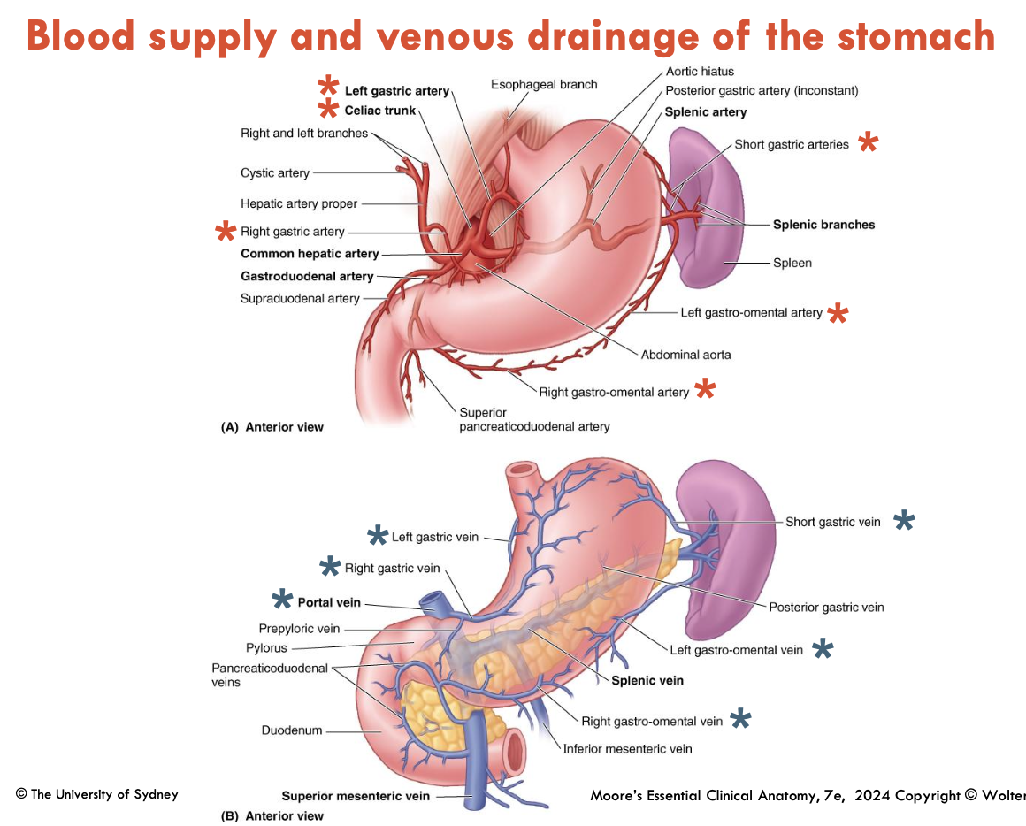
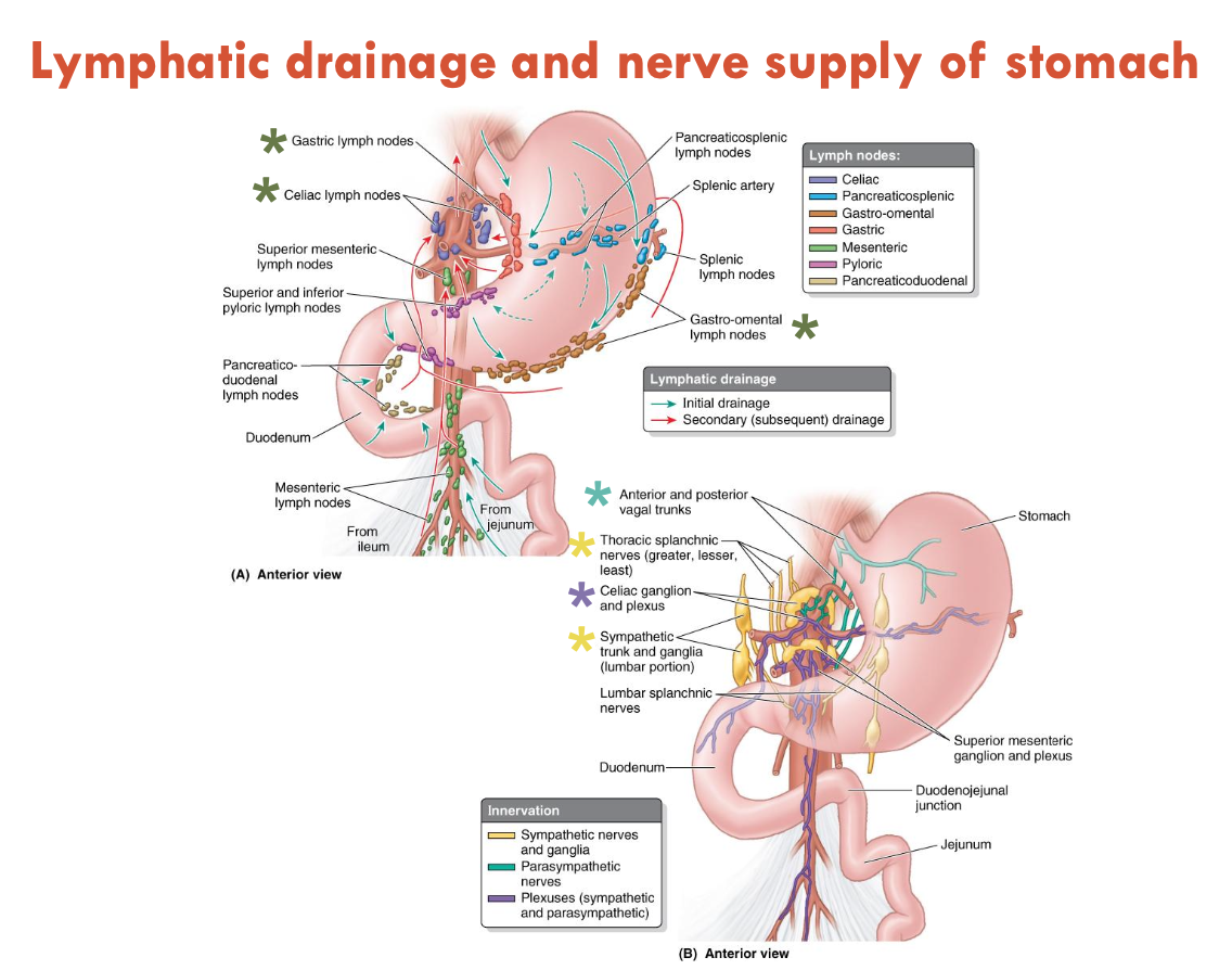
Image of Lymphatic Drainage and Nerve Supply of Stomach

Lymph Nodes

  • gastric lymph nodes

  • coeliac lymph nodes

  • gastro-omental lymph nodes

Nerves

  • Ant. & Post. vagal trunks

  • thoracic splanchnic nerves (greater, lesser, least)

  • coeliac ganglion and plexus

  • sympathetic trunk and ganglia (lumbar)

<p><u>Lymph Nodes</u></p><ul><li><p>gastric lymph nodes</p></li><li><p>coeliac lymph nodes</p></li><li><p>gastro-omental lymph nodes</p></li></ul><p><u>Nerves</u></p><ul><li><p>Ant. &amp; Post. vagal trunks</p></li><li><p>thoracic splanchnic nerves (greater, lesser, least)</p></li><li><p>coeliac ganglion and plexus</p></li><li><p>sympathetic trunk and ganglia (lumbar)</p></li></ul><p></p>