A&P 1 Exam 4
1/82
There's no tags or description
Looks like no tags are added yet.
Name | Mastery | Learn | Test | Matching | Spaced | Call with Kai |
|---|
No analytics yet
Send a link to your students to track their progress
83 Terms
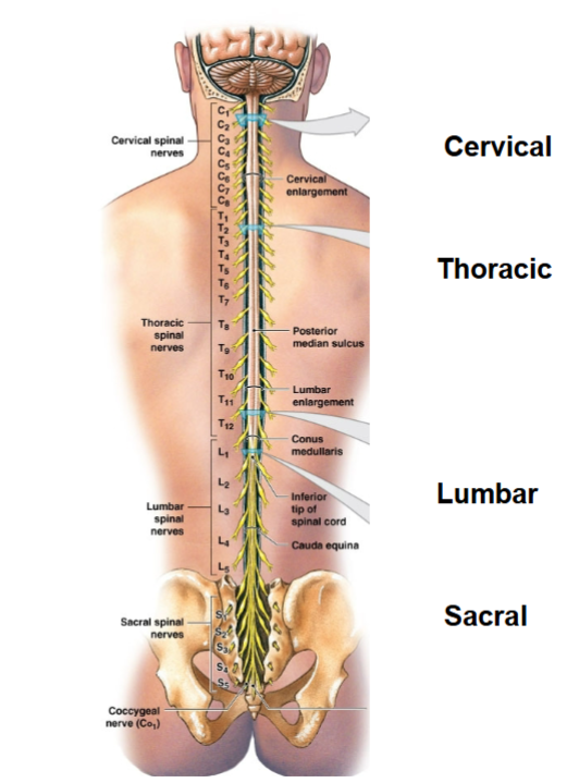
What are the four main regions of the spinal cord, and what key anatomical features do they include?
Cervical (C1-C8): Cervical spinal nerves, cervical enlargement. (neck)
Thoracic (T1-T12): Thoracic spinal nerves, posterior median sulcus. (chest)
Lumbar (L1-L5): Lumbar spinal nerves, lumbar enlargement. (lower back)
Sacral (S1-S5): Sacral spinal nerves, conus medullaris, cauda equina, coccygeal nerve. (sacrum/tail bone)
do not need to know numbers*
What are dermatomes, and how are they organized along the spinal cord?
Dermatomes are areas of skin innervated by specific spinal nerves. *
Cervical (C1-C8): Innervates arms, neck, and back of the head.
Thoracic (T1-T12): Covers the chest and abdomen.
Lumbar (L1-L5): Provides sensation to the lower back and front of the legs.
Sacral (S1-S5): Innervates the back of the legs, buttocks, and groin area.
Spinal cord talks to entire body. Chicken pox virus sleeps in spinal cord, thoracic mostly, wakes up shingles in T3, wraps around dermatome
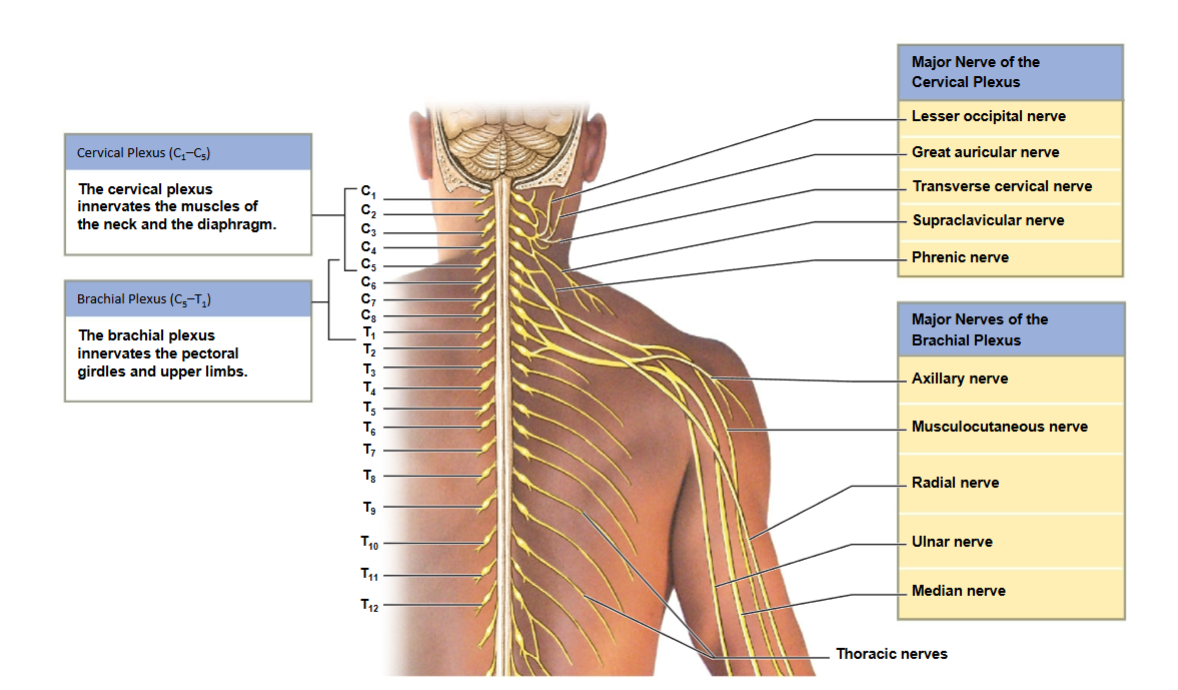
What are the major nerves of the cervical and brachial plexuses, and what areas do they innervate?
Cervical Plexus (C1-C5): Innervates muscles of the neck, head, and diaphragm. Major nerves: Lesser occipital, Great auricular, Transverse cervical, Supraclavicular, Phrenic.
Brachial Plexus (C5-T1): Innervates pectoral girdles and upper limbs, talks to arm. Major nerves: Axillary, Musculocutaneous, Radial, Ulnar, Median.
These nerves are crucial for controlling movements and sensation in the neck, diaphragm, and upper limbs. Plexus is a region of nerves that go to same area.
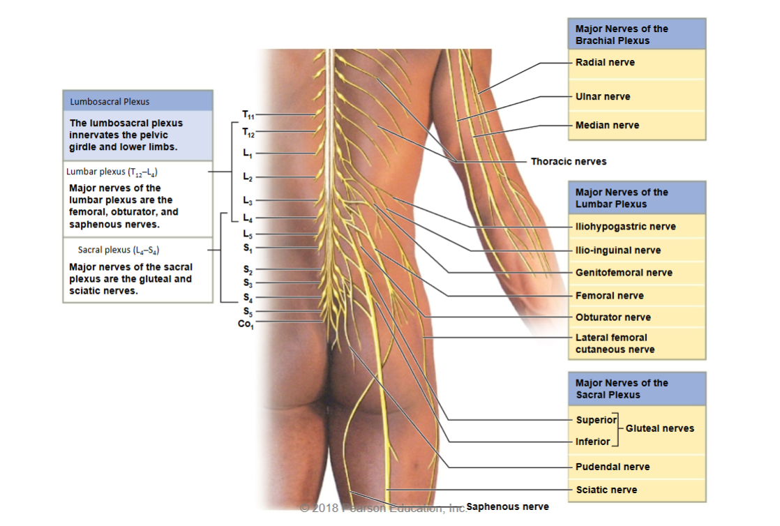
What are the major nerves of the lumbar and sacral plexuses, and what areas do they innervate?
Lumbar Plexus (T12-L4): Innervates pelvic girdle and lower limbs. Major nerves: Iliohypogastric, Ilio-inguinal, Genitofemoral, Femoral, Obturator, Lateral femoral cutaneous, and Saphenous.
Sacral Plexus (L4-S4): Innervates the gluteal region and lower limbs. Major nerves: Superior/Inferior Gluteal, Pudendal, Sciatic (longest)
These nerves control movement and sensation in the pelvic area and legs.
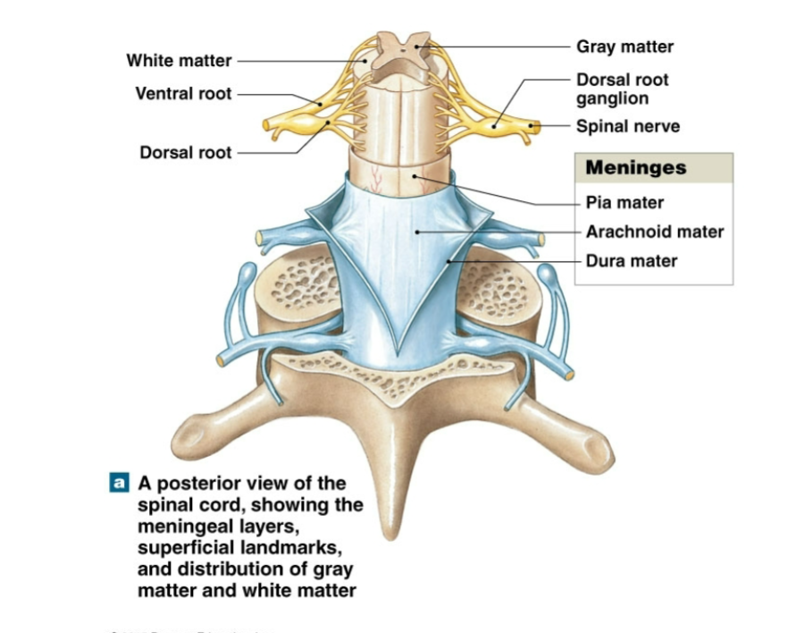
What are the three meningeal layers of the spinal cord, and what are their functions?
Dura Mater: The outermost layer providing strong protection for the spinal cord.
Arachnoid Mater: The middle layer, filled with cerebrospinal fluid to cushion the spinal cord.
Pia Mater: The innermost layer, closely adhering to the spinal cord and nourishing it.
Additionally, the image shows gray and white matter, along with the dorsal and ventral roots, which are essential for nerve signal transmission.
What are the key structures in a spinal cord cross-section?
Vertebral Body - Provides structural support for the spine.
Spinal Cord - Contains nerve fibers that transmit signals between the brain and the body.
Meninges - Protective layers around the spinal cord, epidural, including:
Dura Mater
Arachnoid Mater (spinal tap)
Pia Mater (closest to neurons and nerves)
Subarachnoid Space - Contains cerebrospinal fluid (CSF) that cushions the spinal cord.
Epidural Space - Contains adipose tissue, providing additional cushioning.
Dorsal Root Ganglion - Contains sensory neuron cell bodies.
Ventral Root of Spinal Nerve - Carries motor signals from the spinal cord to the body.
Dorsal Ramus - Supplies nerves to the back muscles and skin.
Ventral Ramus - Supplies nerves to the front and side of the body and limbs.
Rami Communicantes - Connect the spinal nerves to the autonomic nervous system.
Autonomic (Sympathetic) Ganglion - Part of the sympathetic nervous system, involved in "fight or flight" responses.
Dentate Ligament - Stabilizes the spinal cord within the vertebral column.
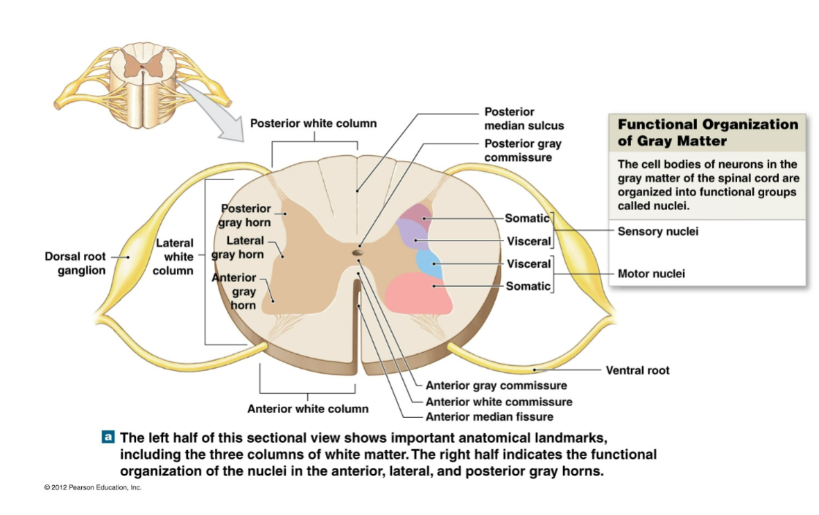
What are the main regions and functional groups in the cross-sectional anatomy of the spinal cord?
Regions of Gray Matter:
Posterior Gray Horn: Contains sensory nuclei (receives somatic and visceral sensory input).
Lateral Gray Horn: Present only in thoracic and lumbar segments; contains visceral motor nuclei.
Anterior Gray Horn: Contains motor nuclei for somatic motor control.
Regions of White Matter:
Posterior White Column: Carries ascending sensory information to the brain.
Lateral White Column: Contains both ascending sensory and descending motor tracts.
Anterior White Column: Carries both motor and sensory tracts, including those that control posture.
Additional Structures:
Dorsal Root Ganglion: Contains cell bodies of sensory neurons.
Ventral Root: Contains axons of motor neurons.
Anterior and Posterior Gray Commissures: Connects the two sides of the gray matter.
Anterior Median Fissure: A groove along the anterior (front) side of the spinal cord.
Posterior Median Sulcus: A groove along the posterior (back) side of the spinal cord.
How does a spinal nerve collect and deliver sensory information to the spinal cord?
Sensory Pathways:
1. Sympathetic Nerve: Carries sensory information from visceral organs.
2. Anterior Ramus: Carries sensory information from the ventrolateral body surface, body wall structures, and limbs.
3. Posterior Ramus: Carries sensory information from the skin and skeletal muscles of the back.
4. Posterior Root of Each Spinal Nerve: Transmits sensory information directly to the spinal cord's sensory nuclei.
Types of Sensations:
Somatic Sensations (Orange): From exteroceptors and proprioceptors, related to body surface and limbs.
Visceral Sensations (Purple): From interoceptors within visceral organs and the body wall.
How does a spinal nerve distribute motor commands to different parts of the body?
Motor Pathways:
1. Anterior Root: Contains axons of somatic and visceral motor neurons originating from motor nuclei in the spinal cord.
2. Spinal Nerve Formation: Spinal nerves form just lateral to the intervertebral foramen, where the anterior and posterior roots unite.
3. Posterior Ramus: Carries somatic and visceral motor fibers to the skin and skeletal muscles of the back.
4. Anterior Ramus: Supplies the ventrolateral body surface, structures in the body wall, and the limbs with motor commands.
5. Sympathetic Ganglion: Contains preganglionic and postganglionic fibers involved in the autonomic (sympathetic) nervous system.
Rami Communicantes:
White Ramus Communicans: Carries preganglionic visceral motor fibers to sympathetic ganglia (only present in T1-L2 regions).
Gray Ramus Communicans: Carries postganglionic fibers to glands, smooth muscles, and adipose tissue, associated with each spinal nerve.
Types of Motor Commands:
Somatic Motor Commands (Pink): Control skeletal muscles.
Visceral Motor Commands (Blue): Control smooth muscles, glands, and internal organs.
What are the five processing patterns of interneurons in neural circuits?
Divergence: A single neuron sends information to multiple neurons, amplifying the signal.
Convergence: Multiple neurons send information to a single neuron, allowing for summation of inputs.
Serial Processing: Information is relayed in a sequential, step-by-step manner from one neuron to another.
Parallel Processing: Information is processed simultaneously along multiple pathways.
Reverberation: Neurons form a feedback loop, which can maintain a signal within the circuit for extended processing or rhythmic activity.
What are divergence and convergence in neural processing, and can you provide an example of each?
Divergence:
Definition: A neural circuit that spreads stimulation to multiple neurons or neuronal pools in the CNS.
Function: Amplifies the signal, allowing a single neuron to influence multiple neurons.
Example: When sensory information from the eyes is transmitted to various brain regions, such as those for visual processing, attention, and coordination, enabling multiple areas to process visual input simultaneously.
Convergence:
Definition: A neural circuit that provides input to a single neuron from multiple sources.
Function: Allows for summation of inputs, enabling the neuron to integrate information from various sources.
Example: Motor neurons receive converging inputs from multiple sources, such as different parts of the brain and spinal cord, which helps coordinate complex movements by integrating signals from various regions.
What are serial processing, parallel processing, and reverberation in neural circuits? Provide an example of each.
Serial Processing:
Definition: A neural circuit where neurons or pools work sequentially, processing information step-by-step.
Example: Reflex arcs, like the knee-jerk reflex, where a sensory neuron activates a motor neuron in a straightforward sequence.
Parallel Processing:
Definition: A neural circuit in which neurons or pools process the same information simultaneously along multiple pathways.
Example: When you step on something sharp, your brain processes the sensation of pain, initiates a withdrawal reflex, and activates a vocal response (“ouch!”) simultaneously in different pathways.
Reverberation:
Definition: A positive feedback loop in a neural circuit that re-stimulates the circuit, maintaining activity.
Example: Breathing rhythm is maintained by reverberating circuits in the brainstem, which help keep the respiratory cycle ongoing without conscious effort.
Review Questions:
Which root receives sensory information?
a) Dorsal/Posterior
b) Ventral/Anterior
Which root sends out motor information?
a) Dorsal/Posterior
b) Ventral/Anterior
Shingles is a viral disease in which the chickenpox virus remains dormant in one region of the spinal cord. When activated, it causes painful rashes. Where would you expect to find the rashes?
Why would grasping my ring and pinky finger help after striking my "funny bone" on a table?
What are the four regions of the brain?
a) Dorsal/Posterior – The dorsal (posterior) root receives sensory information.
b) Ventral/Anterior – The ventral (anterior) root sends out motor information.
Rashes would appear in the dermatomes corresponding to the affected spinal nerve region, as shingles typically follows the path of the infected sensory nerves in the skin. (usually T3-T5)
Grasping the ring and pinky finger can help relieve "funny bone" pain because striking the "funny bone" compresses the ulnar nerve, which supplies sensation to these fingers. Holding them can stimulate the nerve and counteract the tingling or pain sensation by providing competing sensory input, helping to reduce discomfort.
Frontal, Parietal, Occipital, Temporal
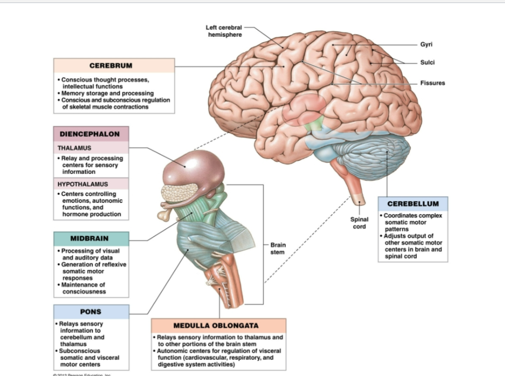
Identify the function of each brain region: Cerebrum, Diencephalon (Thalamus and Hypothalamus), Midbrain, Pons, Medulla Oblongata, and Cerebellum.
Cerebrum: Conscious thought processes, intellectual functions, memory storage and processing, regulation of skeletal muscle contractions. Complex motion and sensory
Diencephalon:
Thalamus: Relay and processing center for sensory information.
Hypothalamus: Controls emotions, autonomic functions, and hormone production.
Midbrain: Processes visual and auditory data, generates reflexive somatic motor responses, maintains consciousness.
Pons: Relays sensory information to cerebellum and thalamus, regulates subconscious somatic and visceral motor centers.
Medulla Oblongata: Relays sensory information to the thalamus, contains autonomic centers for regulating visceral functions.
Cerebellum: Coordinates complex motor coordination, adjusts somatic motor centers in the brain and spinal cord.
Brain stem: most primitive region, works to keep us alive.
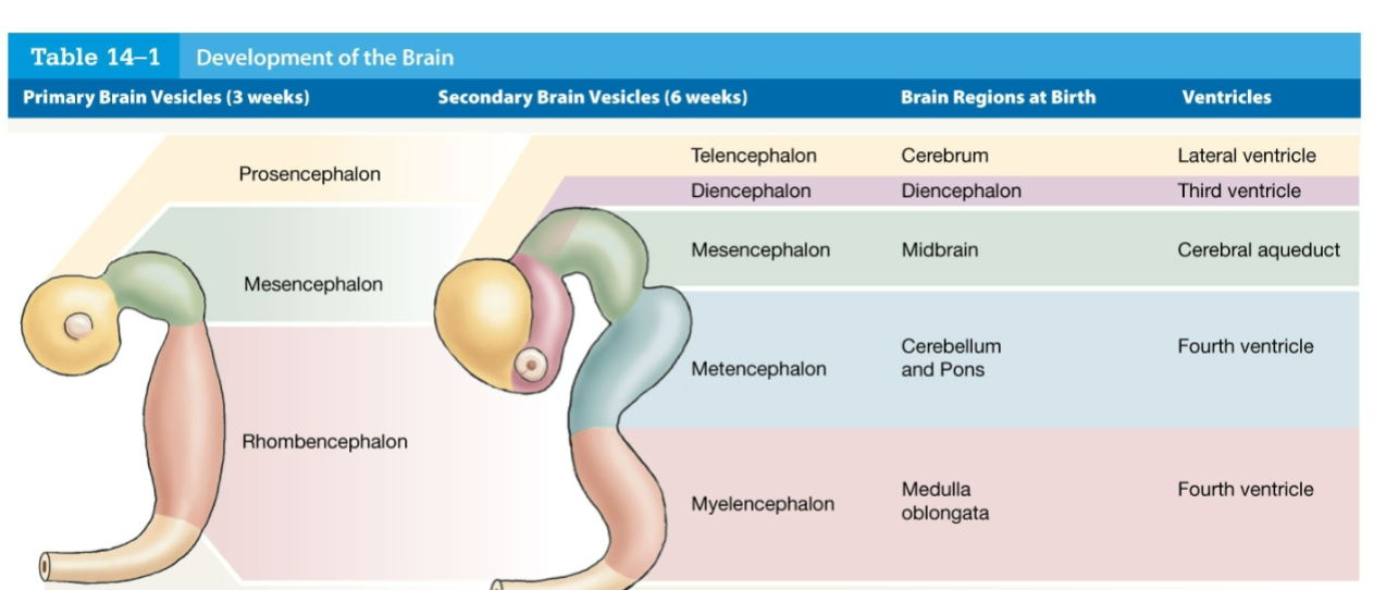
What are the primary and secondary brain vesicles, their corresponding brain regions at birth, and associated ventricles?
Primary Brain Vesicles (3 weeks):
Prosencephalon
Secondary Vesicles (6 weeks): Telencephalon and Diencephalon
Brain Regions at Birth: Cerebrum (Telencephalon), Diencephalon
Ventricles: Lateral ventricle (Telencephalon), Third ventricle (Diencephalon)
Mesencephalon
Secondary Vesicle: Mesencephalon
Brain Region at Birth: Midbrain
Ventricle: Cerebral aqueduct
Rhombencephalon
Secondary Vesicles: Metencephalon and Myelencephalon
Brain Regions at Birth: Cerebellum and Pons (Metencephalon), Medulla oblongata (Myelencephalon)
Ventricle: Fourth ventricle
What are the main functions of cerebrospinal fluid (CSF), its composition, and what cells produce it?
Main Functions of CSF:
Supporting the brain
Cushioning delicate neural structures
Transporting nutrients, chemical messengers, and wastes
Composition: Perfect for neurons
Produced by: Ependymal cells
What are the key characteristics of the Blood Brain Barrier (BBB) and the Blood-CSF Barrier?
Blood Brain Barrier (BBB):
Prevents large molecules from passing into brain tissue
Allows only small molecules to pass through
Comprised of tightly connected endothelial cells
Astrocytes in tight capillaries
Blood-CSF Barrier:
Formed by choroid cells with tight junctions
Has fenestrations in endothelium for selective transport
Separates blood from cerebrospinal fluid (CSF), maintaining the environment for neural activity
What factors are considered when developing a drug to cross the Blood Brain Barrier (BBB)?
Factors to Consider:
Size of the Molecule: Smaller molecules are more likely to pass through the BBB.
Hydrophobicity: Molecules with higher hydrophobicity are more likely to penetrate the BBB.
Molecular Similarity: The drug should resemble molecules that naturally cross the BBB to increase its chances of passing through.
What are the steps involved in a reflex arc?
Arrival of Stimulus and Activation of Receptor – A stimulus activates a receptor.
Activation of a Sensory Neuron – The receptor stimulates a sensory neuron, sending a signal through the dorsal root.
Information Processing in the CNS – The CNS processes the sensory information.
Activation of a Motor Neuron – A motor neuron is activated in response.
Response by Effector – The effector (e.g., muscle) responds, completing the reflex action.
most reflexes are in spinal cord, some in brain stem. knee jerk response goes straight to the spinal cord
How can reflexes be classified based on development, response, complexity of circuit, and processing site?
Development:
Innate Reflexes: Genetically determined
Acquired Reflexes: Learned
Response:
Somatic Reflexes: Control skeletal muscle contractions, include superficial and stretch reflexes
Visceral (Autonomic) Reflexes: Control actions of smooth and cardiac muscles, glands, and adipose tissue
Complexity of Circuit:
Monosynaptic: One synapse
Polysynaptic: Multiple synapses (two to several hundred)
Processing Site:
Spinal Reflexes: Processed in the spinal cord
Cranial Reflexes: Processed in the brain
Describe where the signals go in a monosynaptic reflex:
Name the 4 major regions of the brain and their origins
In a monosynaptic reflex, such as the patellar (knee-jerk) reflex, sensory neurons carry the signal from a receptor (like a muscle spindle) directly to the spinal cord. There, the sensory neuron synapses directly with a motor neuron in the spinal cord without involving an interneuron. The motor neuron then carries the signal back to the muscle, causing it to contract. Goes dorsal to ventral.
The four major regions of the brain are:
Cerebrum: Originates from the telencephalon; responsible for higher brain functions like thought and action.
Diencephalon: Originates from the diencephalon; includes structures like the thalamus and hypothalamus, involved in sensory information processing and autonomic functions.
Brainstem (which includes the midbrain, pons, and medulla oblongata): Originates from the mesencephalon and myelencephalon; controls essential functions such as breathing, heart rate, and sleep cycles.
Cerebellum: Originates from the metencephalon; involved in motor control, balance, and coordination.
What are the three main regions of the brainstem?
Medulla Oblongata – Controls vital functions such as heart rate, breathing, and blood pressure.
Pons – Assists with breathing, communication between different parts of the brain, and sensations such as hearing, taste, and balance.
Midbrain – Coordinates visual and auditory reflexes and controls eye movement.
Medulla Oblongata - Key Structures
Autonomic Centers:
Reticular Formation (being awake and alert)
Cardiovascular Centers (control heart)
Solitary Nucleus (higher order sensory)
Relay Stations:
Olivary Nucleus (green olive shape, input to cerebellum for motor)
Nucleus Cuneatus (upper body propio)
Nucleus Gracilis (lower body propio)
Lateral White Columns
Other Structures:
Olive
Pyramids
Decussation
Respiratory Rhythmicity Center - Function
Located in the medulla oblongata.
Controls the basic rhythm of breathing.
Contains two main groups:
Dorsal Respiratory Group (DRG): Primarily controls inspiration, especially during restful breathing.
Ventral Respiratory Group (VRG): Activates during forced breathing, controlling both inspiration and expiration.
Works with the pons to regulate breathing rate and depth based on body’s needs
The Pons - Key Regions and Functions
White Matter:
Descending Tracts: Carry motor commands from higher centers to nuclei of cranial or spinal nerves.
Ascending Tracts: Carry sensory information from brainstem nuclei to the thalamus.
Transverse Pontine Fibers: Interconnect cerebellar hemispheres.
Faster with Myelin
Gray Matter:
Apneustic and Pneumotaxic Centers: Adjust respiratory rhythmicity centers in the medulla oblongata.
Reticular Formation: Processes incoming sensations and outgoing motor commands.
Nuclei of Cranial Nerves V, VI, VII, VIII: Relay sensory information and issue somatic motor commands.
Other Nuclei/Relay Centers: Relay sensory and motor information to the cerebellum.
Overall isolated, has multiple breathing areas to help adapt to different activities.
Midbrain - Key Regions and Functions
Gray Matter:
Tectum (Roof):
Superior Colliculi: Integrates visual information with other sensory input; initiates reflex responses to visual stimuli. Light changes
Inferior Colliculi: Relays auditory information to the medial geniculate nuclei; initiates reflex responses to auditory stimuli.
Walls and Floor:
Substantia Nigra: Regulates activity in the basal nuclei. makes dopamine, black pigment.
Red Nucleus: Controls upper limb position and background muscle tone subconsciously.
Reticular Formation: Processes incoming sensations and outgoing motor commands; initiates involuntary responses to stimuli and helps maintain consciousness. Forms here first
White Matter:
Cerebral Peduncles: Connect the primary motor cortex with motor neurons in the brain and spinal cord; carry ascending sensory information to the thalamus.
Pineal gland has endocrine tissue and glandular tissue, makes melatonin.
Diencephalon Basics
Two Major Brain Regions:
Thalamus
Hypothalamus
Overall Job: Acts as a relay center, connecting the brain to the rest of the body.
Thalamus - Major Relay Center
Function: The thalamus serves as the brain's major relay center, processing and transmitting information to various areas. Talks to brain
Types of Information Relayed:
Sensory: Takes in sensory inputs before sending them to the cortex.
Motor: Communicates out motor signals between the basal nuclei, cerebellum, and motor cortex.
Memory: Integrates/recalls memory-related signals, connecting to the limbic system and cerebral cortex.
Connections to Brain Regions:
Limbic System: Influences emotional processing.
Frontal Lobes: Involved in higher cognitive functions.
Parietal Lobes and Cingulate Gyrus: Processes sensory integration.
Association Areas of Cortex: Supports complex processing.
Basal Nuclei - Key Structures
Main Components:
Caudate Nucleus: Involved in motor control, learning, and memory.
Putamen: Works with the caudate nucleus to regulate movement and influence learning.
Globus Pallidus: Regulates voluntary movement, working with the putamen and caudate nucleus.
Associated Structures:
Lateral Ventricle: Located above the basal nuclei.
Third Ventricle: Positioned near the thalamus, helps circulate cerebrospinal fluid.
Function: The basal nuclei play a critical role in coordinating and regulating voluntary movements, as well as influencing cognition and emotion.
base of cerebrum sits next/talks to thalamus
Disinhibition in Motor Control
Key Pathway:
Striatum (A): Receives excitatory inputs from the cortex, inhibiting the Globus Pallidus.
Globus Pallidus (B): Normally inhibits the VA/VL complex of the thalamus.
VA/VL Complex of Thalamus (C): When disinhibited (released from inhibition), it activates the motor cortex.
Motor Cortex (D): Sends excitatory signals to lower motor neurons, enabling movement.
Function: This disinhibition pathway allows the motor cortex to be activated indirectly, enabling precise and controlled voluntary movements.
Process Summary: Cortex ➔ Striatum inhibits ➔ Globus Pallidus ➔ Disinhibits Thalamus ➔ Activates Motor Cortex ➔ Movement.
In Parkinson’s disease, disinhibition is disrupted due to the degeneration of dopamine-producing neurons in the substantia nigra, a part of the basal ganglia.
Hypothalamus - Key Nuclei and Functions
Paraventricular Nucleus: Secretes oxytocin; stimulates smooth muscle contractions in the uterus and mammary glands.
Pre-optic Area: Regulates body temperature via control of autonomic centers in the medulla oblongata.
Autonomic Centers:
Sympathetic and Parasympathetic: Control heart rate and blood pressure.
Lateral Tuberal Nuclei: Produces hormones that control the anterior pituitary (adenohypophysis).
Mammillary Bodies: Involved in feeding reflexes like licking and swallowing.
Suprachiasmatic Nucleus: Regulates circadian rhythms.
Supra-optic Nucleus: Secretes antidiuretic hormone (ADH) to restrict water loss by the kidneys.
controls autonomic nervous system, fight or flight. controls endocrine system
Limbic System - Key Components and Functions
Diencephalic Components:
Thalamus: Anterior nuclei, involved in sensory relay and emotion/mood/memory.
Hypothalamus: Hypothalamic nuclei and mammillary bodies, regulate emotions and autonomic functions, stress.
Cerebral Components: (dont know)
Cingulate Gyrus: Superior portion of the limbic lobe, involved in emotion formation and processing.
Parahippocampal Gyrus: Inferior portion, associated with memory encoding and retrieval.
Dentate Gyrus: Posterior portion, plays a role in memory and spatial navigation.
Tracts:
Fornix: Connects hippocampus to the hypothalamus, crucial for memory.
Nuclei:
Amygdaloid Body: Processes emotions, especially fear and pleasure.
Hippocampus: Within dentate gyrus, essential for memory formation and spatial navigation.
Reticular formation is sometimes impacted by depression, struggle to stay awake. Pineal melatonin slows down reticular formation.
Amygdala - Key Functions and Connections
Primary Functions:
Initiates the fight or flight response.
Links emotion to memory, especially with intense or traumatic events.
Connections with Other Brain Regions:
Hypothalamus: Regulates emotions like rage, fear, pain, sexual arousal, and pleasure.
Thalamus: Supports recollective memory, helping process and retrieve emotionally charged memories.
Hippocampus - Key Functions
Primary Function: Essential for memory formation and spatial navigation.
Unique Feature: Contains stem cells that contribute to neurogenesis, the formation of new neurons, which is rare in the adult brain.
Shape and Name: Named after its resemblance to a seahorse
Stem cells become hippocample neurons.
patterns
Decussate, Contralateral, and Ipsilateral - Key Terms
Decussate:
Definition: To cross over from one side of the body or brain to the other.
Example: In the medulla oblongata, nerve fibers decussate, crossing from the left side of the body to the right side of the brain (or vice versa), leading to contralateral control.
Contralateral:
Definition: Refers to control or effects on the opposite side.
Example: The left side of the brain controls the right side of the body, which is contralateral. Injuries on one side of the brain can affect the opposite side of the body.
Ipsilateral:
Definition: Refers to control or effects on the same side.
Example: Reflex pathways may be ipsilateral, meaning a stimulus and response occur on the same side of the body.
Summary:
Decussate: Act of crossing over.
Contralateral: Opposite side control or function.
Ipsilateral: Same side control or function.
Which region of the brainstem is the most primitive?
Which region of the brainstem is most affected by changes in movement?
Which region of the brainstem is tied to our sleeping patterns?
What is the role of the diencephalon?
Medulla Oblongata - Controls essential functions like breathing, heart rate, and blood pressure.
nucleus gracilis, olivary nucleus, nucleus canatis, pons
Pons - Regulates sleep patterns, especially REM sleep, and controls the sleep-wake cycle. Midbrain with pineal gland
Diencephalon - Relay information to the brain, thalamus to brain, hypathalamus mostly to body
What are the main layers of the cerebellar cortex, and what are their functions?
Molecular Layer: The outermost layer, containing dendrites of Purkinje cells and axons of granule cells. It plays a role in processing input and coordination. Picks up signals.
Purkinje Cell Layer: Contains large Purkinje cells, which are the primary output neurons of the cerebellum. These cells send inhibitory signals to deep cerebellar nuclei, essential for motor coordination. Cerebellum only has this.
Granule Layer: The innermost layer, densely packed with granule cells, which receive input from mossy fibers and send excitatory signals to the molecular layer. This layer is crucial for processing sensory and motor information. Glial cells, axons.
cerebellum: instrument, basketball.
What are the key characteristics of Purkinje cells in the cerebellum?
GABA-ergic: Purkinje cells release GABA, an inhibitory neurotransmitter, which helps regulate and fine-tune motor control by inhibiting output from the cerebellar cortex.
Steriogenic: take cholesterol converts it
High Metabolism: Purkinje cells have a high metabolic rate, requiring significant energy to maintain their complex structure and active signaling functions. All those branches require a lot of energy.
Drunkeness ethanol
kills dendritic branches, and blocks activity - causing stumbling
Can you name the major brain regions and their primary functions as shown in the cerebral cortex diagram?
Primary Motor Cortex: Controls voluntary movements.
Primary Sensory Cortex: Processes tactile and proprioceptive information.
Parietal Lobe: Manages touch, spatial awareness, and body positioning.
Temporal Lobe: Processes auditory information, memory, and emotion.
Occipital Lobe: Responsible for visual processing.
Insula: Involved in taste, visceral sensation, and emotions.
Prefrontal Cortex: Handles decision-making and higher cognitive functions.
Gustatory Cortex: Processes taste.
Olfactory Cortex: Manages smell perception.
Speech Center (Broca’s Area): Controls speech production.
General Interpretive Area (Wernicke’s Area): Critical for language comprehension.
corpus callosum: connects left and right side
What are the main white matter structures in the brain, and what are their functions?
Corpus Callosum: Connects the left and right cerebral hemispheres, allowing communication between them.
Anterior Commissure: Connects parts of the temporal lobes across the hemispheres, facilitating communication for certain functions, including olfactory processing.
Posterior Commissure: Involved in the coordination of eye movements and certain reflexes.
Association Fibers: Connect different parts of the same hemisphere, enabling integrated processing within a single hemisphere.
Projection Fibers: Link the cerebral cortex with lower brain areas and the spinal cord, transmitting motor and sensory information.
white matter originates from thalamus signalling
Can you identify the main cortex areas in each lobe of the brain and their functions?
Frontal Lobe:
Primary Motor Cortex: Controls voluntary movements.
Premotor Cortex: Involved in planning movements. 30% of lower face.
Prefrontal Cortex: Responsible for decision-making, personality, and complex cognitive behavior.
Parietal Lobe:
Primary Somatosensory Cortex: Processes touch, temperature, and pain.
Somatosensory Association Cortex: Integrates sensory information for spatial awareness and perception.
Temporal Lobe:
Auditory Cortex: Processes sounds.
Auditory Association Area: Interprets sounds.
Olfactory Cortex: Processes smell information.
Occipital Lobe:
Visual Cortex: Processes visual information.
Visual Association Area: Interprets visual data, aiding in object recognition and spatial processing.
Insula:
Associated with taste, visceral sensations, and emotions.
Gustatory cortex: taste
What is the homunculus, and what do the somatosensory and motor strips represent?
Homunculus: A visual representation of the body mapped onto the brain's somatosensory and motor cortex, showing the relative amount of brain area dedicated to each body part.
Somatosensory Strip: Located in the parietal lobe (postcentral gyrus), it processes sensory information from various body parts. Larger regions (like lips and hands) indicate more sensory receptors and finer sensory discrimination.
Motor Strip: Located in the frontal lobe (precentral gyrus), it controls voluntary movement for each body part. Larger areas represent more motor control, as seen with hands and facial muscles.
Larger organ, more neurons.
What are the functions of the primary association areas in the brain?
Motor Areas:
Primary Motor Cortex: Controls voluntary movements.
Motor Association Area (Premotor Cortex): Plans and coordinates movements.
Frontal Eye Field: Controls voluntary eye movements.
Prefrontal Cortex:
Involved in decision-making, personality, and complex cognitive behavior. Uses recall on past events for decision making. Deja Vu
Broca’s Area: Located in the left hemisphere, responsible for speech production.
Sensory Areas and Related Association Areas:
Primary Somatosensory Cortex: Processes touch, pressure, and pain.
Sensory Association Area: Integrates sensory information for perception.
Wernicke’s Area: Located in the left hemisphere, important for language comprehension.
General Interpretation Area:
Primary Visual Cortex: Processes visual input.
Visual Association Area: Interprets visual information, helping with object recognition.
Primary Auditory Cortex: Processes sound.
Auditory Association Area: Interprets sounds, aiding in language and music comprehension.
Carl and Karen are each having issues with language following
a stroke. Carl is able to produce sounds, but everything he says
is unintelligible. For example, he would say things like “cat
housewife elephant.” Karen is unable to produce words, but she
can understand what you are saying to her. For example, she
will say “bji blad wah” in response to “how are you?”
• Which region is affected in each patient? Why?
Carl: Likely has damage to Wernicke’s area (typically in the left temporal lobe). Wernicke’s area is responsible for language comprehension, and damage here can cause Wernicke’s aphasia, where speech is fluent but lacks meaning and is unintelligible (e.g., "cat housewife elephant").
Karen: Likely has damage to Broca’s area (typically in the left frontal lobe). Broca’s area is responsible for speech production, and damage here can cause Broca’s aphasia, where comprehension is intact, but speech is non-fluent and difficult to produce (e.g., “bji blad wah”).
What are the primary functions associated with the left and right hemispheres of the brain? Hemispheric Lateralization
Left Brain:
Logic, Analysis, Organization
Administration, Math & Science
Knowledge/Facts, Detail-oriented thinking
Right Brain:
Emotion, Intuition, Spirituality
Interpersonal Skills, Art & Music
Belief, Big Picture thinking
What are the basic classifications and functions of cranial nerves?
Cranial Nerves enter the brainstem, similar to spinal cord nerves.
They can be:
Sensory only: Carry sensory information straight to the brain.
Motor only: Control muscles by carrying motor signals.
Mixed: Contain both sensory and motor fibers for dual functionality.
Each cranial nerve connects to a specific region in the brainstem or directly to the brain, serving various sensory and motor functions.
What is the function of Cranial Nerve I (Olfactory)? Where does it synapse?
Olfactory Nerve (I): Responsible for the sense of smell. It is purely sensory with no motor function. It synapses directly on the olfactory region of the cerebrum, allowing for quick processing of dangerous smells (e.g., chemicals or strong odors).
What is the function of Cranial Nerve II (Optic)? Where does it synapse?
Optic Nerve (II): Responsible for vision and is entirely sensory. The nerve travels to the diencephalon, including the optic chiasma and superior colliculi, where visual information is processed.
What is the function of Cranial Nerve III (Oculomotor)? Where does it synapse?
Oculomotor Nerve (III): Controls eye movements and pupil constriction. It is a motor nerve that innervates somatic (up, down, rolling) and visceral muscles of the eye. It synapses in the midbrain in response to visual regulation.
What does Cranial Nerve IV (Trochlear) control? Where does it synapse?
Trochlear Nerve (IV): Controls the superior oblique muscle, allowing for eye rolling movements. It is a motor nerve and the smallest cranial nerve. It synapses in the midbrain.
What is the function of Cranial Nerve V (Trigeminal)? Where does it synapse?
Trigeminal Nerve (V): A mixed nerve with both sensory and motor functions, controlling sensations around the eyes, nose, jaw, and tongue. It synapses in the pons, facilitating facial movement and sensory processing.
What does Cranial Nerve VI (Abducens) control? Where does it synapse?
Abducens Nerve (VI): Controls lateral eye movement (side to side). It is a motor nerve and synapses in the pons.
What is the function of Cranial Nerve VII (Facial)? Where does it synapse?
Facial Nerve (VII): A mixed nerve involved in facial expressions, taste (front 2/3 of the tongue), and proprioception of the face. Sensory information goes to the pons, and motor signals originate from the pons.
What functions are associated with Cranial Nerve VIII (Vestibulocochlear)? Where does it synapse?
Vestibulocochlear Nerve (VIII): Involved in hearing and balance. It synapses in the pons and medulla oblongata, connecting to the reticular formation and motor centers.
What is the function of Cranial Nerve IX (Glossopharyngeal)? Where does it synapse?
Glossopharyngeal Nerve (IX): Controls taste (back 1/3 of the tongue), swallowing, and monitoring blood pressure changes via the carotid artery. It synapses in the medulla oblongata.
What are the functions of Cranial Nerve X (Vagus)? Where does it synapse?
Vagus Nerve (X): A major nerve controlling visceral functions (e.g., heart rate, digestion). It is a mixed nerve and synapses in the medulla oblongata, helping regulate daily autonomic functions.
What does Cranial Nerve XI (Accessory) control? Where does it synapse?
Accessory Nerve (XI): Controls muscles in the neck and upper back, aiding in swallowing and breathing. It synapses in the medulla oblongata.
What is the main function of Cranial Nerve XII (Hypoglossal)? Where does it synapse?
Hypoglossal Nerve (XII): Controls tongue movement. It is a motor nerve, and damage to it may cause the tongue to deviate to one side. It synapses in the medulla oblongata.
Describe the flow of information in the sensory pathway starting from the stimulus to the response.
Depolarization of Sensory Receptor: A stimulus changes the membrane potential of a receptor cell, creating a graded potential.
Action Potential Generation: If the stimulus depolarizes the receptor cell to the threshold, an action potential is generated.
Propagation: Axons of sensory neurons transmit the action potential to the CNS, carrying information about the type of stimulus (touch, temperature, etc.).
CNS Processing: The CNS processes the information at relay synapses. Sensory data may be distributed across the spinal cord and brain centers.
Motor Pathways (Involuntary): Immediate reflexive responses are directed by spinal cord or brainstem centers before reaching the cerebral cortex.
Motor Pathways (Voluntary): Voluntary responses involve processing in the cerebral cortex, moderating the reflexive response.
Perception: Only a small percentage (about 1%) of sensory information is relayed to the somatosensory cortex for conscious perception.
What are receptive fields, and how do they relate to sensation?
Receptive Fields: Areas of the skin where sensory neurons detect stimuli. Each neuron has its own receptive field.
Receptive Field 1 vs. Receptive Field 2: The diagram shows two distinct receptive fields, which are areas of skin served by individual sensory neurons.
Free Nerve Endings: Located in the dermis, these structures detect changes in temperature, pain, and pressure.
Epidermis: The outermost layer of the skin, where some nerve endings extend to detect fine touch.
Concept: Smaller receptive fields allow for more precise detection of stimuli (higher sensitivity), while larger fields provide less detailed information.
List the main types of sensory receptors and describe their functions.
Nociceptors:
Detect pain caused by tissue damage, injury, or potentially harmful stimuli.
Found throughout the body, especially in the skin and internal organs.
Thermoreceptors:
Sense changes in temperature (heat and cold).
Help regulate body temperature and provide sensations of warmth or cold.
Chemoreceptors:
Respond to chemical stimuli, such as changes in blood pH, oxygen levels, or taste/smell molecules.
Involved in taste, smell, and monitoring blood chemistry.
Mechanoreceptors:
Detect mechanical changes like pressure, stretch, vibration, and touch.
Found in the skin, muscles, and inner ear (e.g., for balance and hearing).
What are baroreceptors, and where are they found in the body?
Definition: Baroreceptors are stretch-sensitive mechanoreceptors that detect changes in pressure within organs.
Locations and Functions:
Carotid Sinuses and Aortic Arch:
Detect blood pressure changes and relay information to cardiovascular and respiratory control centers.
Lungs:
Monitor lung expansion and help regulate respiratory rate via the respiratory rhythmicity centers.
Digestive Tract:
Sense the volume of contents and trigger reflexive movements along the tract.
Colon:
Detect the volume of fecal material, triggering the defecation reflex.
Bladder Wall:
Monitor the volume of urine, initiating the urination reflex when full.
What are chemoreceptors, and where are they located in the body?
Chemoreceptors: Sensory receptors that respond to chemical changes, such as changes in pH, CO₂, and O₂ levels.
Locations and Functions:
Medulla Oblongata (Respiratory Centers):
Sensitive to changes in pH and CO₂ in cerebrospinal fluid.
Trigger reflexive adjustments in the depth and rate of breathing.
Carotid Bodies:
Located at the bifurcation of the carotid arteries.
Detect changes in blood pH, CO₂, and O₂ levels.
Communicate via Cranial Nerve IX (Glossopharyngeal Nerve).
Adjust respiratory and cardiovascular activity.
Aortic Bodies:
Located along the aortic arch.
Monitor blood pH, CO₂, and O₂ levels.
Signal changes via Cranial Nerve X (Vagus Nerve).
Help regulate respiratory and cardiovascular responses
What are the two main somatosensory pathways, and what types of sensations do they process?
Dorsal Column System:
Processes fine touch, pressure, and proprioception (body position).
Pathway:
First-Order Neuron: Carries sensory information from receptors to the spinal cord.
Second-Order Neuron: Travels from the spinal cord to the brainstem (crosses over), then to the thalamus.
Third-Order Neuron: Sends the signal from the thalamus to the somatosensory cortex for processing.
Nucleus Gracilis (lower body) and Nucleus Cuneatus (upper body) are involved in processing.
Anterolateral (Spinothalamic) System:
Detects pain, temperature, and light touch.
Pathway:
First-Order Neuron: Transmits signals from the receptor to the spinal cord.
Second-Order Neuron: Crosses over in the spinal cord and ascends to the thalamus.
Third-Order Neuron: Relays the information from the thalamus to the somatosensory cortex.
Key Difference:
The dorsal column pathway is responsible for fine, detailed sensations, while the anterolateral system is associated with broader sensations like pain and temperature.
What are the main motor pathways in the nervous system, and what do they control?
Corticospinal Pathway:
Divided into the Lateral Corticospinal Tract and Anterior Corticospinal Tract.
Lateral Corticospinal Tract: Controls precise, voluntary movements of the limbs (crosses over at the medulla).
Anterior Corticospinal Tract: Controls movements of the trunk and proximal muscles (crosses over at the spinal cord level).
Lateral Pathway:
Includes the Rubrospinal Tract, which is involved in involuntary control of upper limb movements, particularly fine motor skills.
Medial Pathway:
Comprises several tracts:
Reticulospinal Tracts (Medial and Lateral): Regulate reflexive movements and posture.
Tectospinal Tract: Coordinates head and eye movements in response to visual and auditory stimuli.
Vestibulospinal Tract: Maintains balance and posture by controlling muscle tone in response to head movements.
Key Points:
The corticospinal pathway is responsible for voluntary motor control.
The lateral and medial pathways handle involuntary and reflexive movements for coordination and balance.
Describe the corticospinal pathway and its main components.
Corticospinal Pathway: Also known as the pyramidal tract, it is responsible for voluntary control of skeletal muscles.
Pathway Overview:
Primary Motor Cortex: Upper motor neurons originate here, represented by the motor homunculus, indicating control over specific body parts.
Corticobulbar Tract: Fibers descend from the cortex to the brainstem, connecting to cranial nerve nuclei to control facial muscles.
Cerebral Peduncle (Midbrain): Descending axons pass through this structure, forming part of the corticospinal tract.
Medulla Oblongata: Axons cross to the opposite side (decussate) at the pyramids of the medulla.
Lateral Corticospinal Tract: Controls fine motor movements of the limbs (crosses at the medulla).
Anterior Corticospinal Tract: Controls trunk muscles (crosses at the spinal cord level).
Spinal Cord: Lower motor neurons in the anterior horn transmit signals to skeletal muscles for movement.
Key Points:
Upper Motor Neurons: Start in the cortex and descend through the brainstem/spinal cord.
Lower Motor Neurons: Connect to skeletal muscles, carrying out motor commands.
Decussation: Crossing of axons allows one hemisphere of the brain to control the opposite side of the body.
Describe the pathway of a sensory signal from perception to response.
Sensory Signal Perception:
Example: Touching a hot surface (pain/thermal sensation).
Nociceptors (pain receptors) in the skin detect the intense heat stimulus.
Transmission to Spinal Cord:
The first-order sensory neuron carries the signal from the nociceptor through peripheral nerves to the spinal cord.
The signal enters the dorsal root of the spinal cord and synapses with a second-order neuron.
Pathway to the Brain:
The second-order neuron decussates (crosses over) in the spinal cord and ascends through the anterolateral (spinothalamic) pathway.
It reaches the thalamus, where it synapses with a third-order neuron.
Processing in the Brain:
The third-order neuron transmits the signal from the thalamus to the somatosensory cortex for processing and perception of the pain sensation.
Motor Response:
The brain sends a response signal via upper motor neurons in the corticospinal tract.
The signal travels down through the brainstem, decussates (if in the lateral corticospinal tract), and reaches the spinal cord.
Effector Activation:
The lower motor neuron in the anterior horn of the spinal cord carries the signal to the effector muscle (e.g., biceps).
The muscle contracts, causing you to withdraw your hand from the hot surface.
What are the main divisions of the nervous system, and what are their functions?
Central Nervous System (CNS):
Comprises the brain and spinal cord.
Processes and integrates sensory information, coordinates motor output, and is responsible for higher functions like reasoning and memory.
Peripheral Nervous System (PNS):
Includes all the nerves outside the CNS.
Divided into:
Somatic Nervous System: Controls voluntary movements of skeletal muscles.
Autonomic (Visceral) Nervous System: Regulates involuntary functions like heart rate, digestion, and respiration.
Sympathetic Division: Activates 'fight or flight' responses (e.g., increased heart rate).
Parasympathetic Division: Promotes 'rest and digest' activities (e.g., decreased heart rate).
Enteric Nervous System (ENS):
Often called the 'second brain,' it controls the functions of the gastrointestinal tract independently of the CNS.
Intracardiac Nervous System:
Specialized nerve network within the heart, helping to regulate cardiac function and response to changes in blood flow and pressure.
Compare the somatic and autonomic nervous system pathways.
Somatic Nervous System (SNS):
Controls voluntary movements of skeletal muscles.
Pathway:
Upper Motor Neurons: Start in the primary motor cortex and descend through the brainstem.
Lower Motor Neurons: Located in the spinal cord or brainstem; send signals directly to skeletal muscles.
Effectors: Skeletal muscles.
Function: Precise, conscious control of muscle movements.
Autonomic Nervous System (ANS):
Regulates involuntary functions of visceral organs.
Pathway:
Preganglionic Neurons: Originate in the brainstem or spinal cord; project to autonomic ganglia.
Ganglionic Neurons: Located in autonomic ganglia; send signals to visceral effectors.
Effectors: Smooth muscle, cardiac muscle, glands, and adipocytes.
Divisions:
Sympathetic Division: Prepares the body for 'fight or flight' responses (e.g., increased heart rate).
Parasympathetic Division: Promotes 'rest and digest' activities (e.g., slowed heart rate).
Key Differences:
Somatic Pathway: Single neuron from CNS to skeletal muscle.
Autonomic Pathway: Two-neuron chain (preganglionic and ganglionic) to visceral effectors.
What does the sympathetic division innervate during a 'fight or flight' response, and what might it inhibit?
Innervation:
Heart: Increases heart rate and contractility to pump more blood.
Lungs: Dilates bronchioles for increased airflow.
Skeletal Muscles: Increases blood flow to provide more oxygen and nutrients.
Adrenal Glands: Stimulates the release of adrenaline (epinephrine) to enhance the body's response.
Pupils: Dilate to enhance vision.
Sweat Glands: Activates sweat production for temperature regulation.
Inhibition:
Digestive System: Reduces blood flow and slows down digestion (inhibits peristalsis).
Urinary System: Relaxes the bladder, inhibiting urination.
Salivary Glands: Decreases saliva production, leading to dry mouth.
Key Concept:
The sympathetic division prepares the body for immediate physical activity, prioritizing survival by diverting energy away from non-essential functions like digestion.
Describe the structure and innervation of the sympathetic division of the autonomic nervous system.
Sympathetic Division: Also called the thoracolumbar division, as it originates from the thoracic (T1-T12) and lumbar (L1-L2) regions of the spinal cord.
Structure:
Preganglionic Fibers: Arise from the spinal cord and enter the sympathetic chain ganglia, which run parallel to the spinal cord.
Postganglionic Fibers: Extend from the ganglia to target organs.
Key Ganglia and Nerves:
Cervical Ganglia: Innervate the eye (dilates pupils) and salivary glands.
Cardiac and Pulmonary Plexuses: Innervate the heart (increase heart rate) and lungs (bronchodilation).
Celiac Ganglion: Innervates the stomach, liver, gallbladder, spleen, and pancreas, reducing digestive activity.
Superior and Inferior Mesenteric Ganglia: Control functions of the intestines, decreasing motility and secretion.
Adrenal Medulla: Directly stimulated by preganglionic fibers to release adrenaline (epinephrine) into the bloodstream.
Functions:
Activates 'fight or flight' responses: Increases heart rate, dilates bronchioles, and mobilizes energy.
Inhibits non-essential functions: Slows digestion and urination.
Key Points:
The sympathetic chain ganglia allow the widespread, simultaneous activation of multiple organs.
The splanchnic nerves (greater, lesser, and lumbar) play a critical role in conveying signals to visceral organs.
What is the sympathetic chain ganglia, and what is its function in the sympathetic nervous system?
Sympathetic Chain Ganglia: A series of interconnected autonomic ganglia located on both sides of the spinal cord. These ganglia form part of the sympathetic division of the autonomic nervous system (ANS).
Components:
Preganglionic Neurons: Originate from the spinal cord and travel via the white ramus communicans to the sympathetic chain ganglia.
Ganglionic Neurons (Postganglionic Fibers): Emerge from the ganglia via the gray ramus communicans and innervate target organs.
Functions:
Visceral Effectors: Innervates organs like the heart, lungs, and blood vessels, initiating 'fight or flight' responses (e.g., increased heart rate, bronchodilation).
Innervation Patterns:
Some postganglionic fibers exit via spinal nerves to innervate skin, blood vessels, and sweat glands.
Other fibers form splanchnic nerves that target visceral organs in the thoracic cavity.
Key Points:
The white ramus communicans carries preganglionic fibers into the ganglia (myelinated).
The gray ramus communicans carries postganglionic fibers out of the ganglia (unmyelinated).
Both the left and right sides of the body have a sympathetic chain, allowing for coordinated activation of the sympathetic response.
What are collateral ganglia, and what role do they play in the sympathetic nervous system?
Collateral Ganglia: Autonomic ganglia located outside the sympathetic chain, involved in innervating visceral organs in the abdominopelvic cavity.
Structure and Pathway:
Preganglionic Fibers: Travel from the spinal cord through the white ramus communicans and enter the sympathetic chain. Instead of synapsing there, they continue as splanchnic nerves to reach the collateral ganglia.
Ganglionic Neurons: Synapse within the collateral ganglia and give rise to postganglionic fibers.
Postganglionic Fibers: Extend from the collateral ganglia to innervate visceral organs such as the intestines, liver, stomach, kidneys, and pancreas.
Key Collateral Ganglia:
Celiac Ganglion: Innervates the stomach, liver, gallbladder, pancreas, and spleen.
Superior Mesenteric Ganglion: Innervates the small intestine and parts of the large intestine.
Inferior Mesenteric Ganglion: Innervates the distal large intestine, kidneys, bladder, and reproductive organs.
Functions:
Regulate digestion, urinary functions, and blood flow to abdominal organs.
Provide targeted control of visceral organs during 'fight or flight' responses, slowing digestion and increasing blood flow to muscles.
What is the role of the adrenal medullae in the sympathetic nervous system?
Adrenal Medullae: Specialized structures of the sympathetic nervous system located in the adrenal glands, sitting on top of the kidneys.
Pathway:
Preganglionic Fibers: Travel directly from the spinal cord to the adrenal medullae.
Endocrine Cells (Specialized Ganglionic Neurons): Act as postganglionic neurons but function as endocrine cells.
Function:
Instead of synapsing with an effector organ, the endocrine cells secrete neurotransmitters directly into the bloodstream.
Neurotransmitters Released:
Epinephrine (Adrenaline): About 80% of the secretion, increases heart rate, dilates airways, and mobilizes energy.
Norepinephrine (Noradrenaline): About 20%, enhances the 'fight or flight' response by increasing blood pressure and blood flow to muscles.
Key Points:
The adrenal medullae function as a direct extension of the sympathetic nervous system, amplifying the 'fight or flight' response by releasing hormones that affect the entire body.
The release of epinephrine and norepinephrine causes prolonged systemic effects, such as increased alertness, heart rate, and metabolic rate.
What are sympathetic varicosities, and what role do they play in neurotransmitter release?
Sympathetic Varicosities:
Definition: Swellings along the length of postganglionic fibers in the sympathetic nervous system that release neurotransmitters.
Found in the autonomic nerve fibers that innervate smooth muscle cells.
Function:
Neurotransmitter Release: Varicosities contain vesicles filled with norepinephrine (NE). These vesicles release NE into the extracellular space, affecting multiple smooth muscle cells simultaneously.
Wide Distribution: Unlike typical synapses, varicosities allow for a more diffuse release of neurotransmitters, enhancing the spread of the sympathetic response.
Structure:
Postganglionic Fiber: An unmyelinated nerve fiber that forms varicosities.
Mitochondria: Present in varicosities, providing energy for neurotransmitter release.
Schwann Cell Cytoplasm: Surrounds the nerve fiber, offering structural support.
Key Points:
Varicosities enable broad and simultaneous activation of smooth muscles, enhancing the 'fight or flight' response.
The release of norepinephrine by varicosities causes vasoconstriction, increased heart rate, and other sympathetic effects.
What does the parasympathetic division innervate during 'rest and digest,' and what might it inhibit?
Parasympathetic Division:
Promotes the 'rest and digest' state, conserving energy and supporting bodily maintenance activities.
Innervates:
Heart: Decreases heart rate and contractility.
Digestive System: Stimulates digestive activity (increases peristalsis and secretion of digestive enzymes).
Lungs: Constricts bronchioles to reduce airflow (less oxygen demand at rest).
Salivary Glands: Increases saliva production for digestion.
Bladder: Contracts the bladder for urination.
Eyes: Constricts pupils and accommodates the lens for near vision.
Inhibits:
Sympathetic Activity: Counters 'fight or flight' responses by reducing the effects of the sympathetic division.
Adrenal Glands: Decreases secretion of adrenaline, lowering overall stress response.
Metabolic Processes: Reduces glucose release from the liver and slows down the breakdown of energy stores.
Key Concept:
The parasympathetic division supports relaxation, digestion, and energy conservation, opposing the sympathetic 'fight or flight' responses.
What are the main components of the parasympathetic division, and which organs does it innervate?
Parasympathetic Division: Also known as the craniosacral division because it originates from cranial nerves and sacral segments of the spinal cord.
Key Components:
Cranial Nerves:
Oculomotor Nerve (III): Innervates the eye muscles, causing pupil constriction and lens accommodation.
Facial Nerve (VII): Controls the lacrimal (tear) glands and salivary glands.
Glossopharyngeal Nerve (IX): Stimulates the parotid salivary gland for increased saliva production.
Vagus Nerve (X): The primary nerve of the parasympathetic division, innervating the heart, lungs, digestive organs (stomach, liver, pancreas, intestines), and more.
Pelvic Nerves:
Arise from the sacral spinal cord (S2-S4).
Innervate the lower intestines, bladder, and reproductive organs (e.g., uterus, penis, scrotum).
Functions:
Heart: Slows heart rate.
Lungs: Constricts bronchioles, reducing airflow.
Digestive Organs: Stimulates digestion, increases motility, and enhances secretion of digestive enzymes.
Urinary System: Promotes bladder contraction for urination.
Key Concept:
The parasympathetic division supports 'rest and digest' activities, conserving energy and promoting bodily functions like digestion and excretion.
What are the key differences between the sympathetic and parasympathetic pathways, and which neurotransmitters do they use?
Sympathetic Pathway:
Origin: Preganglionic neurons arise from the thoracic and lumbar spinal cord.
Preganglionic Fiber: Releases acetylcholine (ACh) at the sympathetic ganglion.
Postganglionic Fiber: Usually releases norepinephrine (NE), targeting organs like the heart, lungs, and blood vessels.
Adrenal Medulla: Acts as a modified sympathetic ganglion, releasing epinephrine (adrenaline) and norepinephrine into the bloodstream, amplifying the 'fight or flight' response.
Parasympathetic Pathway:
Origin: Preganglionic neurons arise from the brainstem (cranial nerves) and sacral spinal cord.
Preganglionic Fiber: Releases acetylcholine (ACh) at the parasympathetic ganglion.
Postganglionic Fiber: Also releases acetylcholine (ACh), targeting organs like the heart, digestive organs, and bladder.
Effect: Promotes 'rest and digest' responses, such as reducing heart rate and stimulating digestion.
Key Differences:
Neurotransmitters:
Sympathetic: Primarily uses norepinephrine (NE) and sometimes epinephrine for widespread activation.
Parasympathetic: Uses acetylcholine (ACh) for localized effects.
Functions:
Sympathetic: Activates 'fight or flight' responses.
Parasympathetic: Activates 'rest and digest' responses.
What are the types of adrenergic alpha receptors, and how do they differ in their effects?
Adrenergic Receptors: Respond to epinephrine and norepinephrine.
Alpha 1 Receptors:
Sensitive to: Norepinephrine.
Effect: Excitatory; causes vasoconstriction, raising blood pressure.
Alpha 2 Receptors:
Sensitive to: Both epinephrine and norepinephrine.
Effect: Inhibitory; found on preganglionic fibers, reduces signaling in postganglionic fibers.
Describe the types of adrenergic beta receptors and their effects.
Beta 1 Receptors:
Sensitive to: Epinephrine and norepinephrine.
Effect: Excitatory; increases heart rate and contractility.
Beta 2 Receptors:
Sensitive to: Epinephrine.
Effect: Inhibitory; relaxes smooth muscles in the lungs, dilating airways.
Beta 3 Receptors:
Location: Found in adipose tissue.
Effect: Increases fat metabolism.
Describe the types of cholinergic receptors found in the parasympathetic division and their effects.
Cholinergic Receptors in Parasympathetic Division:
Nicotinic Receptors: Found at the synapse between pre- and postganglionic neurons; excitatory.
Muscarinic Receptors: Located on target organs; can be excitatory or inhibitory depending on the response needed.
Neurotransmitter: Acetylcholine (ACh) is the main neurotransmitter for both preganglionic and postganglionic neurons.
What are nitroxidergic receptors, and where are they found?
Nitroxidergic Receptors:
Found in skeletal muscles and the brain.
Function: Relax tissues to increase blood flow.
Part of the sympathetic division of the autonomic nervous system.
How do cholinergic receptors function in the sympathetic division?
Cholinergic Receptors:
Respond to acetylcholine (ACh).
Nicotinic Receptors: Activated by preganglionic neurons using acetylcholine, affecting both sympathetic and parasympathetic divisions.
Effect in Sympathetic Division: Stimulates sweat gland secretion.