Histology Lecture #16: The Cardiovascular System
 Call Kai
 Learn
Learn Practice Test
 Spaced Repetition
 Match
Match Flashcards
 Knowt Play
1/155

There's no tags or description
Looks like no tags are added yet.
Name | Mastery | Learn | Test | Matching | Spaced | Call with Kai | Chat |
|---|
No analytics yet
Send a link to your students to track their progress
156 Terms
The heart propels blood
What is the function of the heart in the cardiovascular system?
Arteries are efferent vessels that branch off and have thick walls.
What are the characteristics of arteries?
Capillaries are the smallest vessels connecting arteries and veins; they are the sites of exchange between blood and tissues and form a thin, permeable microvasculature network.
What are capillaries and what is their function?
Veins are afferent vessels formed by the merging of venules; they carry blood back to the heart to be pumped again.
What are veins and what do they do?
1. Endocardium
2. Myocardium
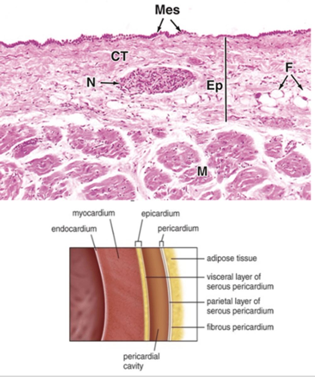
3. Epicardium
What are the three layers of the heart wall?
Endothelium, connective tissue, and smooth muscle tissue.
What tissues make up the endocardium? (3)
Cardiac muscle tissue and connective tissue.
What tissues make up the myocardium? (2)
Mesothelium, loose connective tissue, and abundant blood vessels and nerves.
What tissues make up the epicardium? (2)
- Thin endothelium
- Loose connective tissue
What type of epithelium and connective tissue lines the Inner layer of the endocardium?
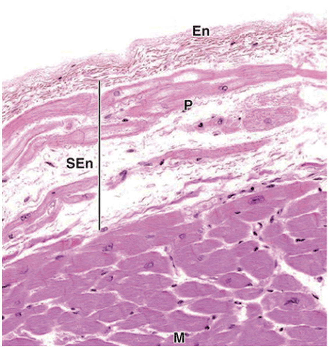
The middle myoelastic layer; it contains smooth muscle and connective tissue.
What is the middle layer of the endocardium called, and what does it contain?
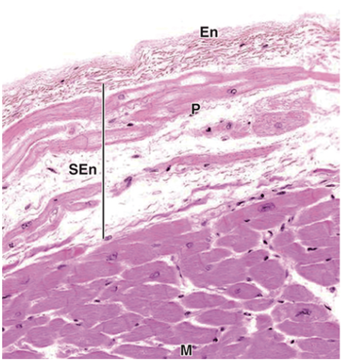
Connective tissue continuous with the connective tissue of the myocardium.
What is the subendothelial layer of the endocardium composed of?
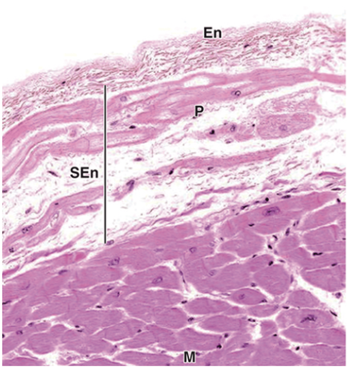
Contains the conducting system of the heart (ex. SA, AV, Bundle of his, purkinje fibers)
What special structure does the endocardium contain?***
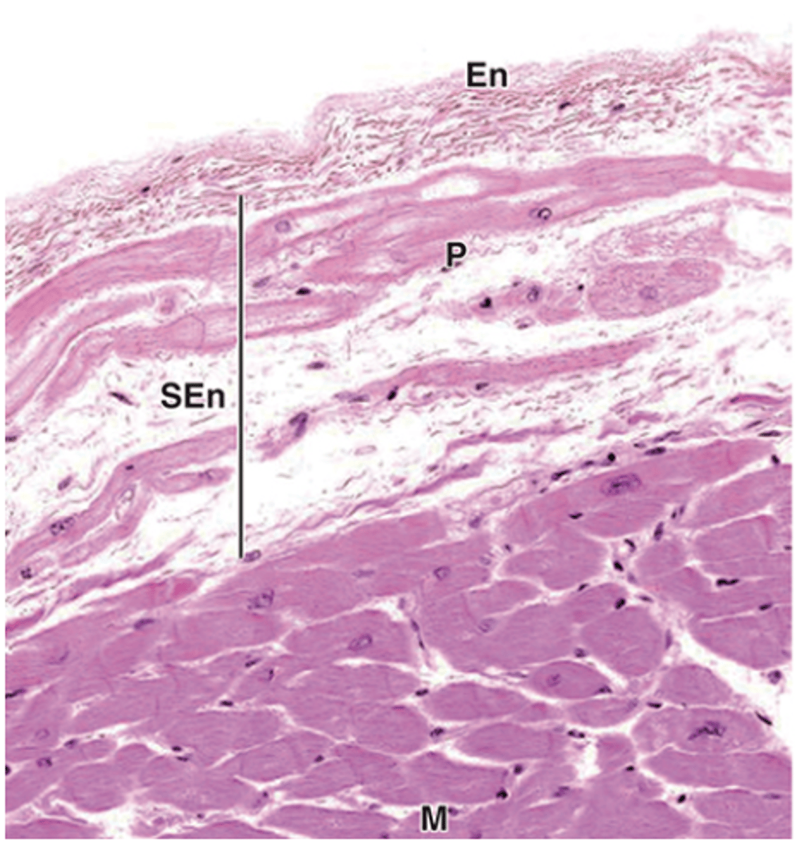
Atrial Purkinje fibers are closer to the endothelium in the ventricle
Where are atrial Purkinje fibers located relative to the endothelium?
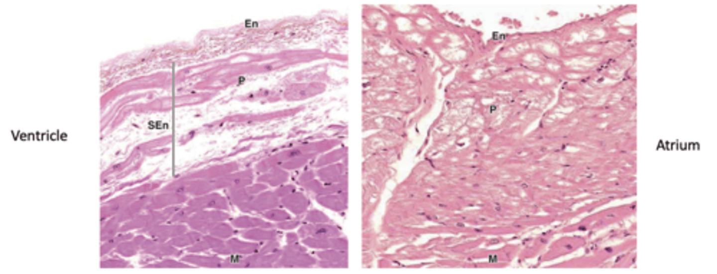
Atrial Purkinje fibers intermingle with the contractile fibers of the myocardium.
How do atrial Purkinje fibers differ in their relationship with contractile fibers compared to ventricular Purkinje fibers?
Modified cardiac muscle fibers specalized in conduction
What are Purkinje fibers?
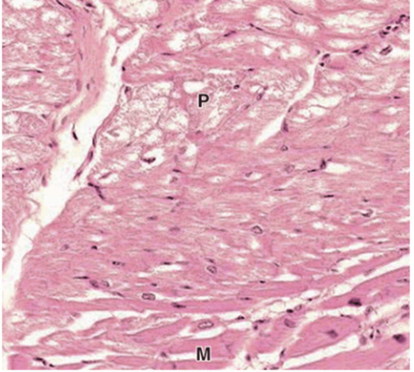
They are joined by intercalated discs.
How are Purkinje fibers connected to each other?
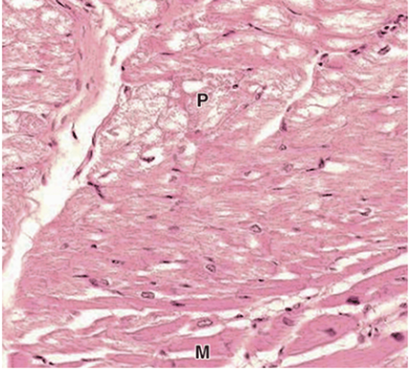
•Cytoplasm filled with glycogen
What fills the cytoplasm of Purkinje fibers?
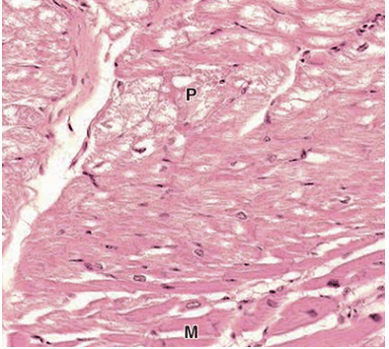
They are displaced peripherally.
Where are the myofibrils located in Purkinje fibers?
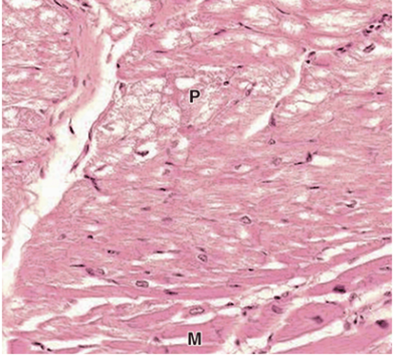
They stain paler.
How do Purkinje fibers appear compared to contractile cardiac muscle fibers?
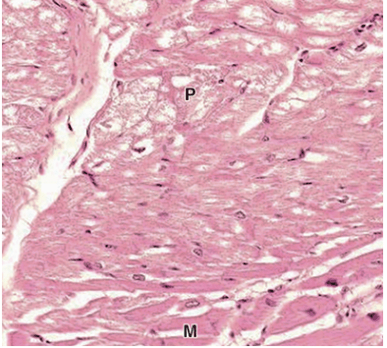
The myocardium.
What is the main functional component of the heart?
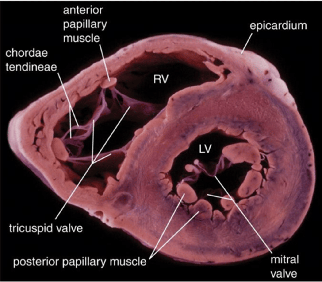
Cardiac muscle tissue with a spiral arrangement of fibers.
What type of tissue makes up the myocardium?
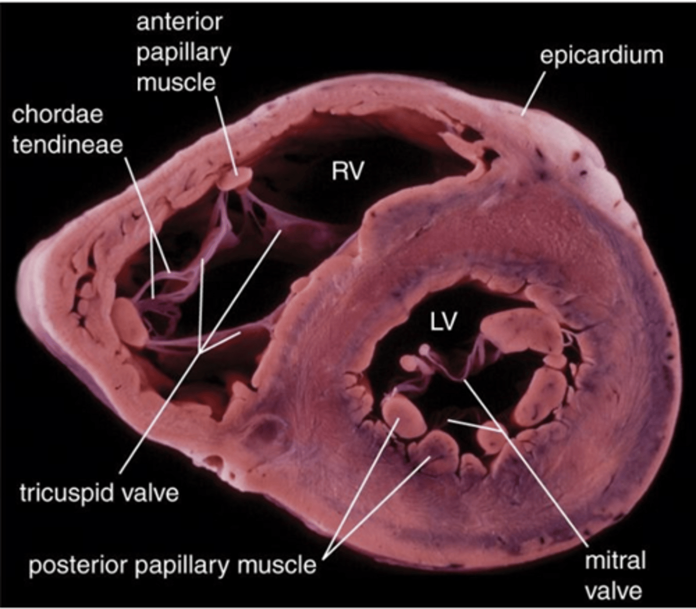
It is thinner in the atria, thicker in the ventricles, and thickest in the left ventricle.
How does the thickness of the myocardium vary in different chambers of the heart?
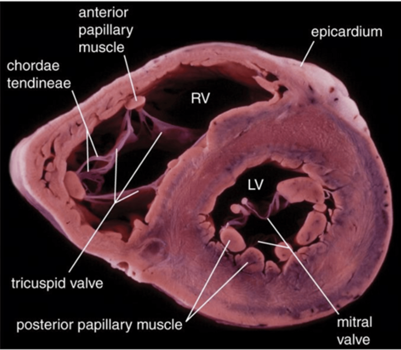
Connective tissue.
Besides cardiac muscle, what other type of tissue is found in the myocardium?
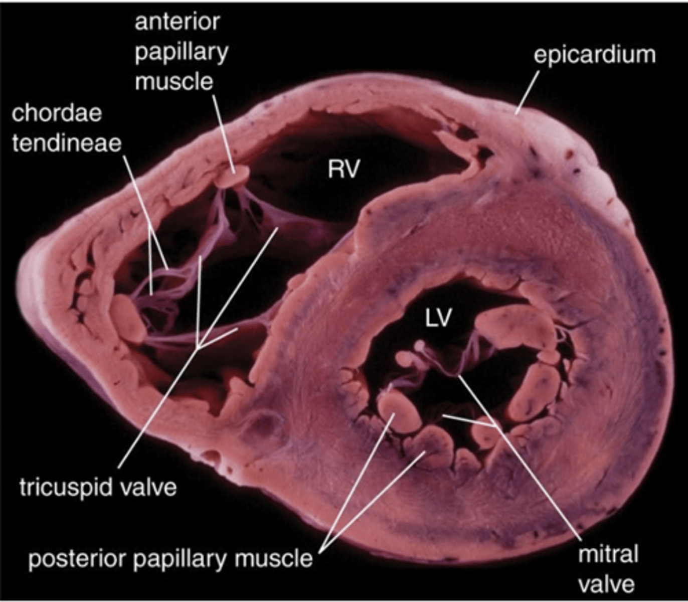
Simple squamous mesothelium and loose connective tissue
What type of epithelium forms the surface of the epicardium and what types of tissue are found?
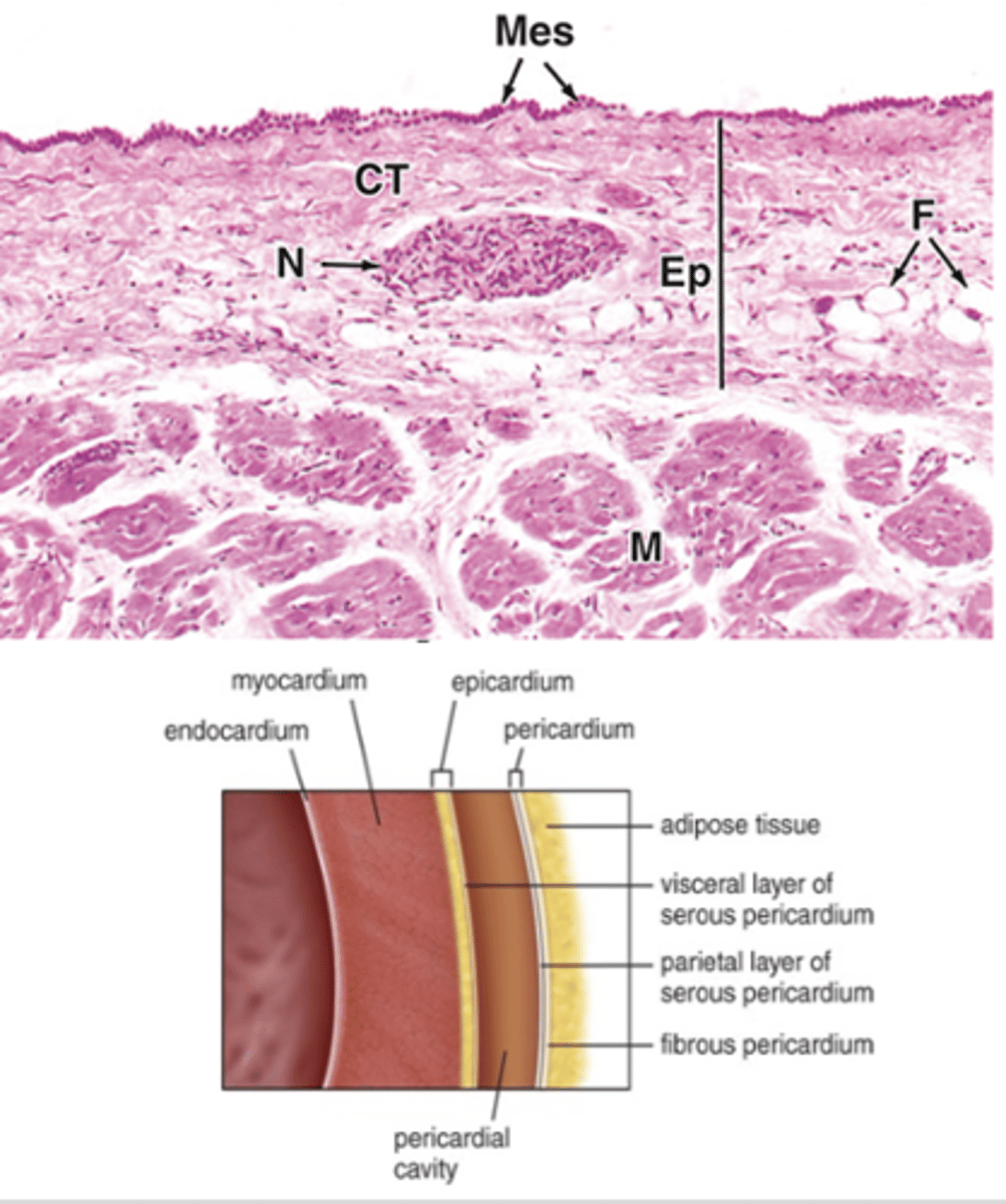
Blood vessels and nerves.
What structures are contained within the epicardium?
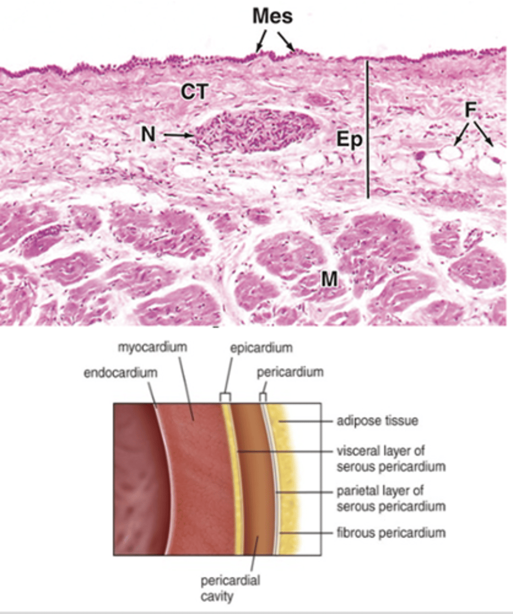
Reflected back at great vessels
Where is the epicardium reflected back?
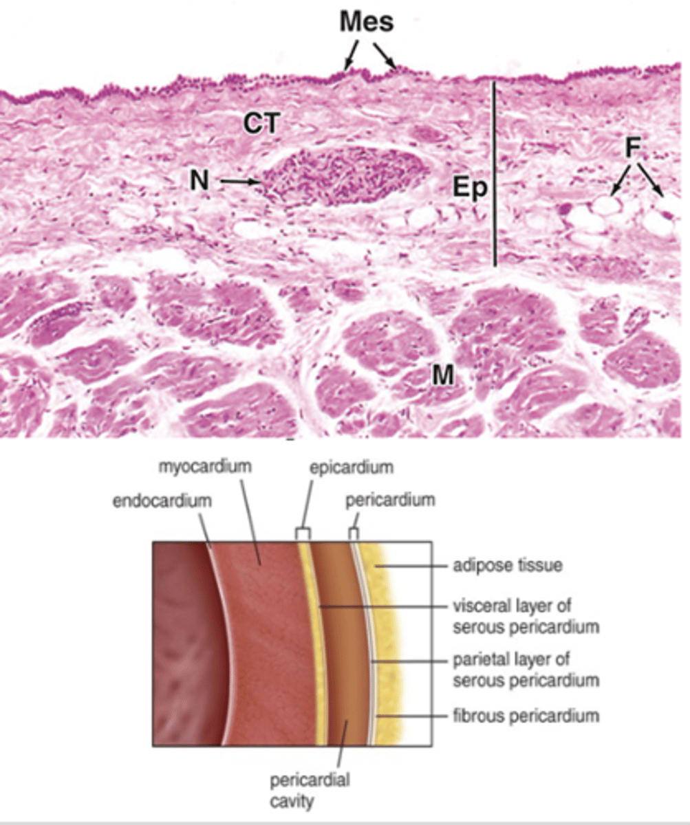
They are continuous with each other.
How are the parietal pericardium and epicardium related?
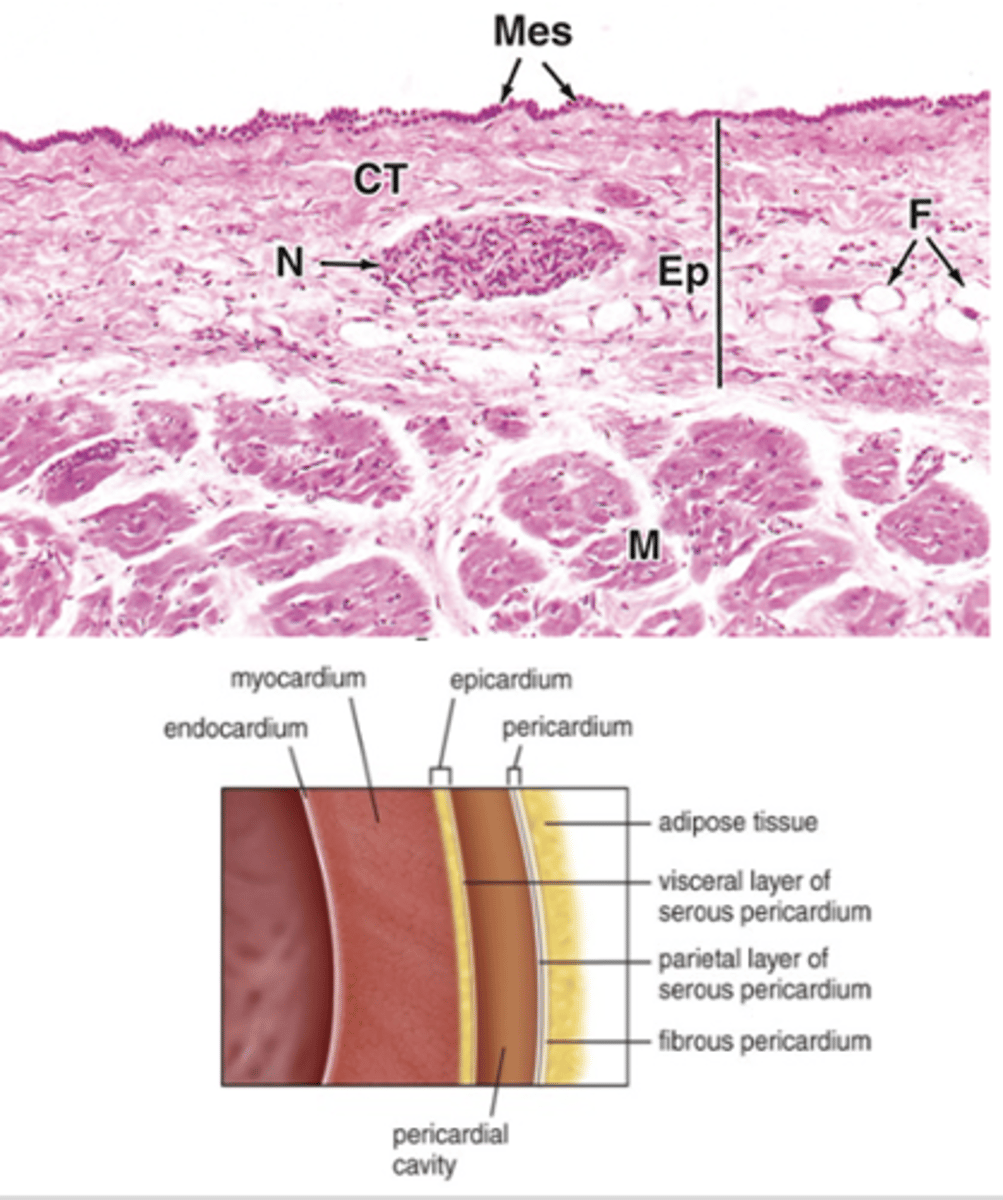
The pericardial cavity, lined by mesothelium of the pericardial layers.
What lies between the parietal pericardium and the epicardium?
Dense irregular connective tissue.
What type of connective tissue makes up the cardiac skeleton?***
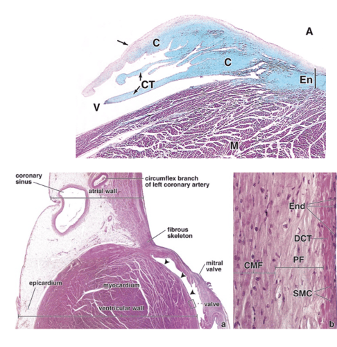
In the interatrial and interventricular septa, and it surrounds the valves.
Where is the cardiac skeleton located?
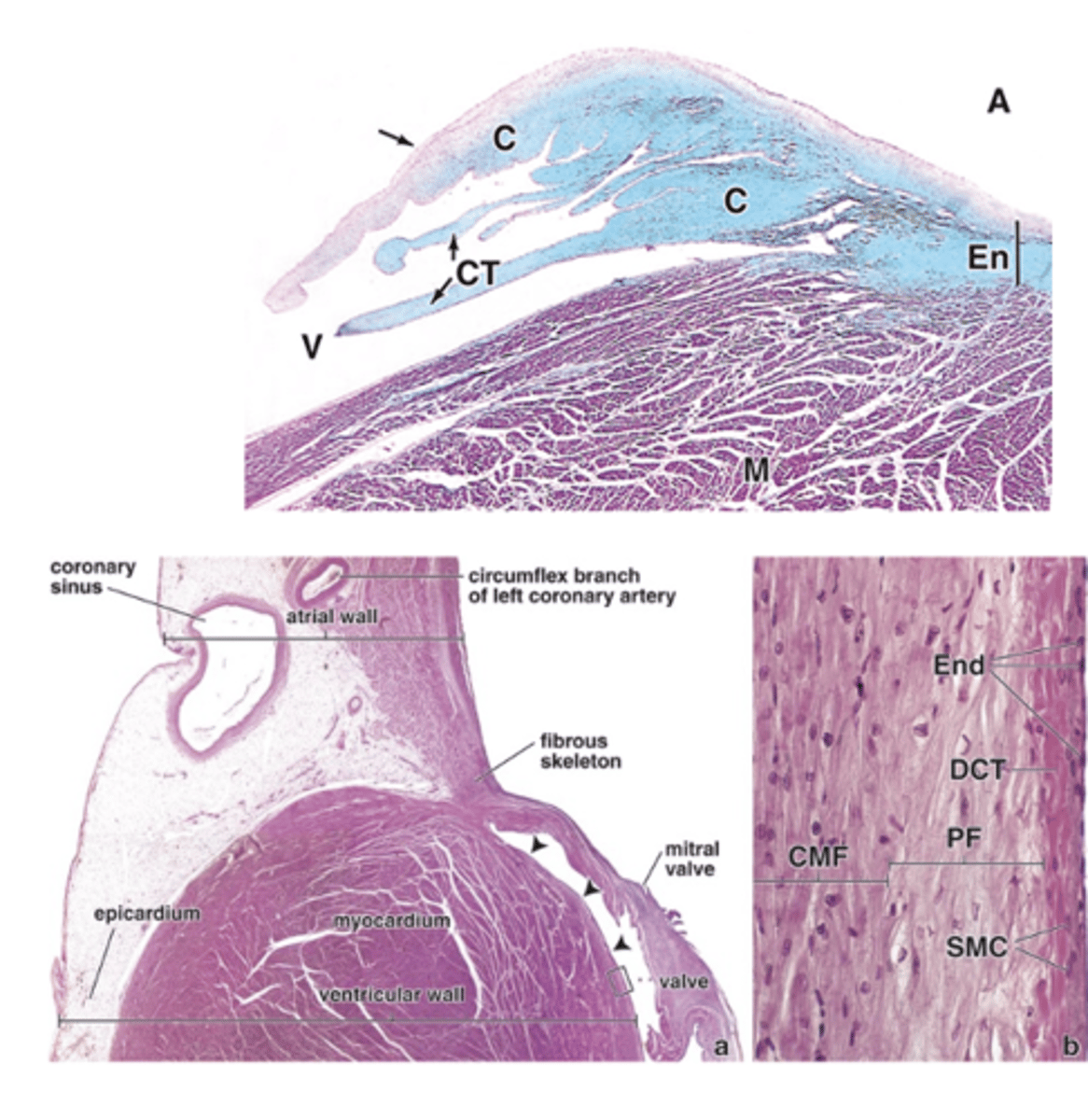
It extends to the valve cusps and the chordae tendineae.
To what structures does the cardiac skeleton extend?
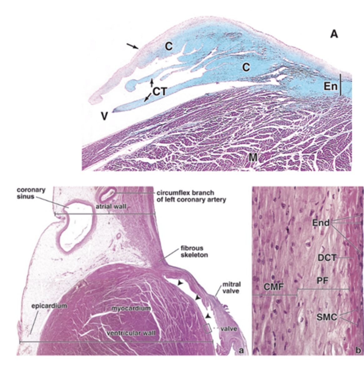
- Anchors and supports the valves
- Provides firm points of insertion for cardiac muscle
- Acts as electrical insulation between atria and ventricles
- Helps coordinate the heartbeat
What are the main functions of the cardiac skeleton? (4)
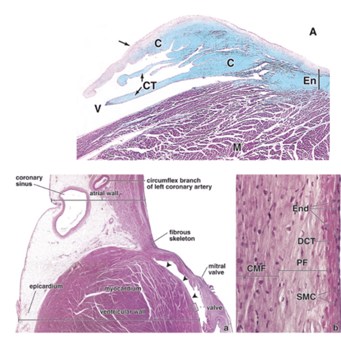
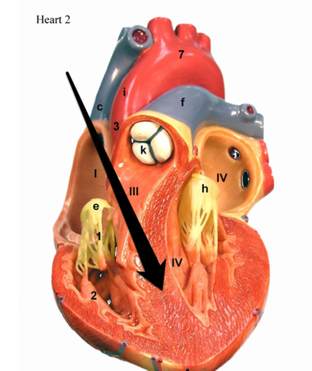
The interventricular septum.
What separates the ventricles of the heart?
Cardiac muscle, except in the membranous part, which is connective tissue.
What type of tissue makes up most of the interventricular septum?
Endothelium.
What lines the interventricular and Interartiral septum?
The interatrial septum.
What separates the atria of the heart?
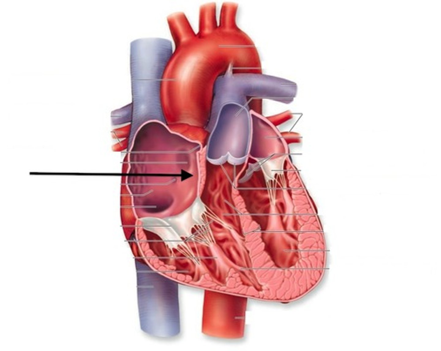
It is thinner.
How does the interatrial septum compare in thickness to the interventricular septum?
Cardiac muscle at the center and areas of connective tissue.
What types of tissue are found in the interatrial septum?
Dense connective tissue that surrounds their orifices.
To what are the heart valves attached?
Fibrosa, spongiosa, and ventricularis.
What are the three layers of the heart valves?
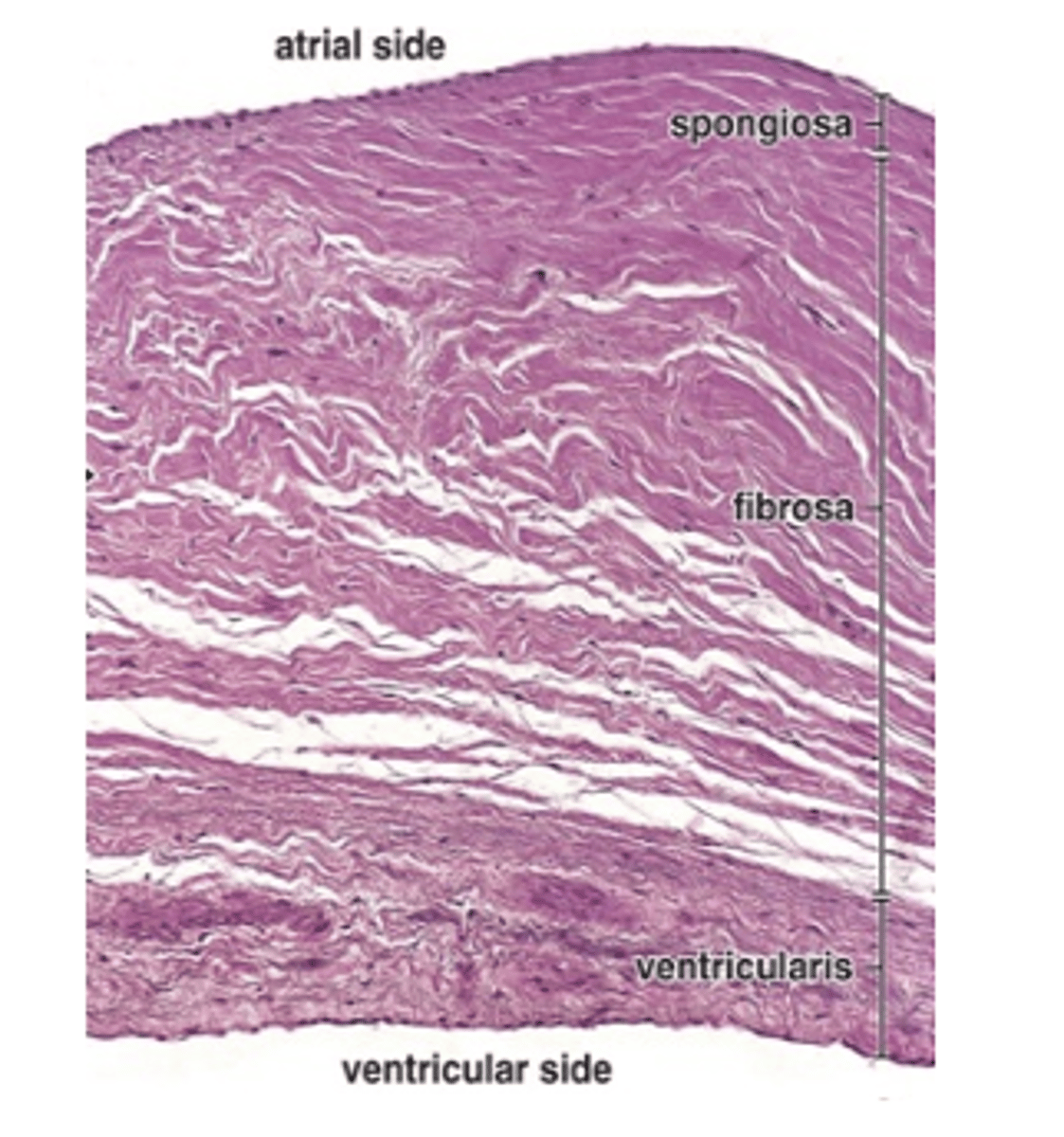
It forms the core of the valve and is composed of extensions of the dense connective tissue of the cardiac skeleton.
What is the fibrosa layer of the heart valve composed of and what is its function?
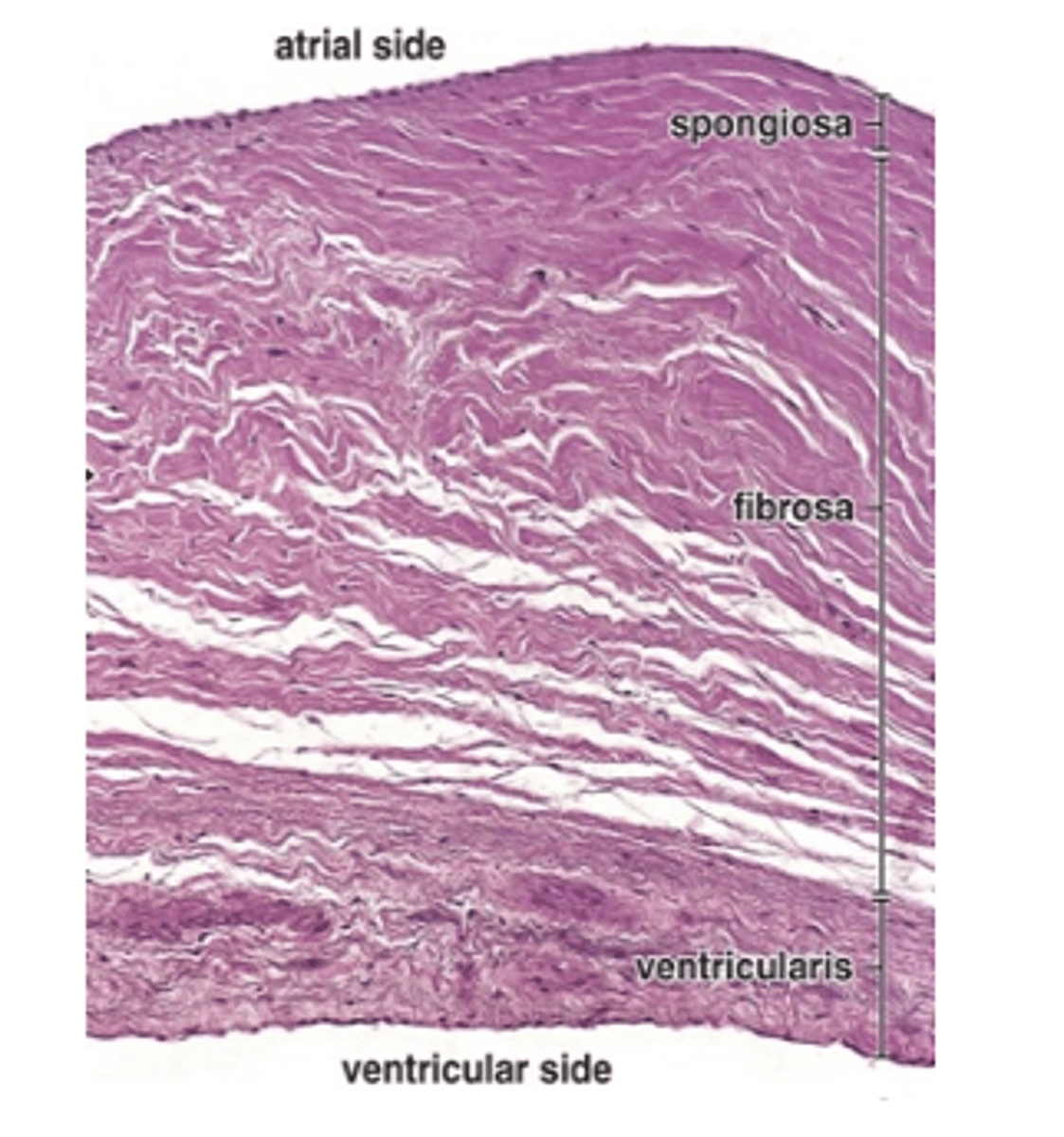
On the atrial or blood vessel side of the valve.
Where is the spongiosa layer located in the heart valves?
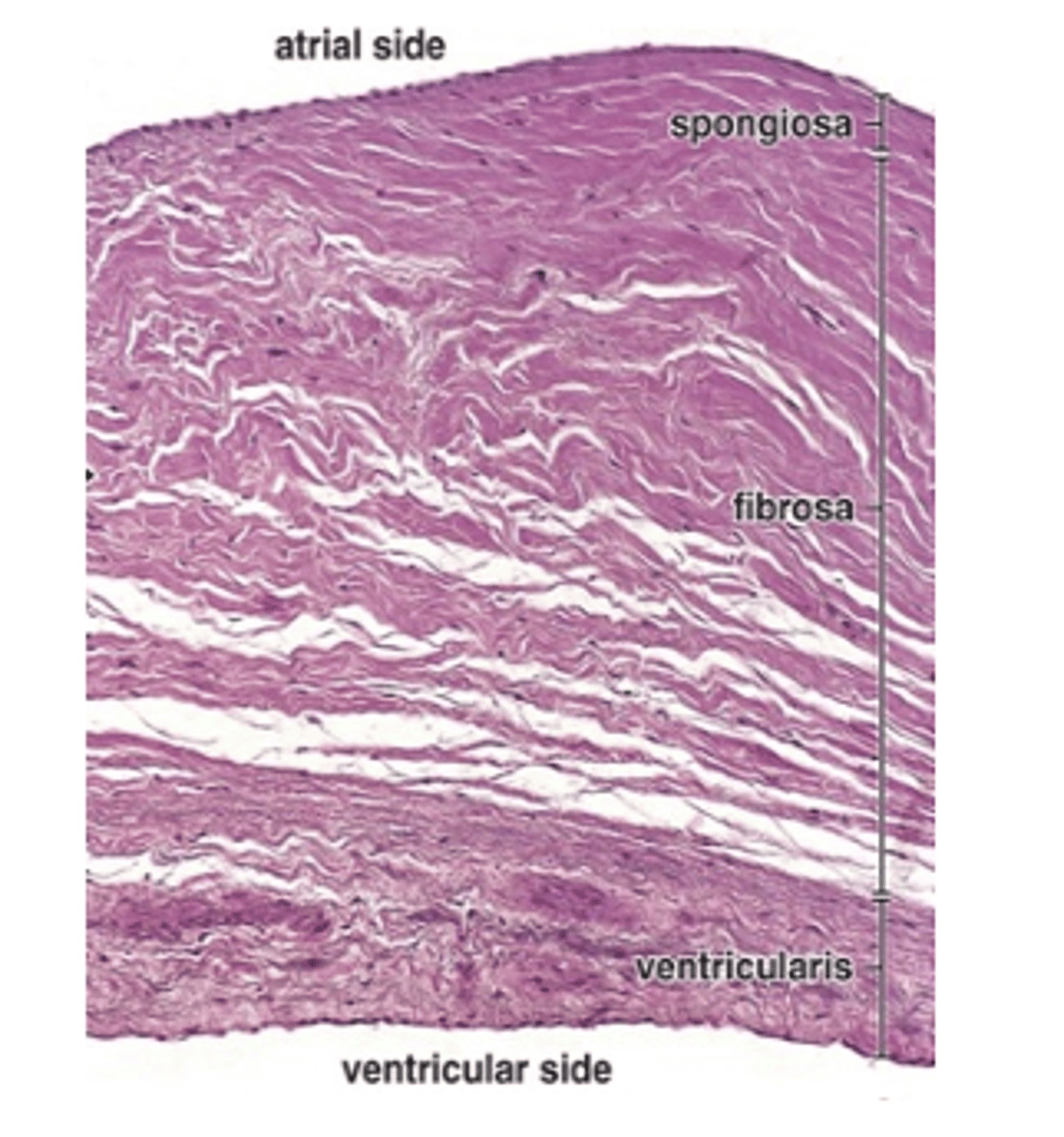
•Tricuspid and bicuspid valves: auricularis
•Aortic and pulmonary valves: arterialis
What is the spongiosa layer called in the tricuspid and bicuspid valves and in the aortic and pulmonary valves?
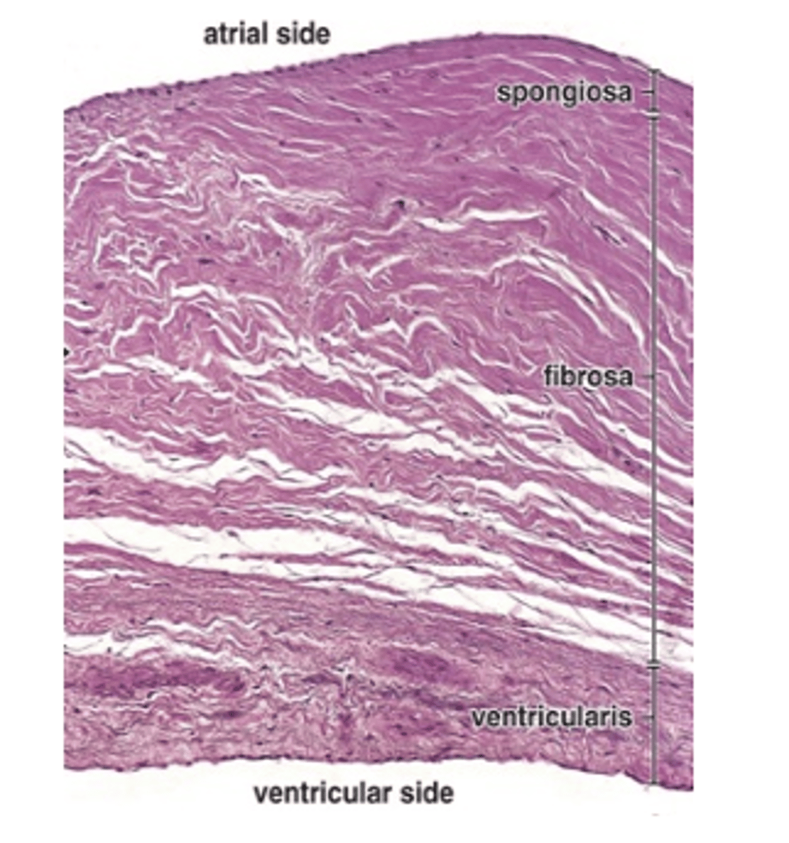
Loose connective tissue with collagen and elastic fibers and a large number of proteoglycans.
What type of tissue and components are found in the spongiosa layer of the heart valves
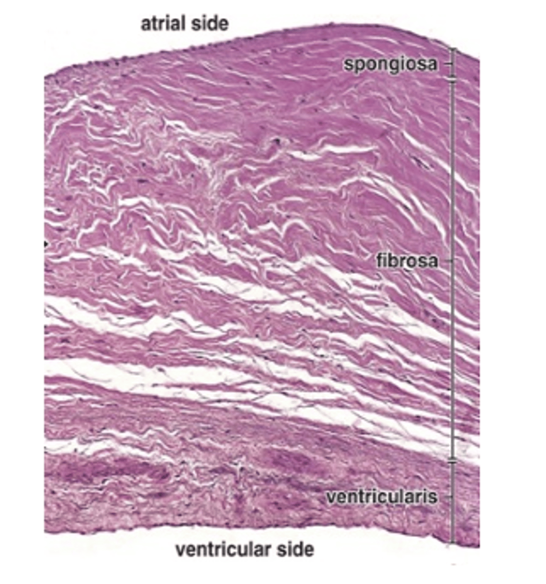
Adjacent to the ventricular or atrial surface of the valves.
Where is the ventricularis layer located in the heart valves?
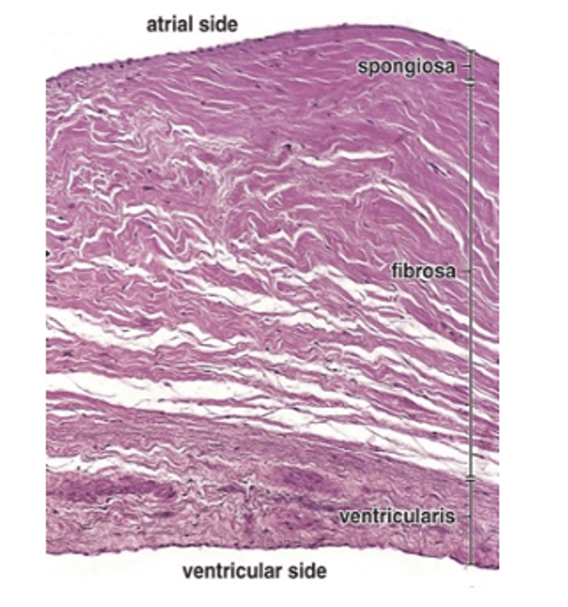
Endothelium.
What covers the ventricularis layer of the heart valves?
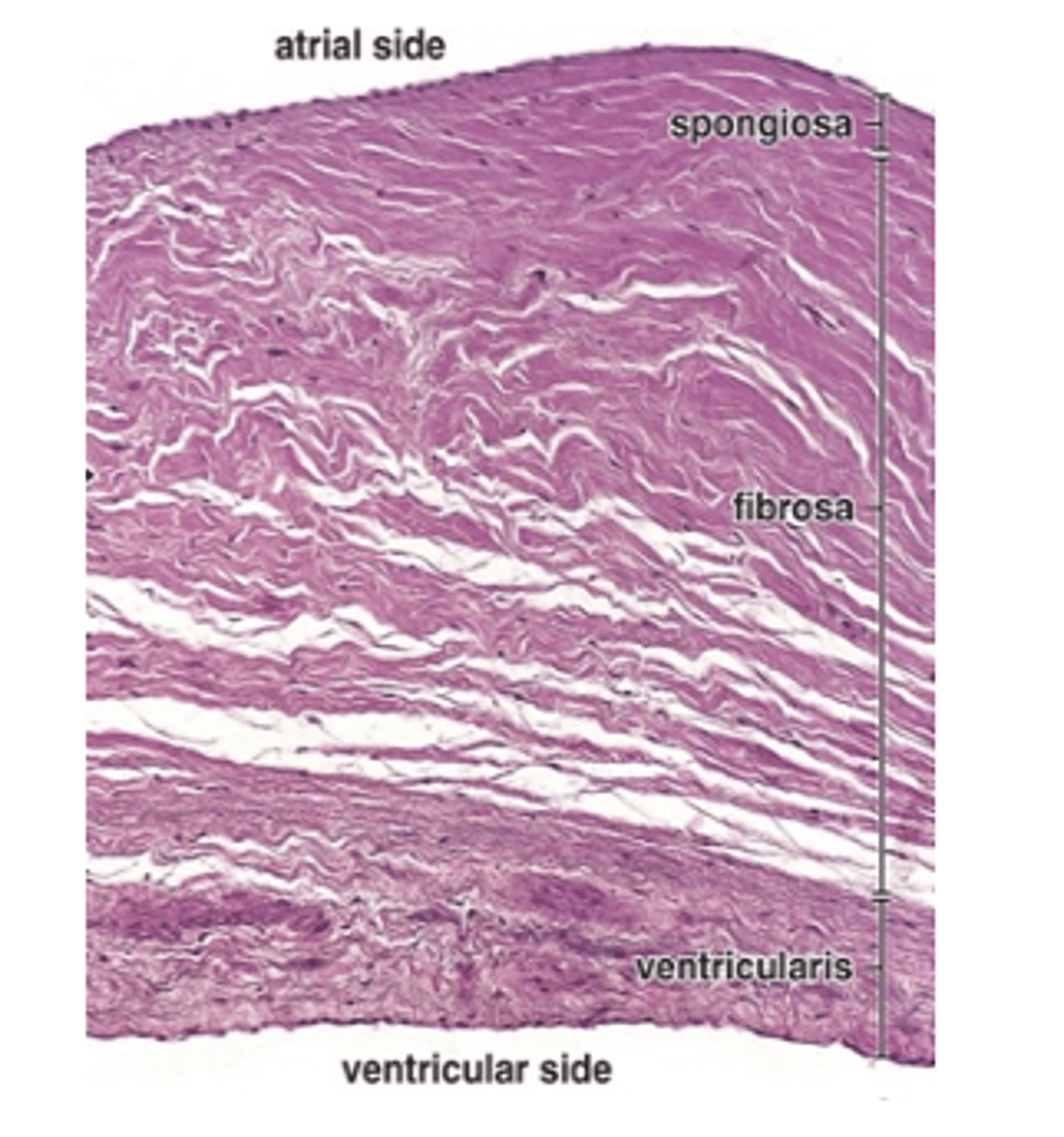
Dense connective tissue with many layers of elastic fibers.
What types of tissue are found in the ventricularis layer of the heart valves?
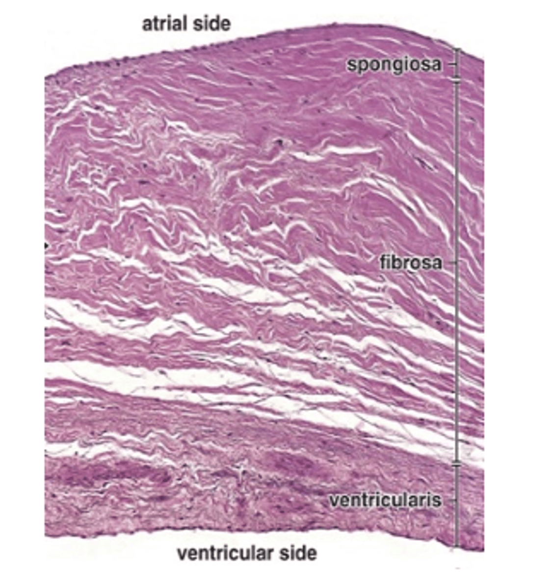
The chordae tendineae.
With what structures is the ventricularis layer continuous in atrioventricular valves?
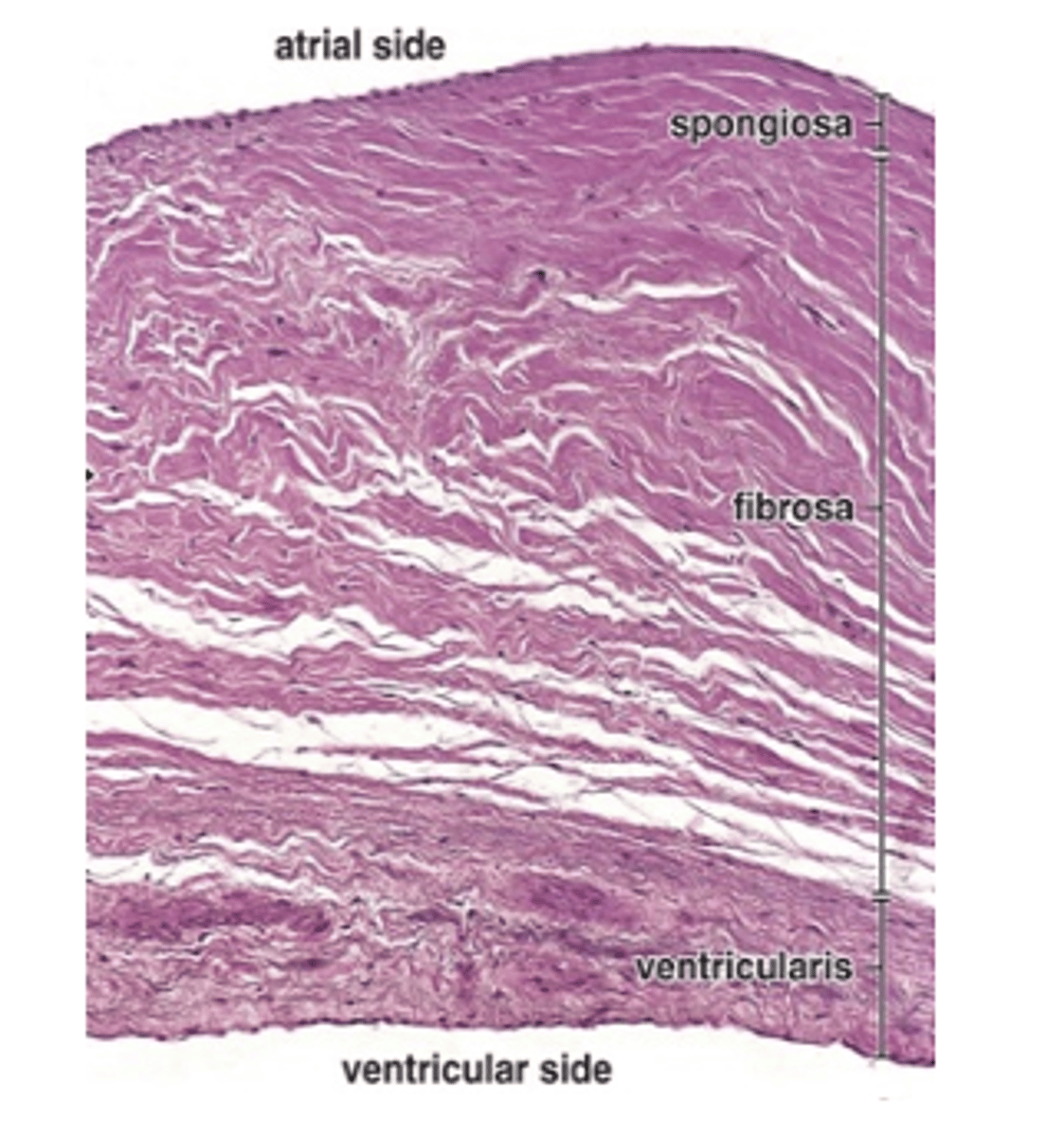
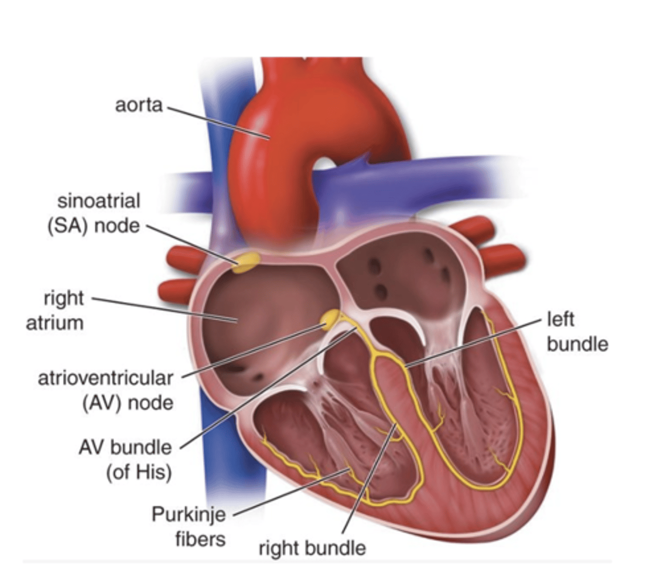
It is a network of modified cardiac muscle cells located in the subendocardial layer adjacent to the myocardium.
What is the intrinsic conducting system of the heart and where is it located?
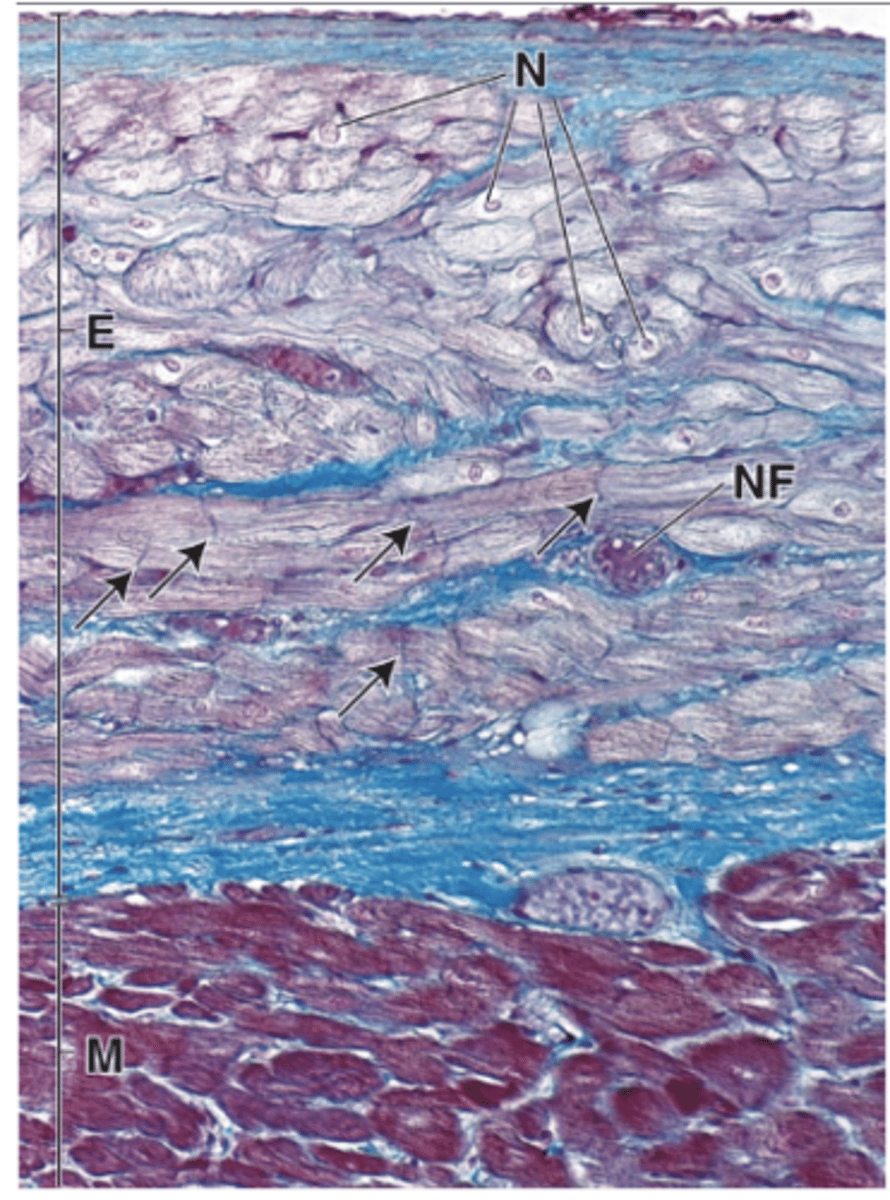
- 6-7 mm³ mass of small cardiac muscle cells
- Fewer myofibrils
- Fewer intercalated discs
What are the characteristics of the SA node?
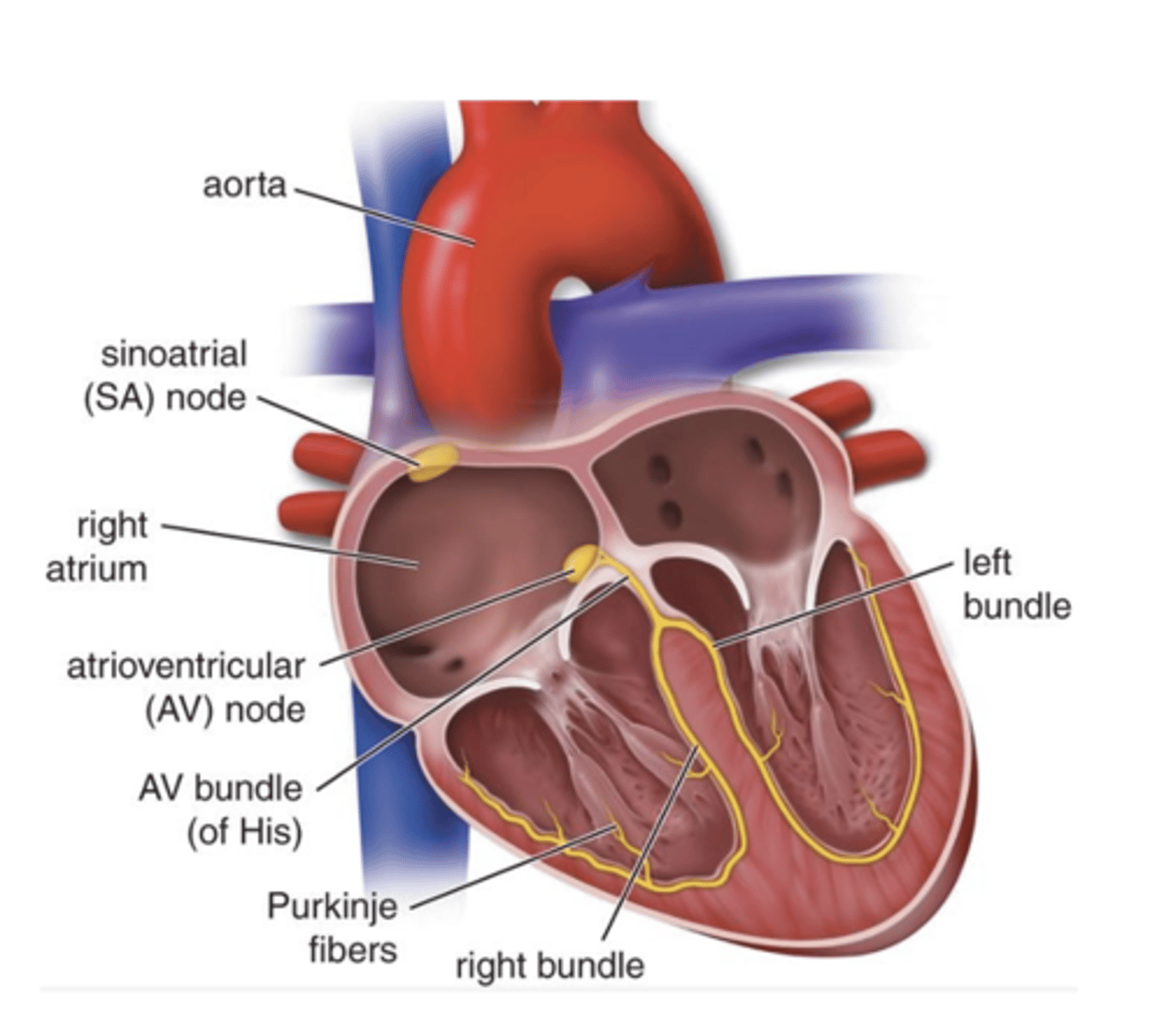
The AV bundle consists of conducting muscle fibers that pass through an opening in the cardiac skeleton into the interventricular septum.
What is the AV bundle and where does it pass?
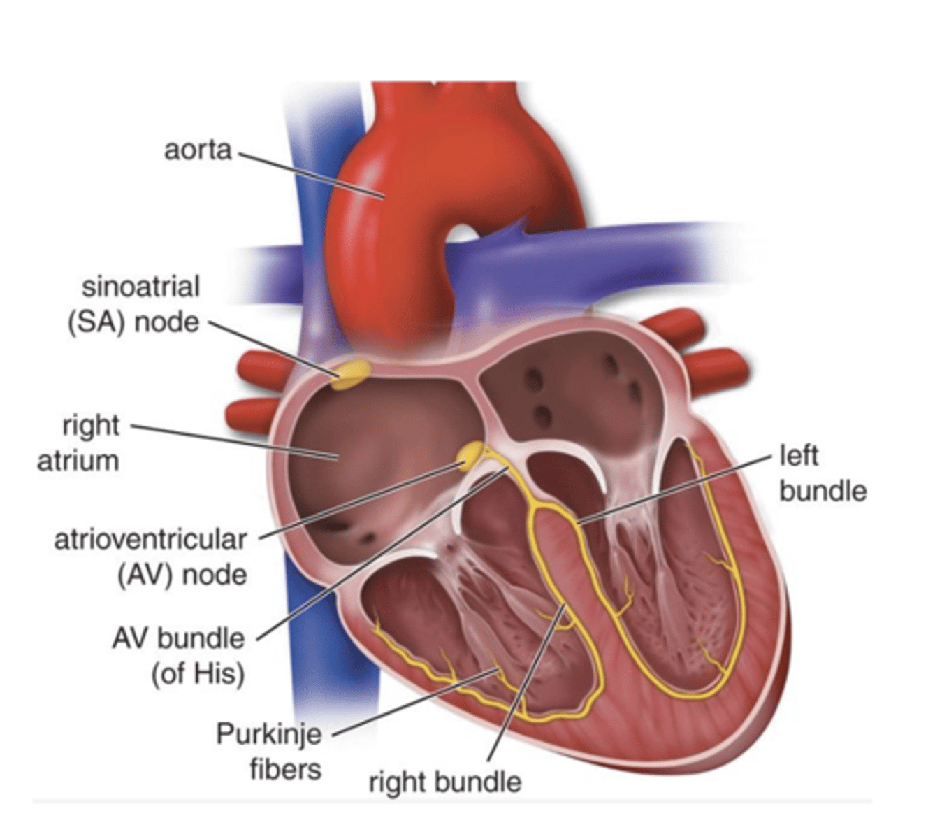
They arise from the AV bundle as it branches within the interventricular septum.
What do the right and left bundle branches arise from?
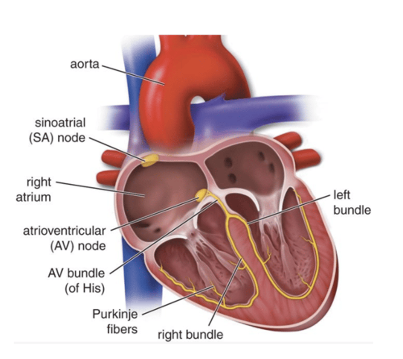
Purkinje fibers are in the subendocardial layer and contact myocardial muscle fibers through intercalated discs.
Where are Purkinje fibers located and how do they connect to the myocardium?
Endothelium
What tissue lines the walls of all blood vessels?
1. Endothelium
2. Smooth muscle tissue
3. Connective tissue
What are the three main tissue layers of vascular walls?
Mechanical factors (pressure) and metabolic factors (local tissue needs)
What factors determine the amount and arrangement of smooth muscle and connective tissue in blood vessels? (2)
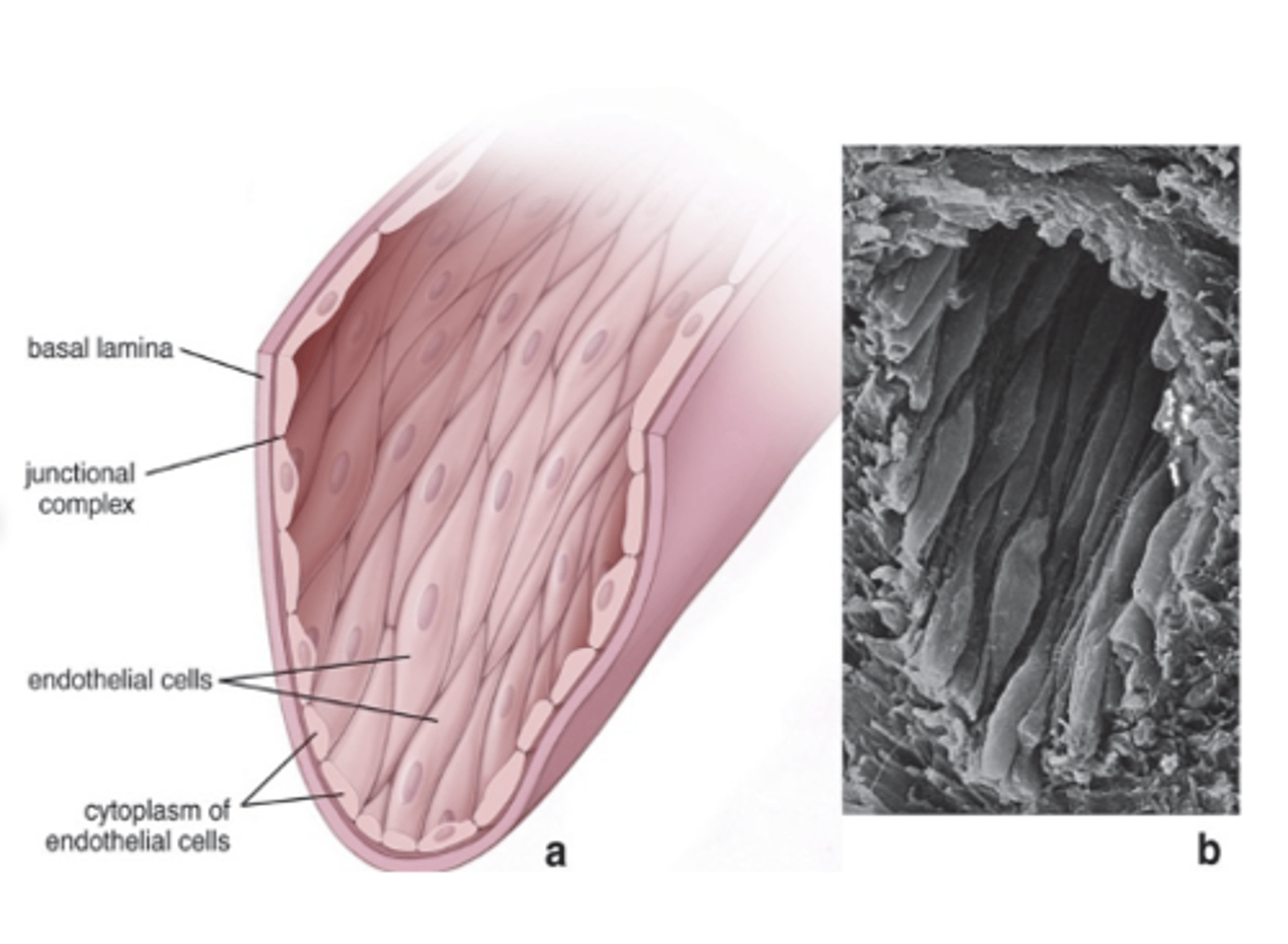
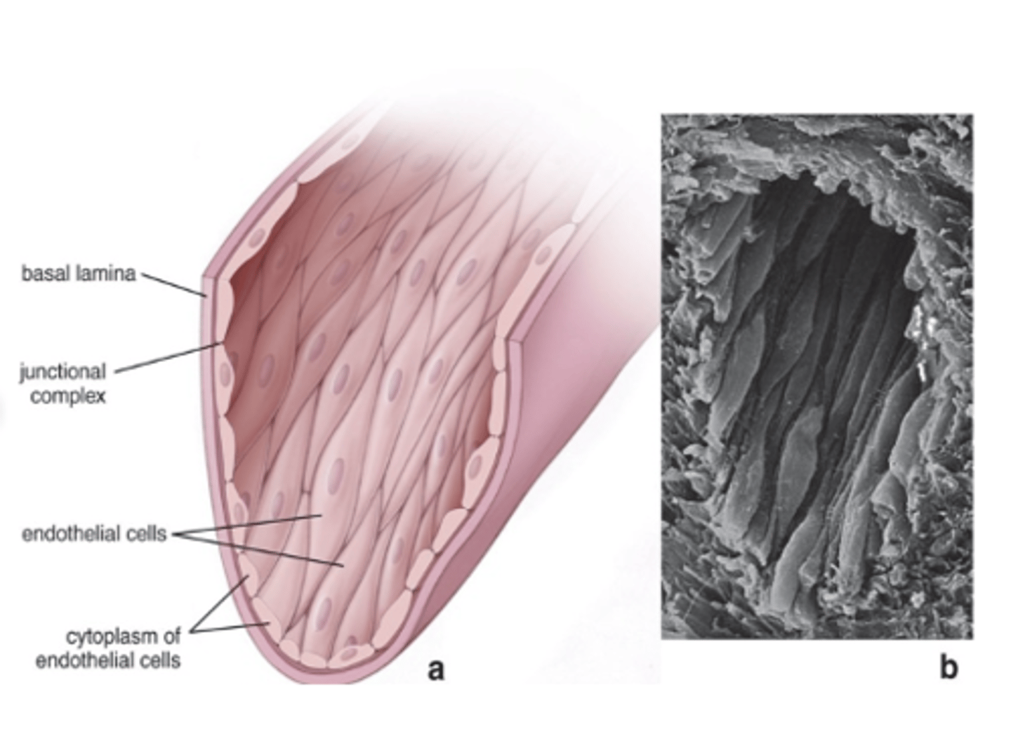
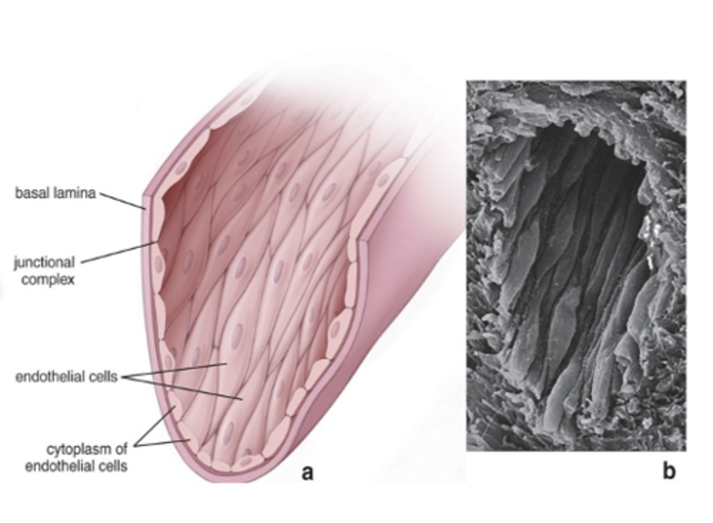
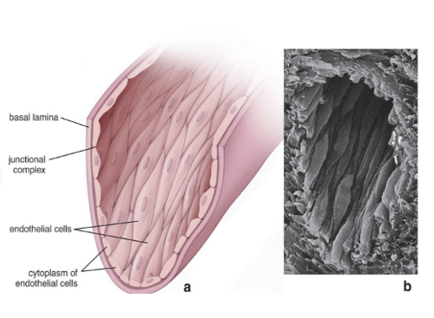
Capillaries are composed of only endothelium.
What is the structural composition of capillaries?
- Squamous
- Polygonal
- Elongated
- Long axis in the direction of blood flow
What shapes can endothelial cells have?
It forms a semipermeable barrier, together with the basal lamina, mediating and monitoring bidirectional exchange of molecules.
What is the main structural role of the endothelium?
It has antithrombotic activity.
How does the endothelium contribute to blood clot prevention?
By locally regulating vascular tone and blood flow.
How does the endothelium regulate vascular function?
It participates in local immune and inflammatory responses and secretes growth factors.
In what immune and repair processes does the endothelium participate?
Found in arteries and veins.
Where are smooth muscle fibers found in the vascular system?***
In a helical arrangement in layers.
How are smooth muscle fibers arranged in blood vessel walls?
Arterioles and small arteries have many more gap junctions, facilitating coordinated Vasoconstriction and vasodilation
Which vessels have more gap junctions in their smooth muscle, and why is this important?
In the subendothelial layer and outer covering; they provide structural support.
Where are collagen fibers located in blood vessels, and what is their role?
They provide resiliency during expansion under pressure.
What is the function of elastic fibers in blood vessels?
Elastin forms parallel lamellae between smooth muscle layers
How is elastin arranged in larger arteries?
Proteoglycans and hyaluronate; their amount varies according to physical and metabolic properties of the vessel.
Which components of connective tissue affect vessel permeability, and how does their amount vary?
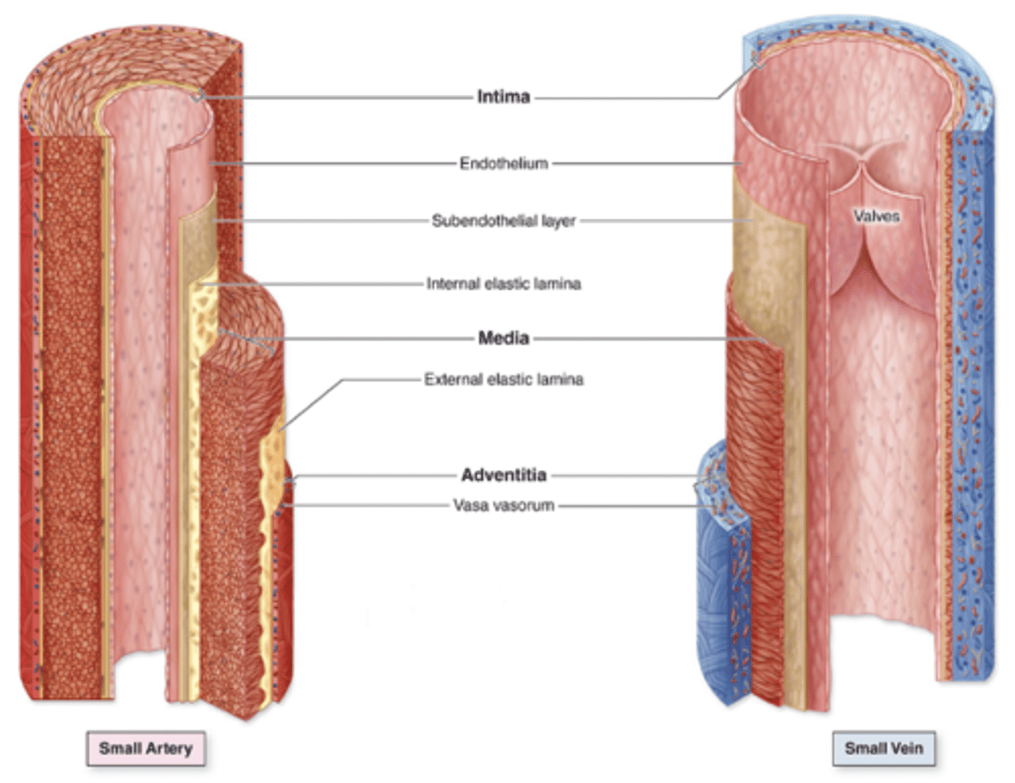
Endothelium and connective tissue; in veins, it is folded to form valves.
What is the composition of the tunica intima?
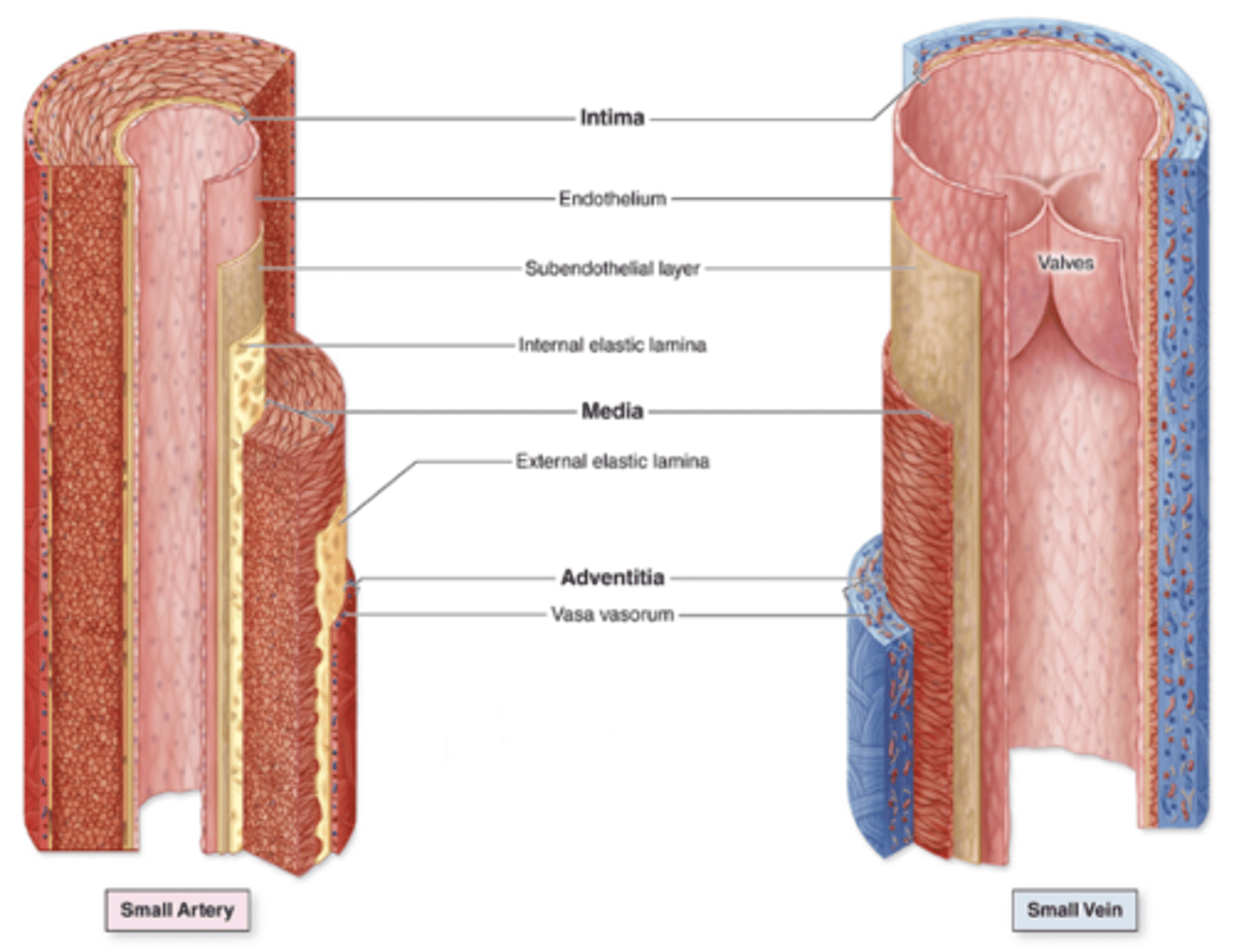
Smooth muscle and other varying components; thicker in arteries than in veins***
What is the composition of the tunica media and how does its thickness compare in arteries and veins?***
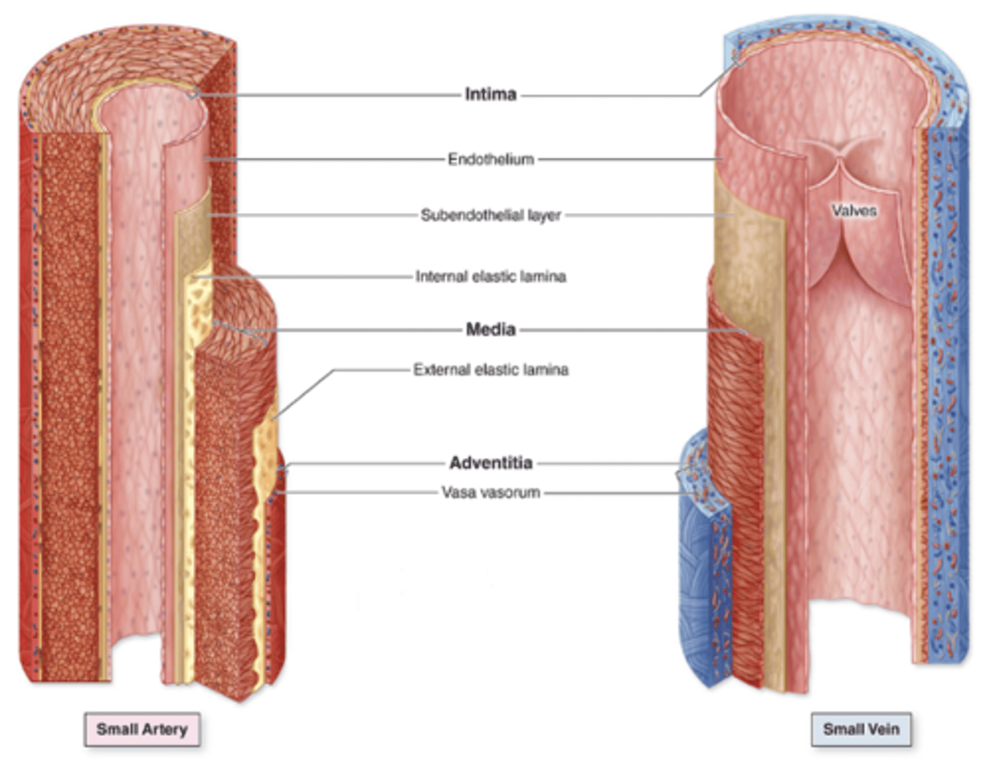
Dense irregular connective tissue; thicker in veins than in arteries.***
What is the composition of the tunica adventitia and how does its thickness compare in arteries and veins?***
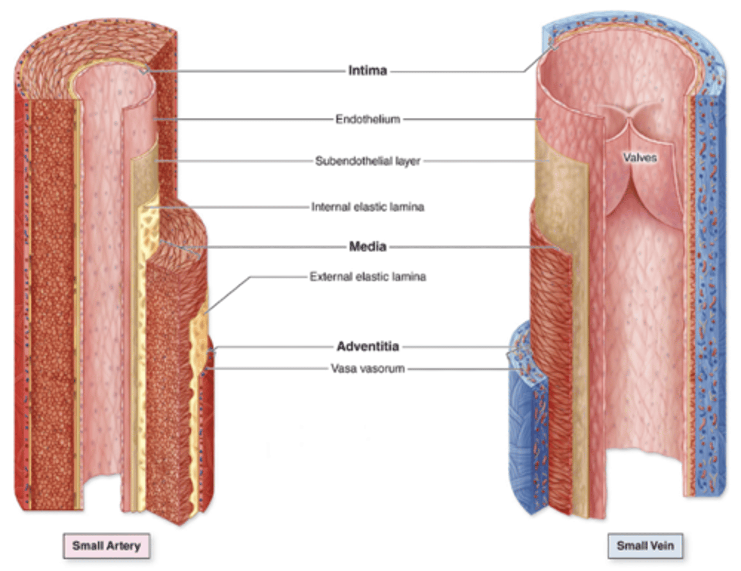
Only endothelium and its basal lamina.
What is the structural composition of capillaries?
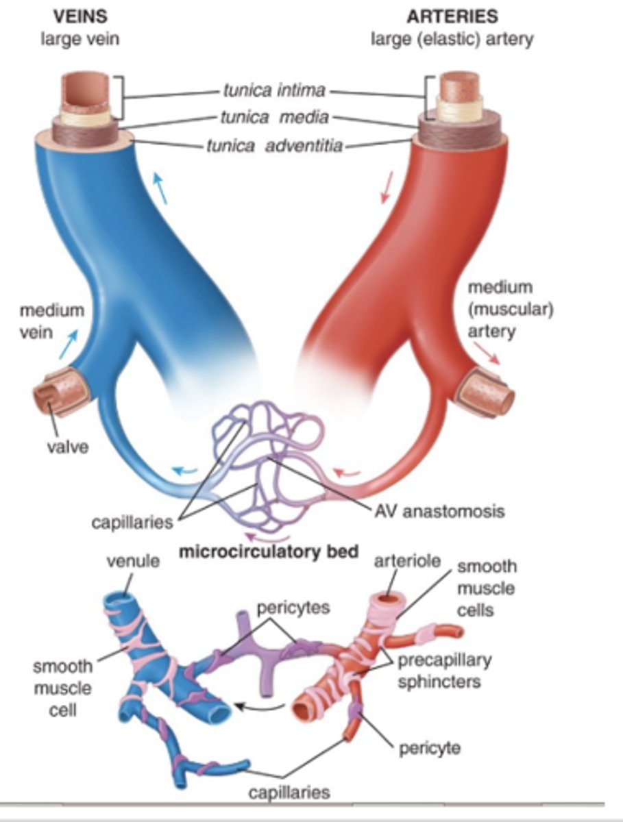
Pericytes.
What cells are found at intervals around the basal lamina of capillaries?
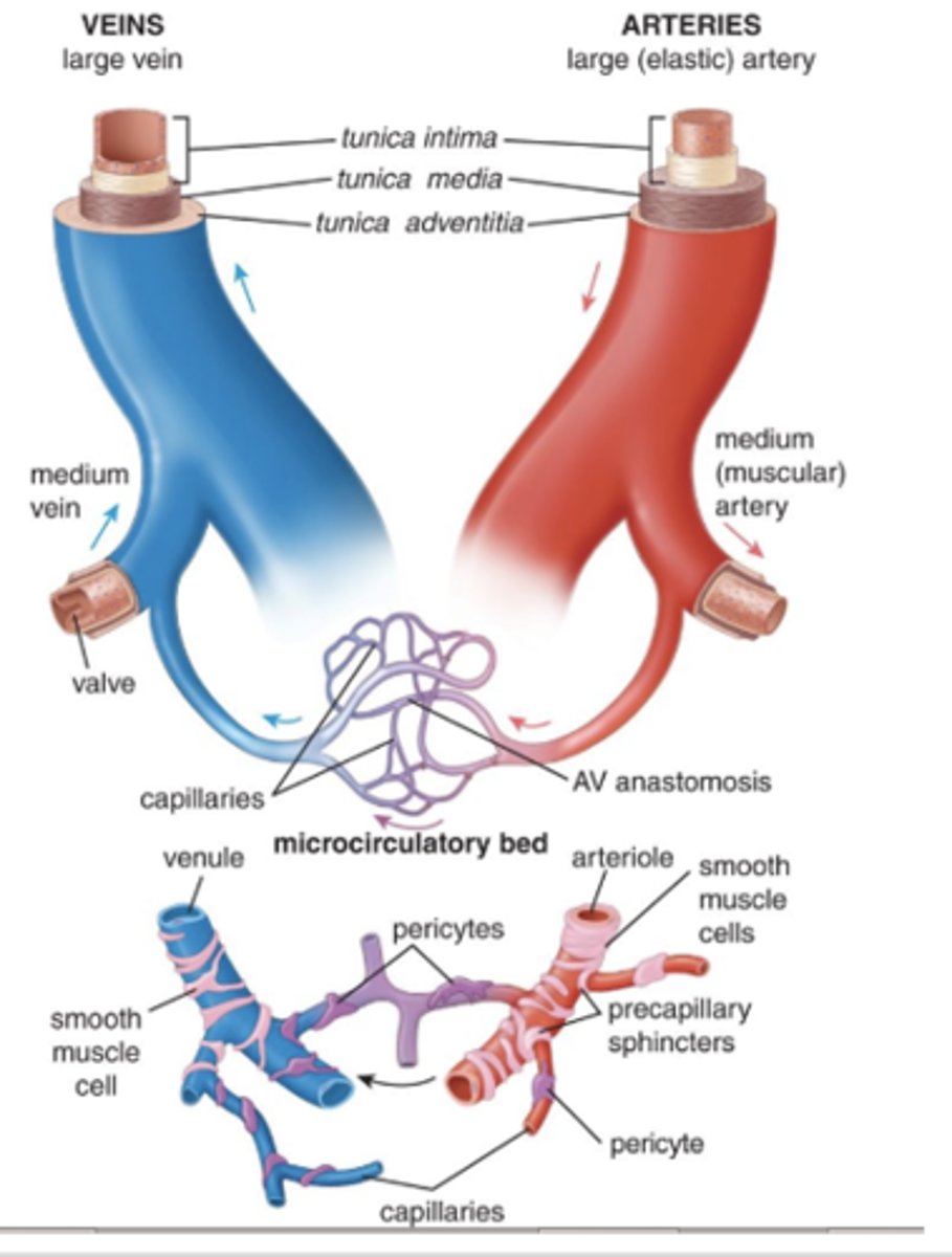
Endothelium, thin basal lamina, and thin subendothelial layer.
What are the main components of the tunica intima?
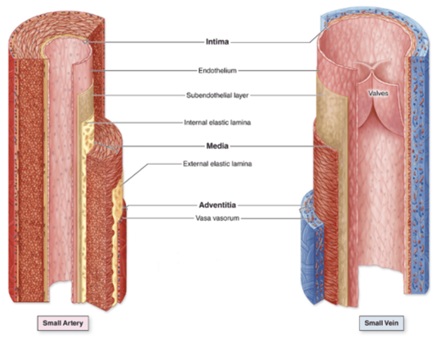
Loose connective tissue, sometimes with smooth muscle.
What can the subendothelial layer of the tunica intima contain?
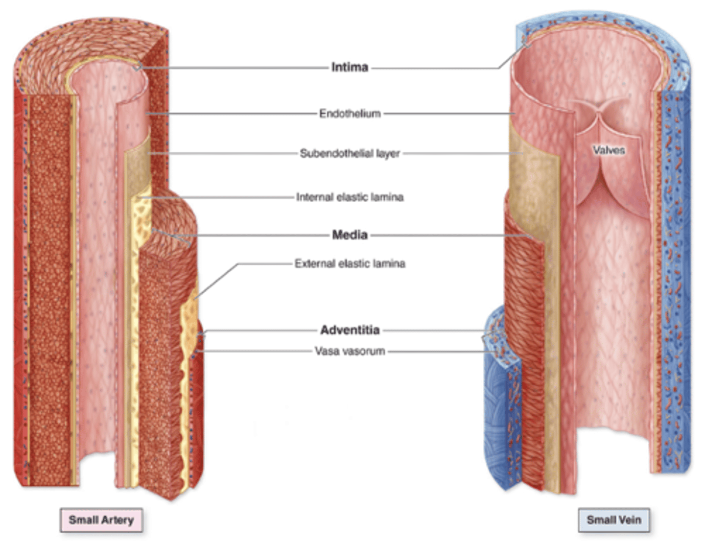
Found in
1. arteries
2. arterioles
3. largest veins
its fenestrations allow nutrient diffusion.
Where is the internal elastic lamina found, and what is its function?***
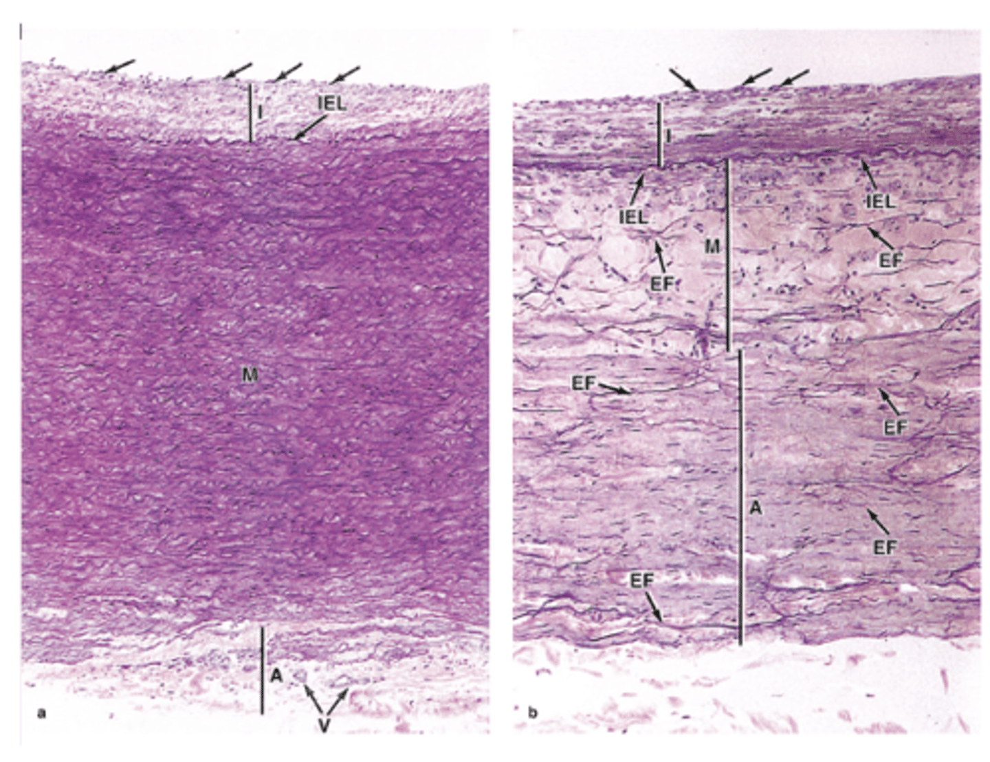
Concentric, helically arranged layers.
How are smooth muscle fibers arranged in the tunica media?***
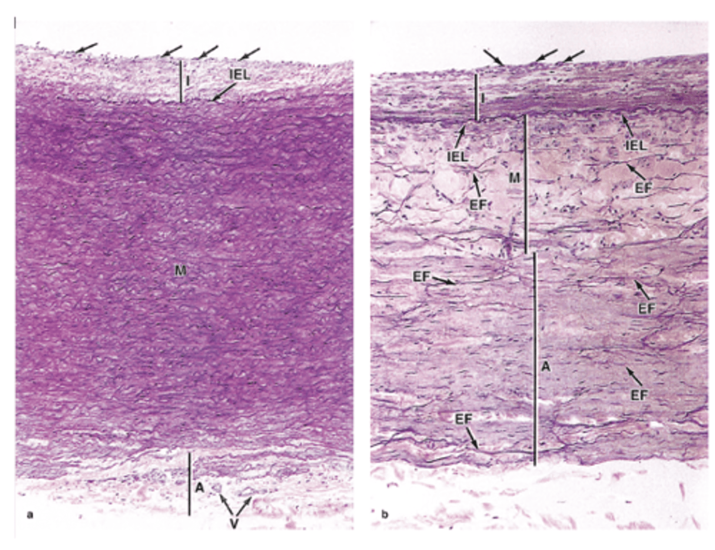
Variable amounts of elastic fibers and elastic lamellae, reticular fibers, and proteoglycans (produced by the muscle fibers).
What other components can be found in the tunica media besides smooth muscle?
They are fenestrated to allow diffusion.
What is special about the elastic lamellae in the tunica media?
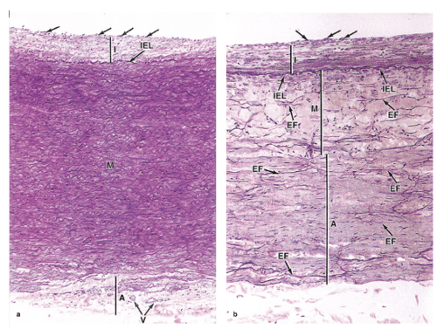
Only in arteries.
In which vessels is the tunica media prominent with elastic lamellae?***
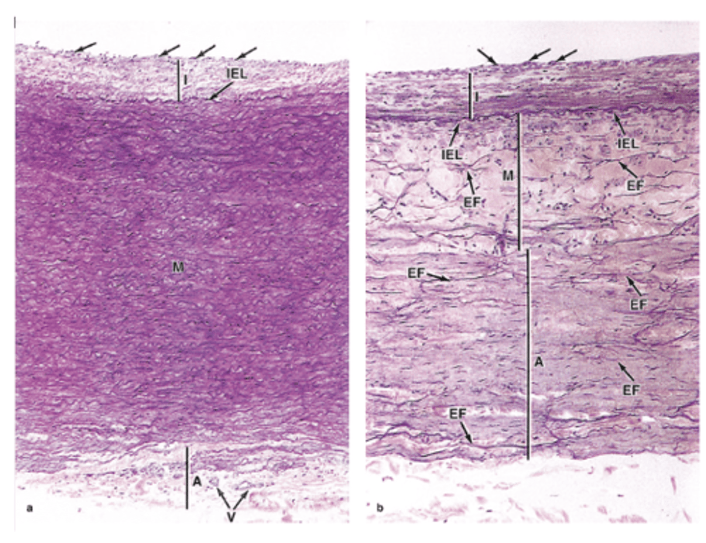
The external elastic lamina.
What separates the tunica media from the tunica adventitia?
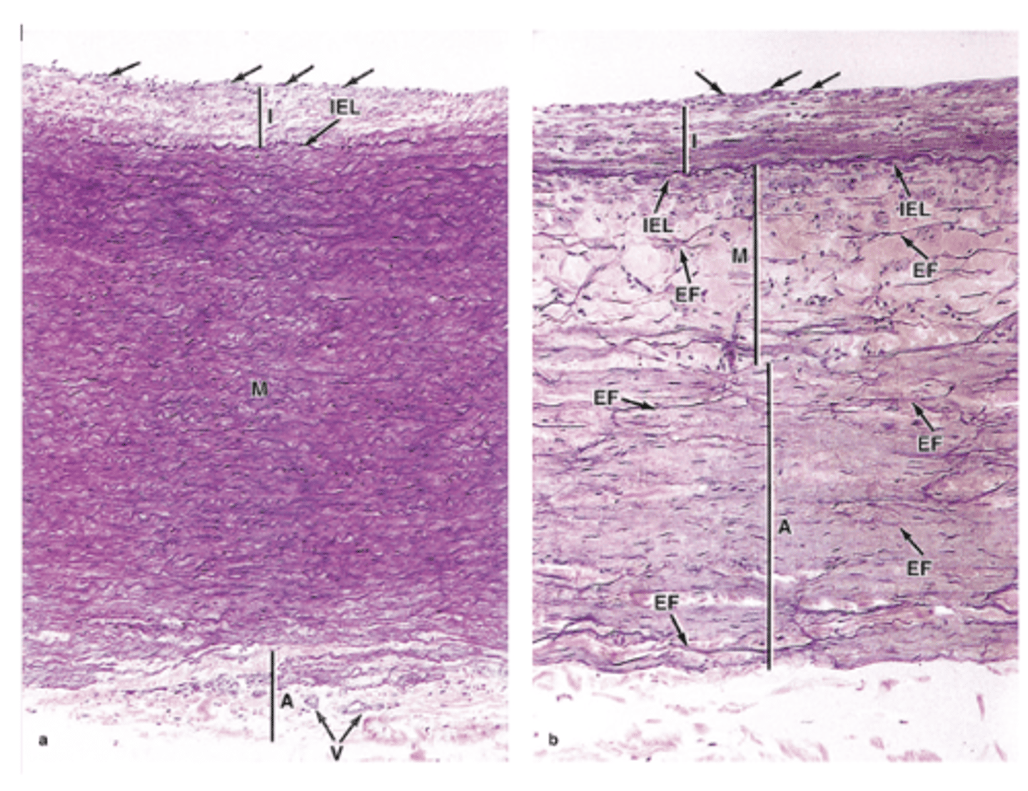
Thicker and contains much more elastin in arteries.
How does the tunica media in arteries compare to veins?
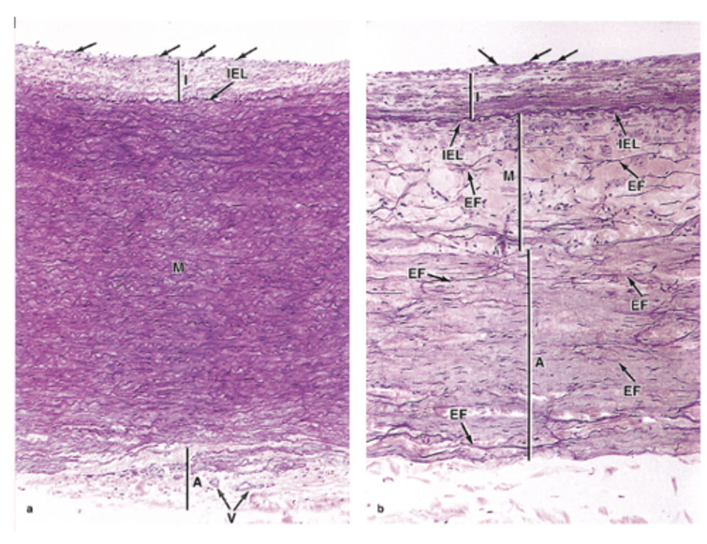
Connective tissue, primarily type I collagen and elastic fibers.
What is the main composition of the tunica adventitia?
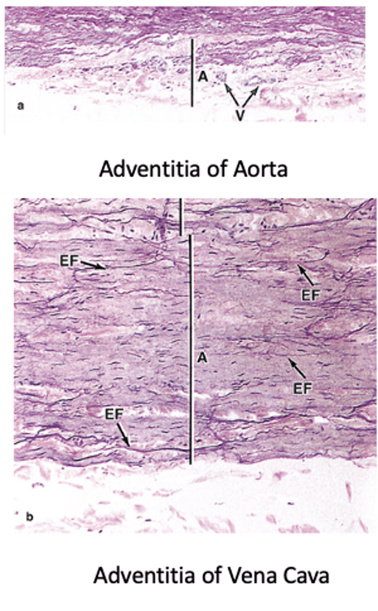
It is continuous with and bound to the stroma of the organ.
How is the adventitia related to surrounding tissues?
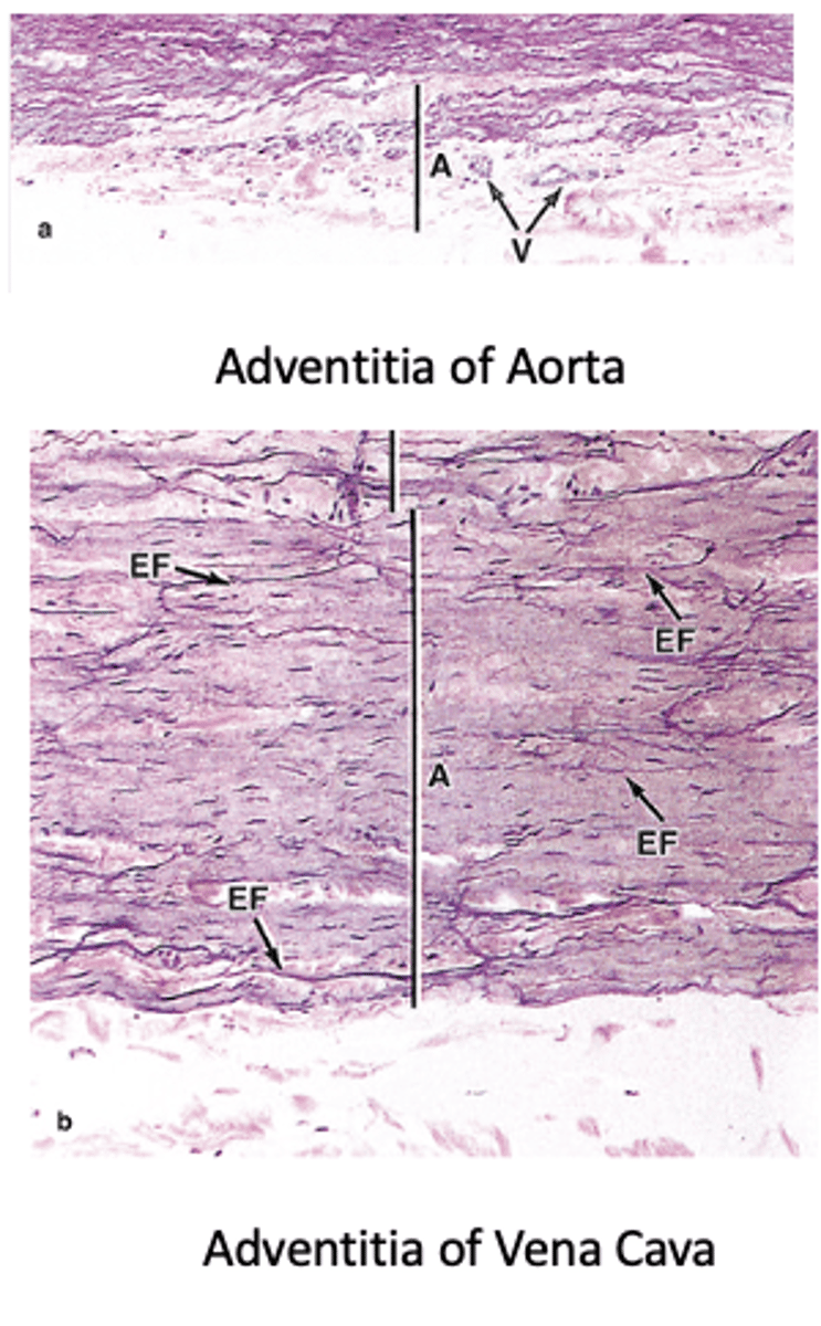
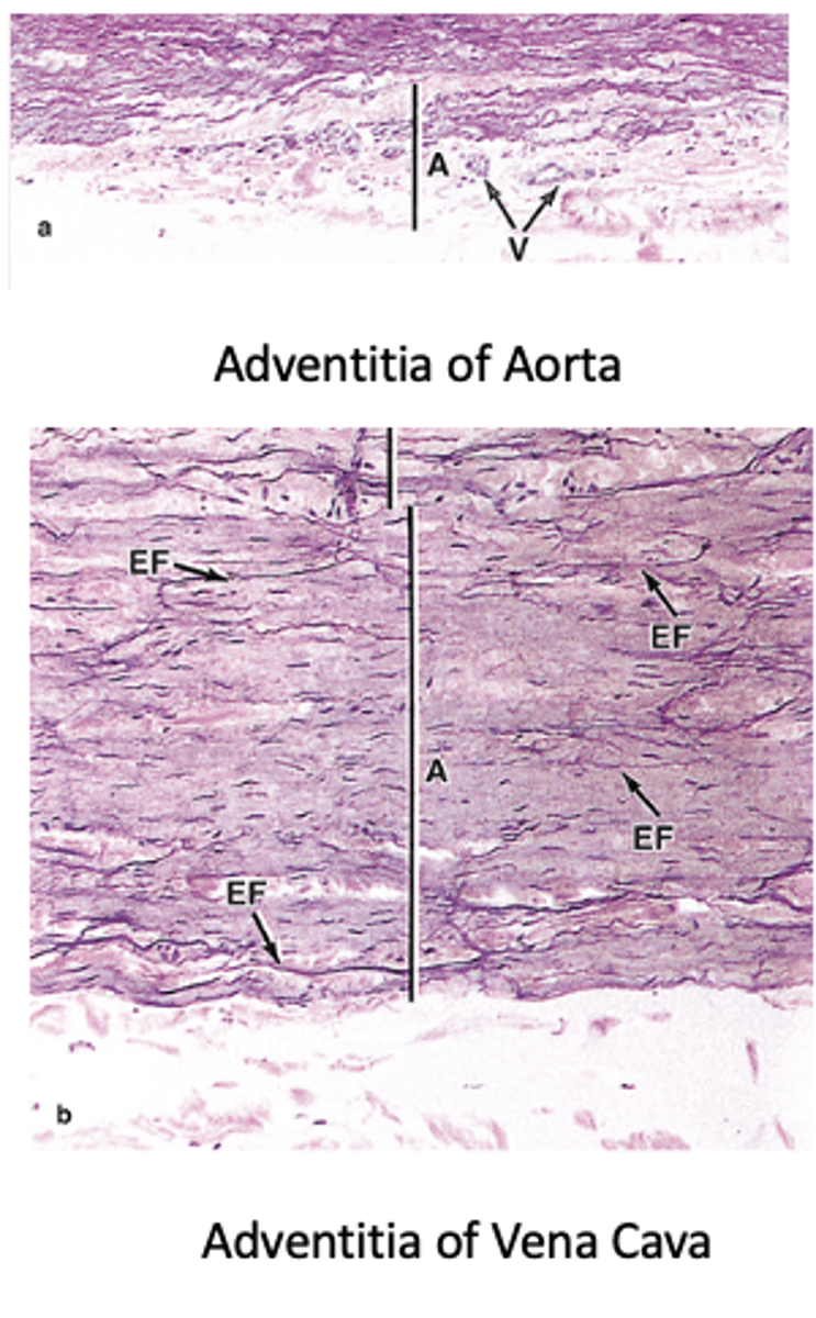
Thinner in arteries and increasingly thicker in venules and veins.
How does the thickness of the adventitia compare between arteries and veins?****
Vasa vasorum (small vessels) and nervi vasorum (nerves); they provide nutrition to layers far from the intima in large vessels.
What structures are found in the adventitia of large arteries and veins, and what is their function?*** (2)
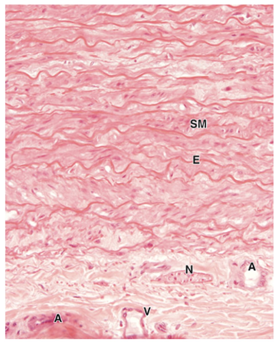
Well-developed and thick, consisting of an endothelial lining with basal lamina and a subendothelial layer.
How is the tunica intima of elastic (conducting) arteries described?
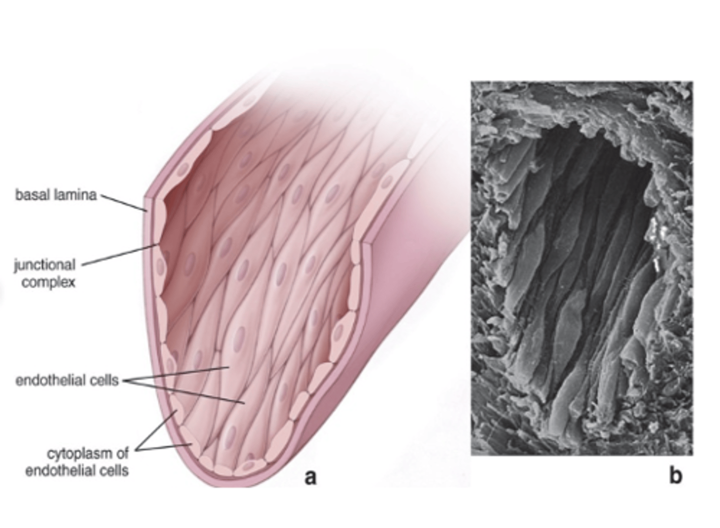
By tight junctions and gap junctions.
How are endothelial cells in elastic arteries (conducting arteries) connected? (2)
Rod-like, electron-dense inclusions in endothelial cell cytoplasm containing von Willebrand factor and P-selectin.
What are Weibel-Palade bodies and where are they found?
Binds coagulation factor VIII and mediates platelet adhesion.
What is the function of von Willebrand factor in elastic arteries (conducting arteries)?
Acts as a cell-adhesion molecule for neutrophil-endothelial recognition.
What is the function of P-selectin in elastic arteries (conducting arteries)?
- Abundant smooth muscle cells
- Secrete extracellular ground substance, and collagen and elastin
- Occasional macrophages
What does the subendothelial layer of elastic arteries (conducting arteries) contain? (3)
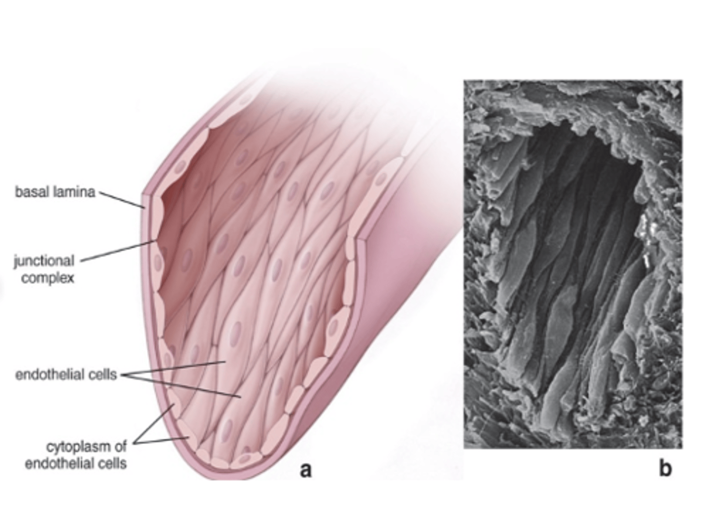
The internal elastic membrane.
What elastic feature is present in the tunica intima of elastic arteries (conducting arteries)?
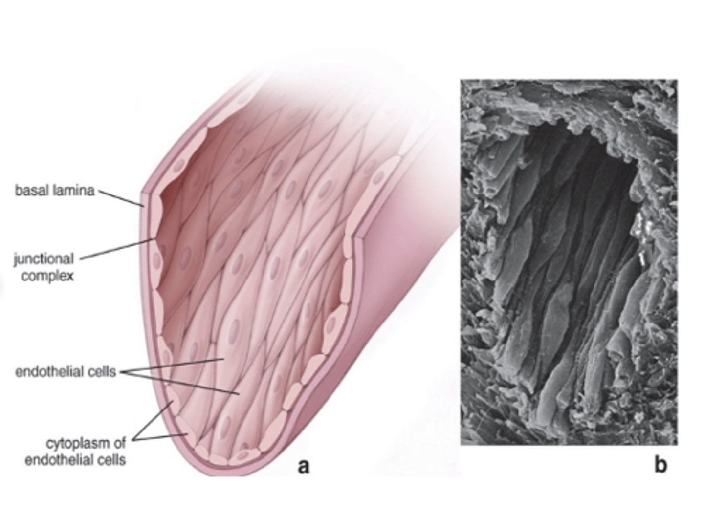
Thick, with fenestrated elastic lamellae alternating with layers of smooth muscle fibers.
How is the tunica media of elastic arteries structured?
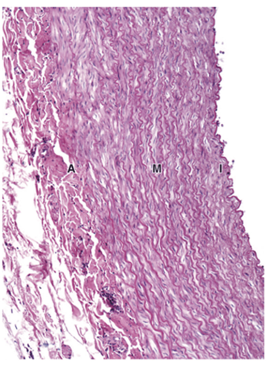
About 50 elastic lamellae; number increases with hypertension.***
Approximately how many elastic lamellae are found in the aorta, and what affects their number?***
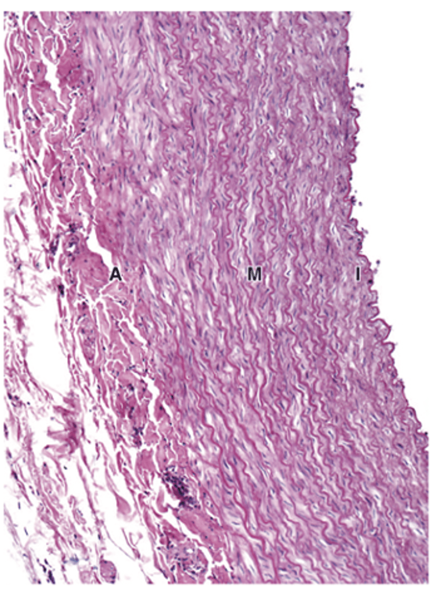
They are more well-defined than the elastic lamellae of other arterial media.
How do the elastic lamellae in the tunica media of elastic arteries compare to those in other arteries?
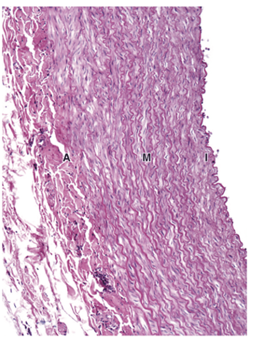
The external elastic membrane.
What separates the tunica media from the tunica adventitia in elastic arteries?
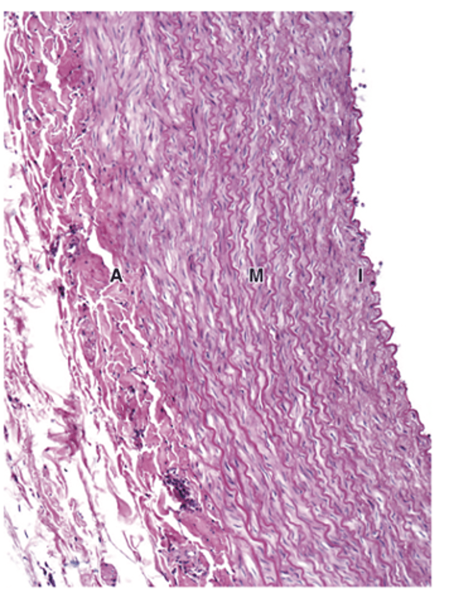
Adventitia is thinner than the media.
How does the adventitia of elastic arteries compare in thickness to the media?
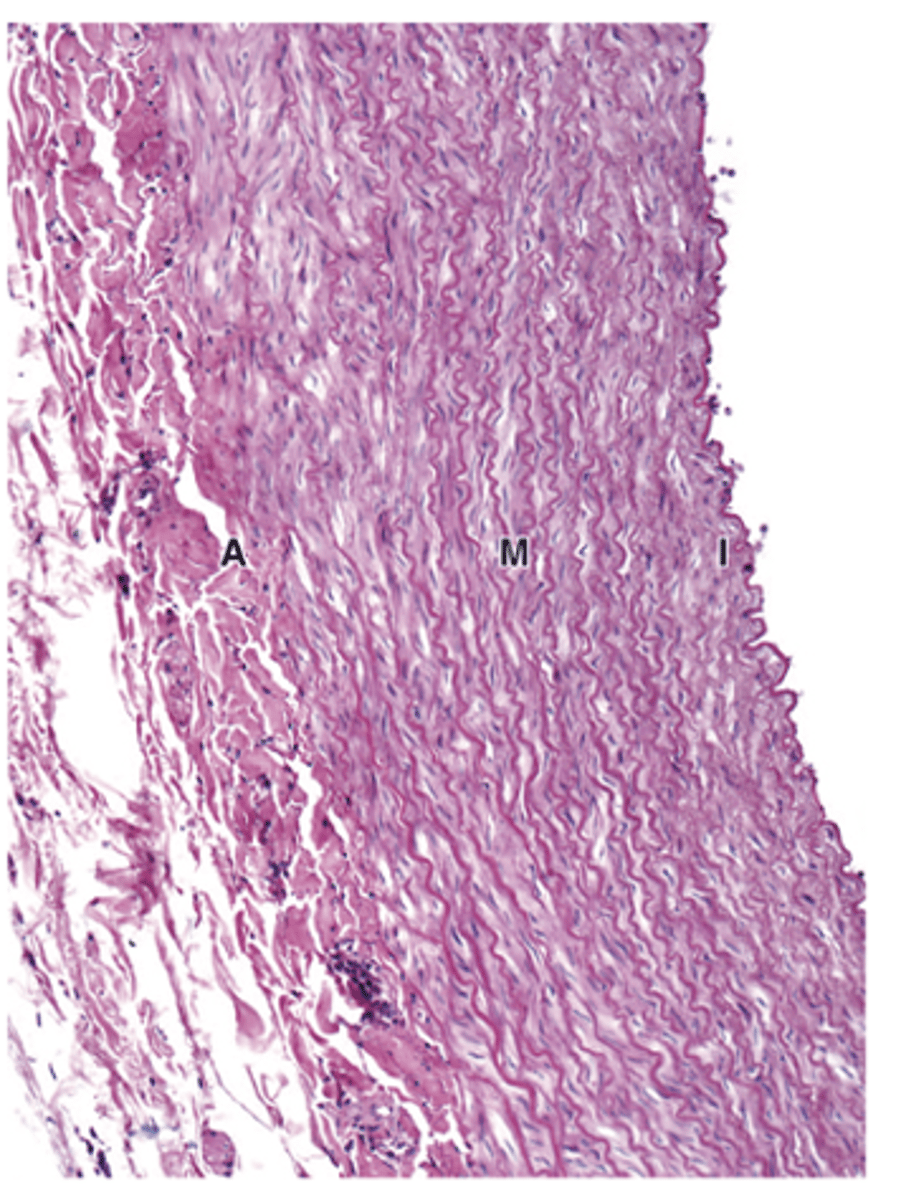
Collagen and elastic fibers forming an elastic network, with fibroblasts and macrophages as main cells.
What are the main components of the adventitia in elastic arteries?