4. Visual Fields in Glaucoma - Management of Glaucoma Summer 2026
 Call Kai
 Learn
Learn Practice Test
 Spaced Repetition
 Match
Match Flashcards
 Knowt Play
1/185

There's no tags or description
Looks like no tags are added yet.
Name | Mastery | Learn | Test | Matching | Spaced | Call with Kai | Chat |
|---|
No analytics yet
Send a link to your students to track their progress
186 Terms
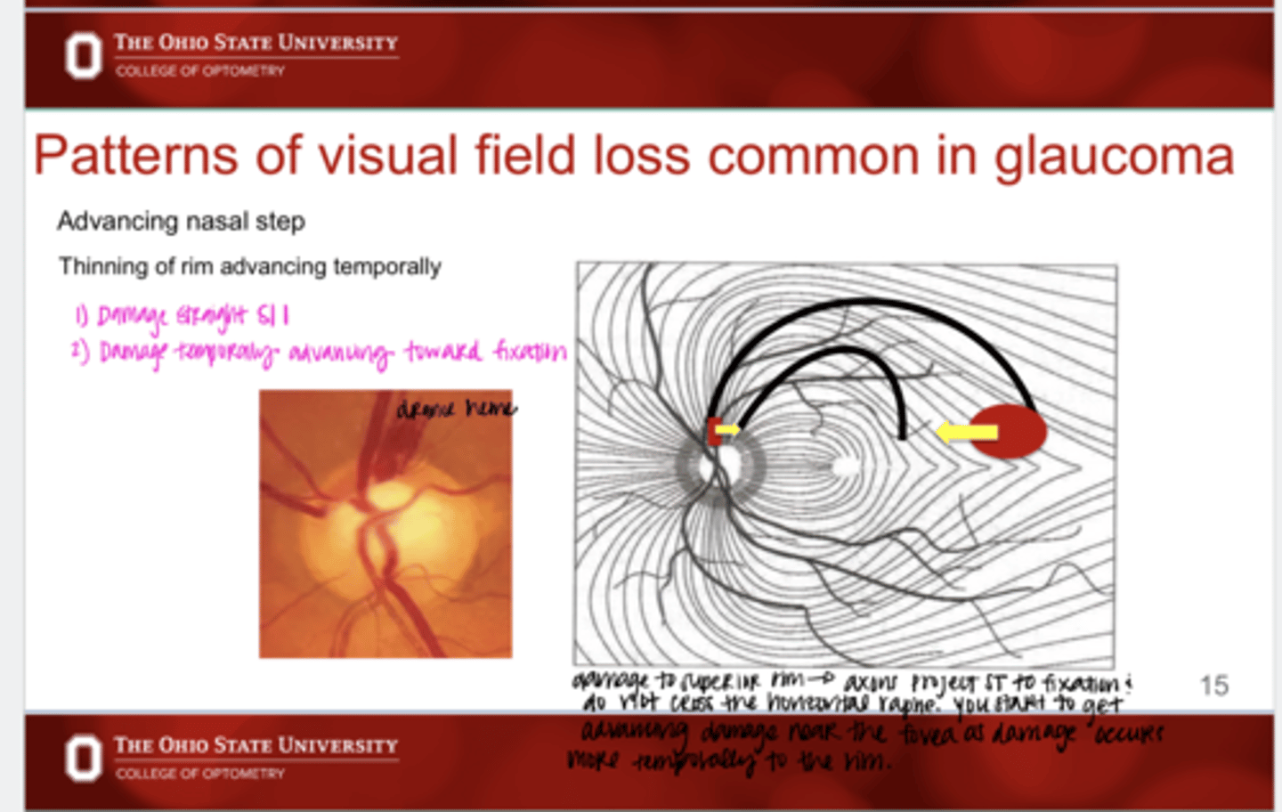
What is the characteristic appearance of an advancing nasal step VF defect?
thinning of the rim advancing temporally
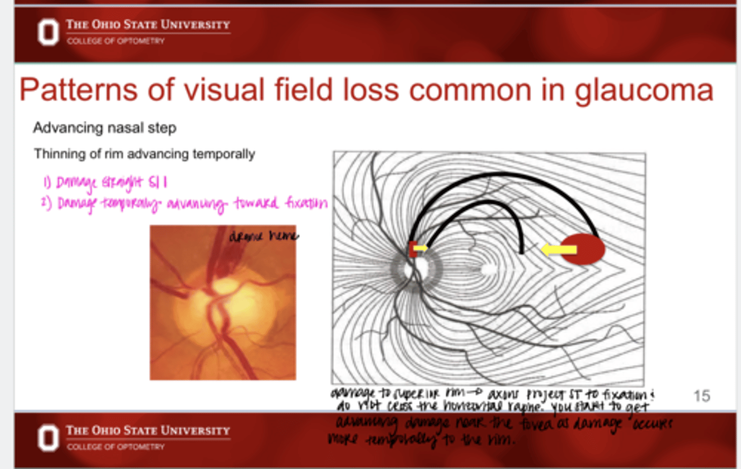
Advancing nasal step is from damage to the ______ rim
superior/inferior
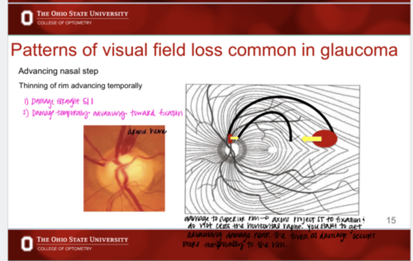
You start to get advancing damage near the fovea as damage occurs more ______ on the ONH rim
temporally
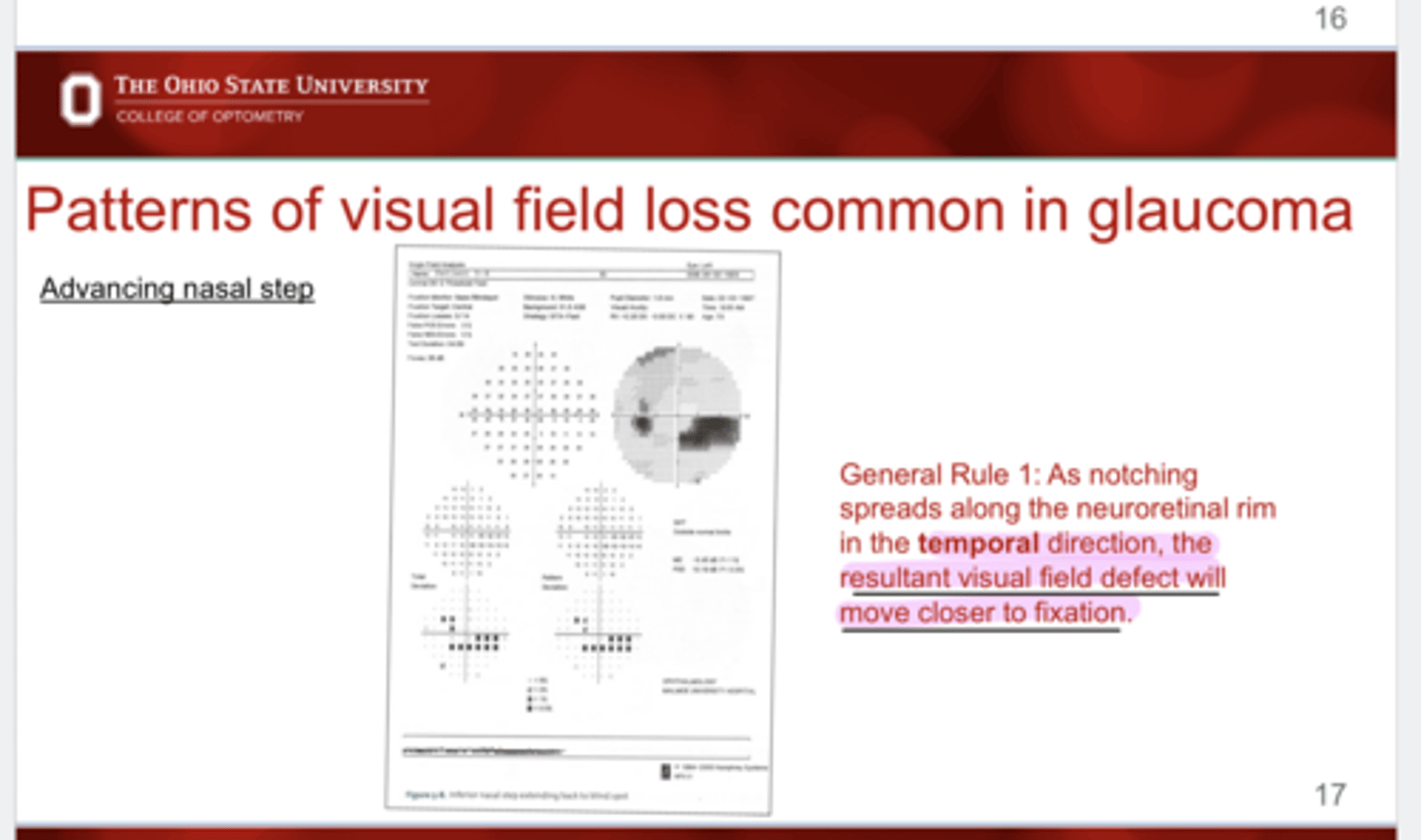
As notching spreads along the neuroretinal rim in the _____ direction, the resultant visual field defect will move closer to fixation
temporally
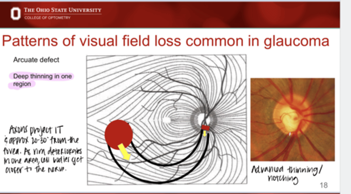
What is the characteristic appearance of a arcuate defect on VF?
deep thinning in one region
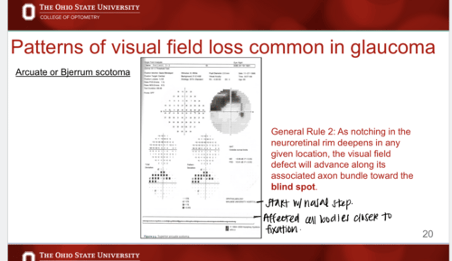
As notching of the reuroretinal rim deepens in any given location, the VF defect will advance where?
along its associated axon bundle toward the blind spot
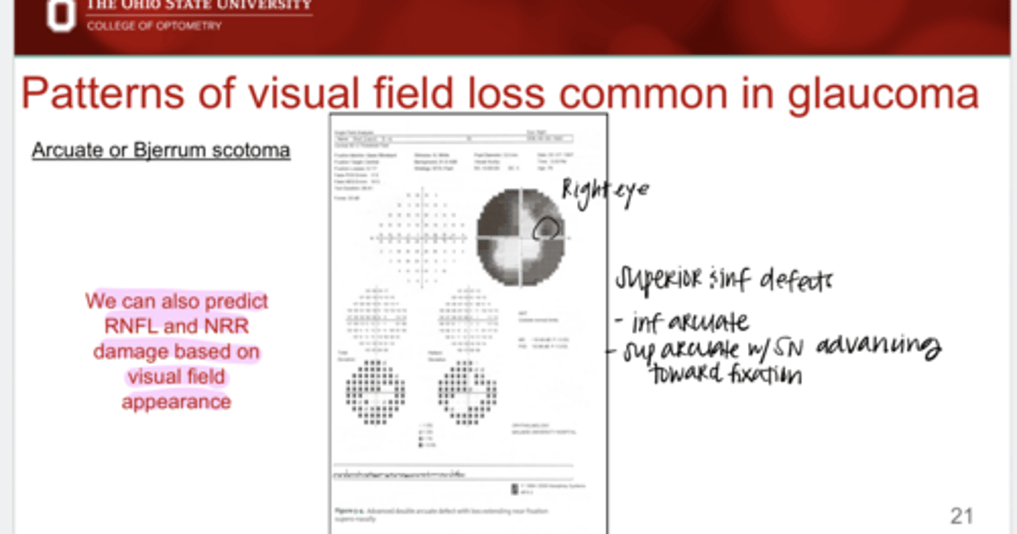
True or False:
We can predict RNFL and NRR appearance and damage based on VF damage
True
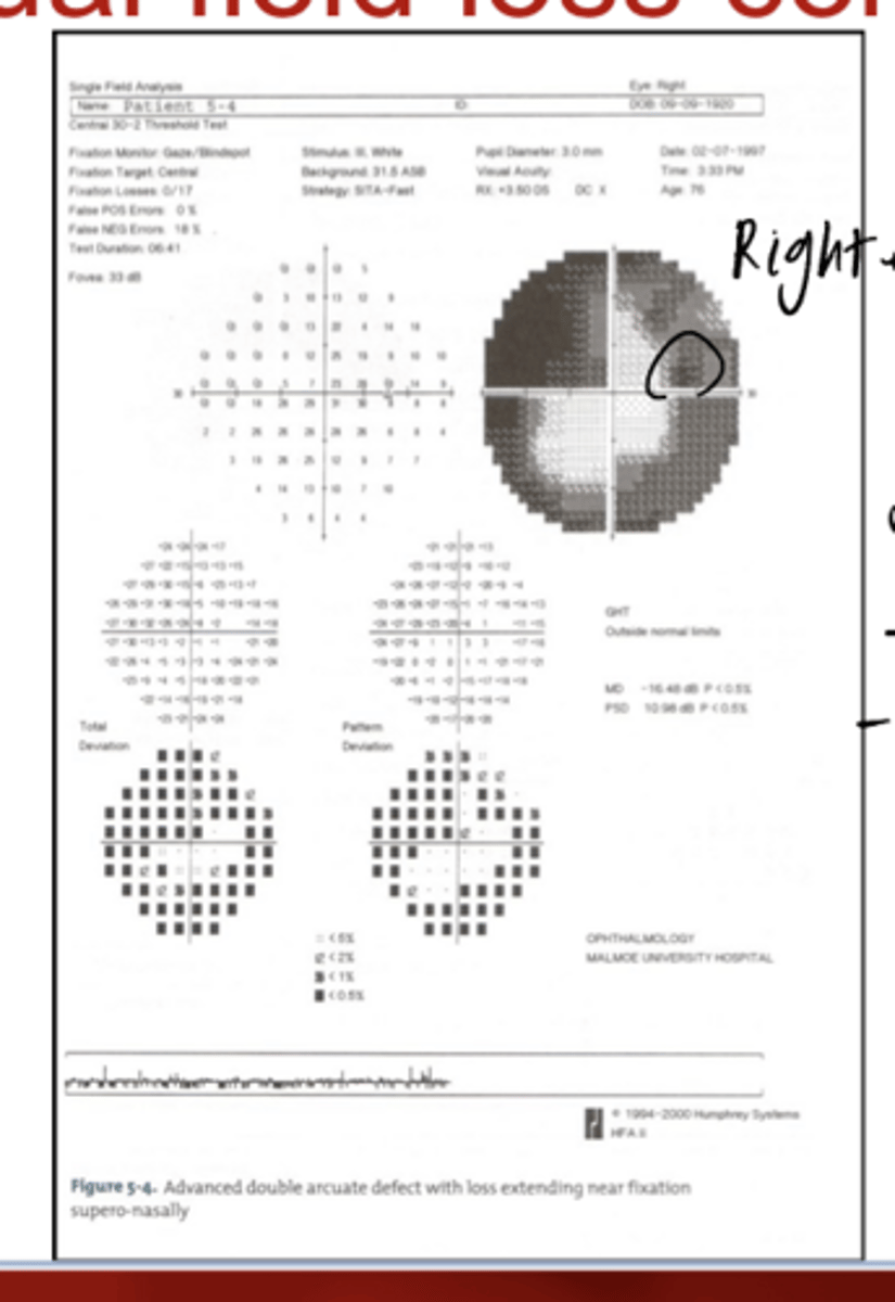
Describe the VF deficits in the pic
-superior and inferior arcuate defects
-superior arcuate defect is advancing towards fixation
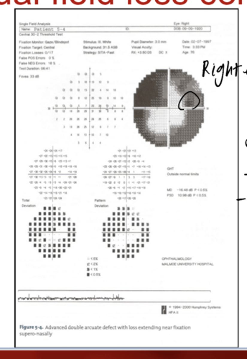
A defect on superior VF will represent the ______ nerve
inferior
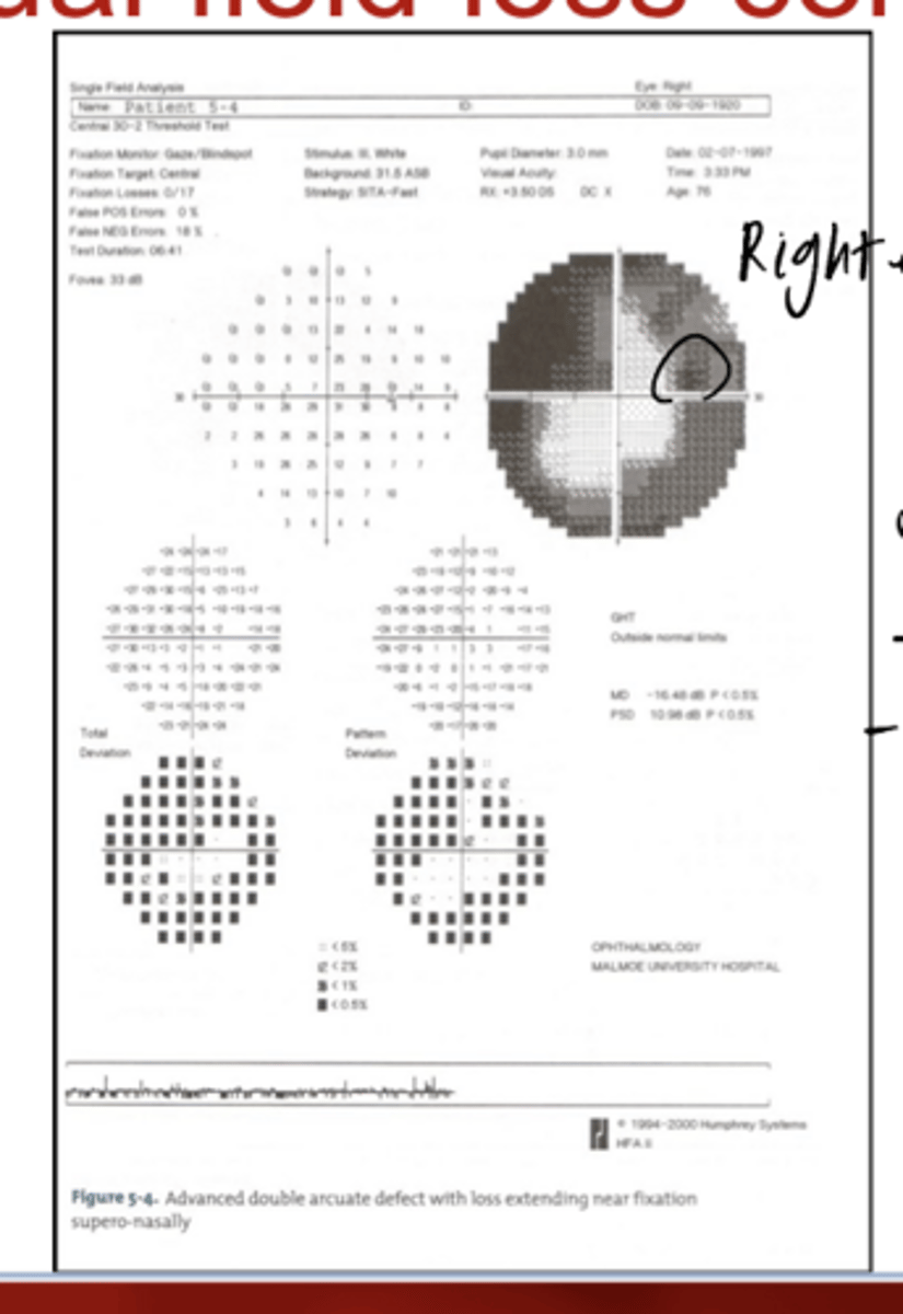
A defect on inferior VF will represent the ______ nerve
superior
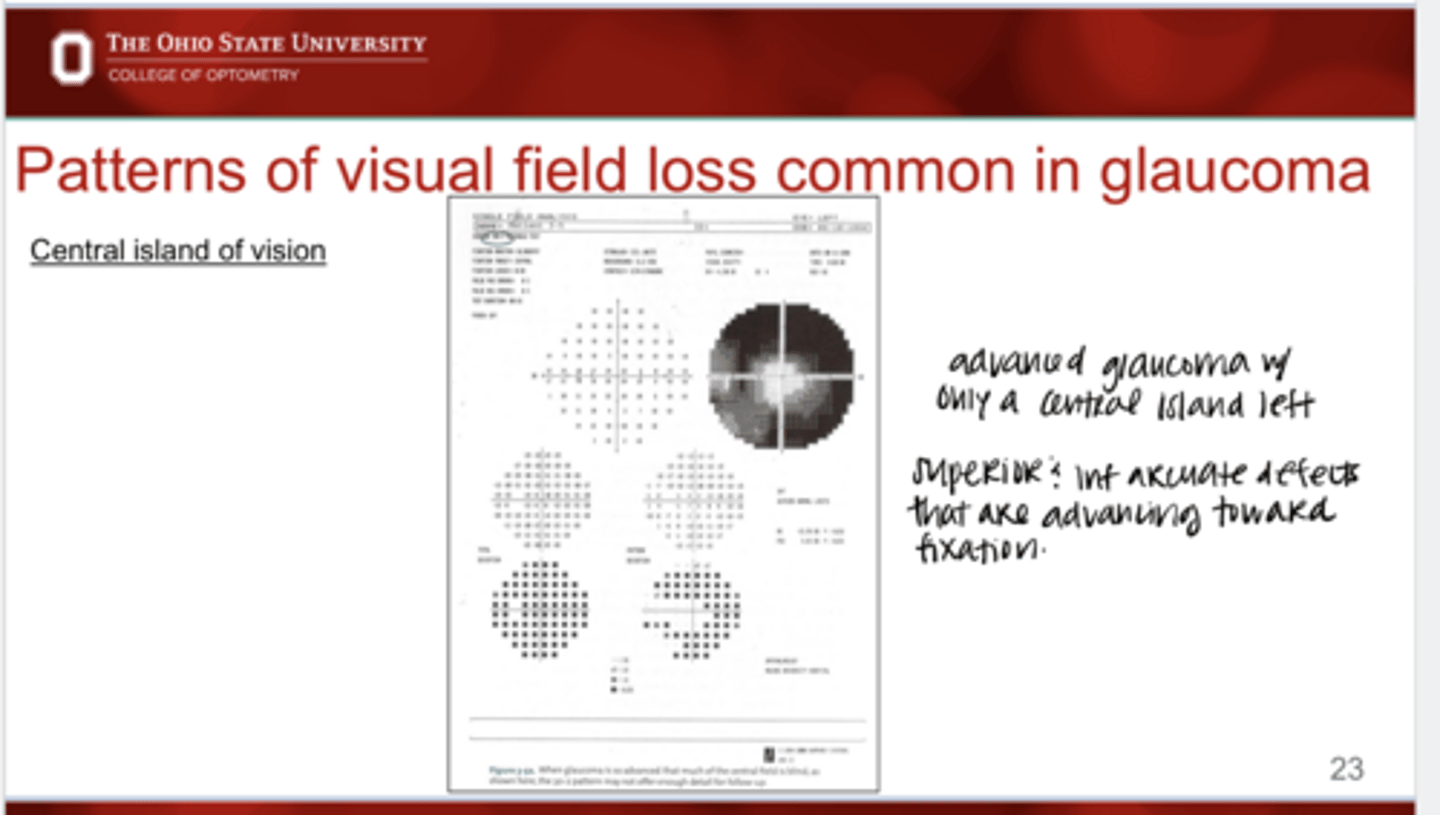
What defect is present in the following pic?
Central island of vision left -- superior and inferior arcuate defects that are both advancing towards fixation
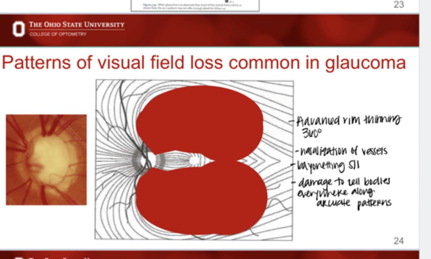
Describe the following ONH
-advanced rim thinning 360º
-nasalization of vessels
-bayonetting of vessels S/I
-Damage to the cell bodies everywhere along arcuate patterns
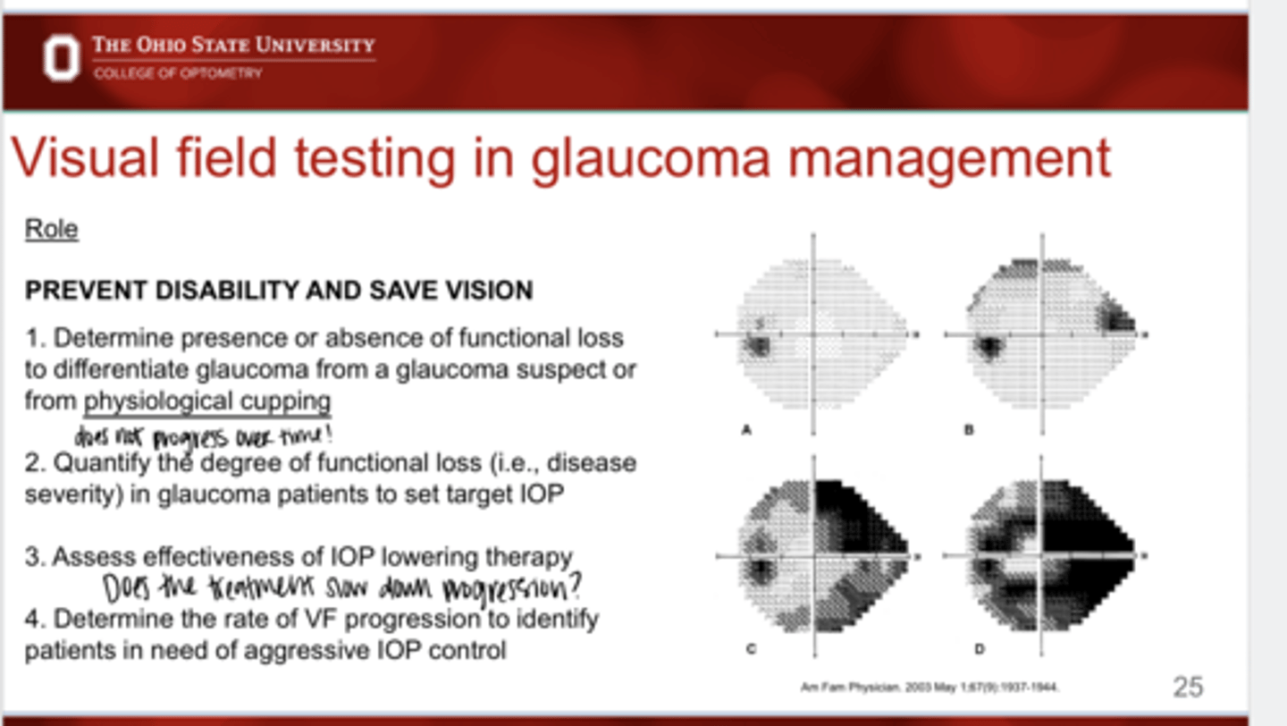
What is the role of VF testing in glaucoma management?
1) Determine presence or absence of functional loss to differentiate glaucoma from a glaucoma suspect or from psychological cupping
2) Quantify the degree of functional loss in glaucoma patients to set target IOP
3) Assess effectiveness of IOP lowering therapy
4) Determine the rate of VF progression to identify patients in need of aggressive IOP control
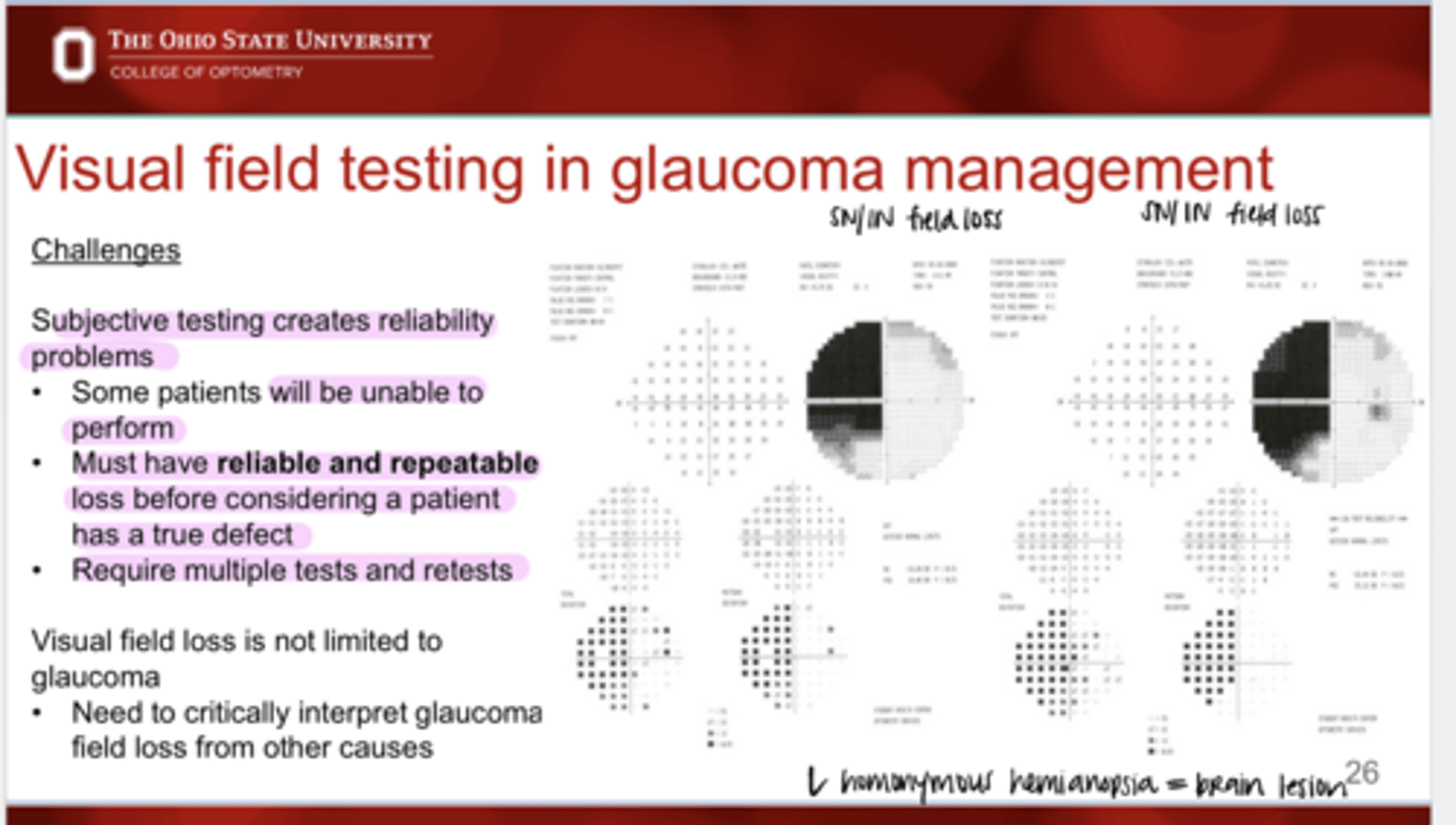
Challenges of VF Testing
_______ testing creates reliability problems
subjective
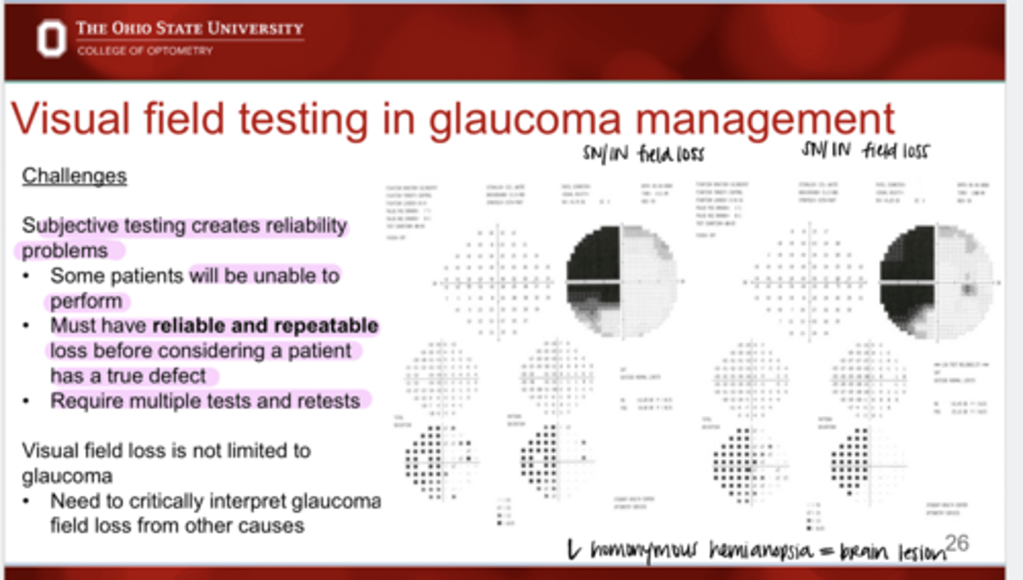
Challenges of VF Testing
Must have ____ and _____ loss before considering a patient has a true defect
reliable, repeatable
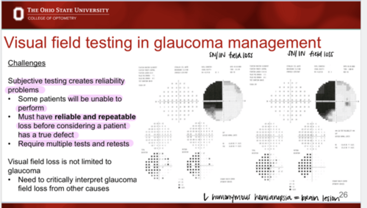
Challenges of VF Testing
Does VF testing require multiple tests and retests?
Yes
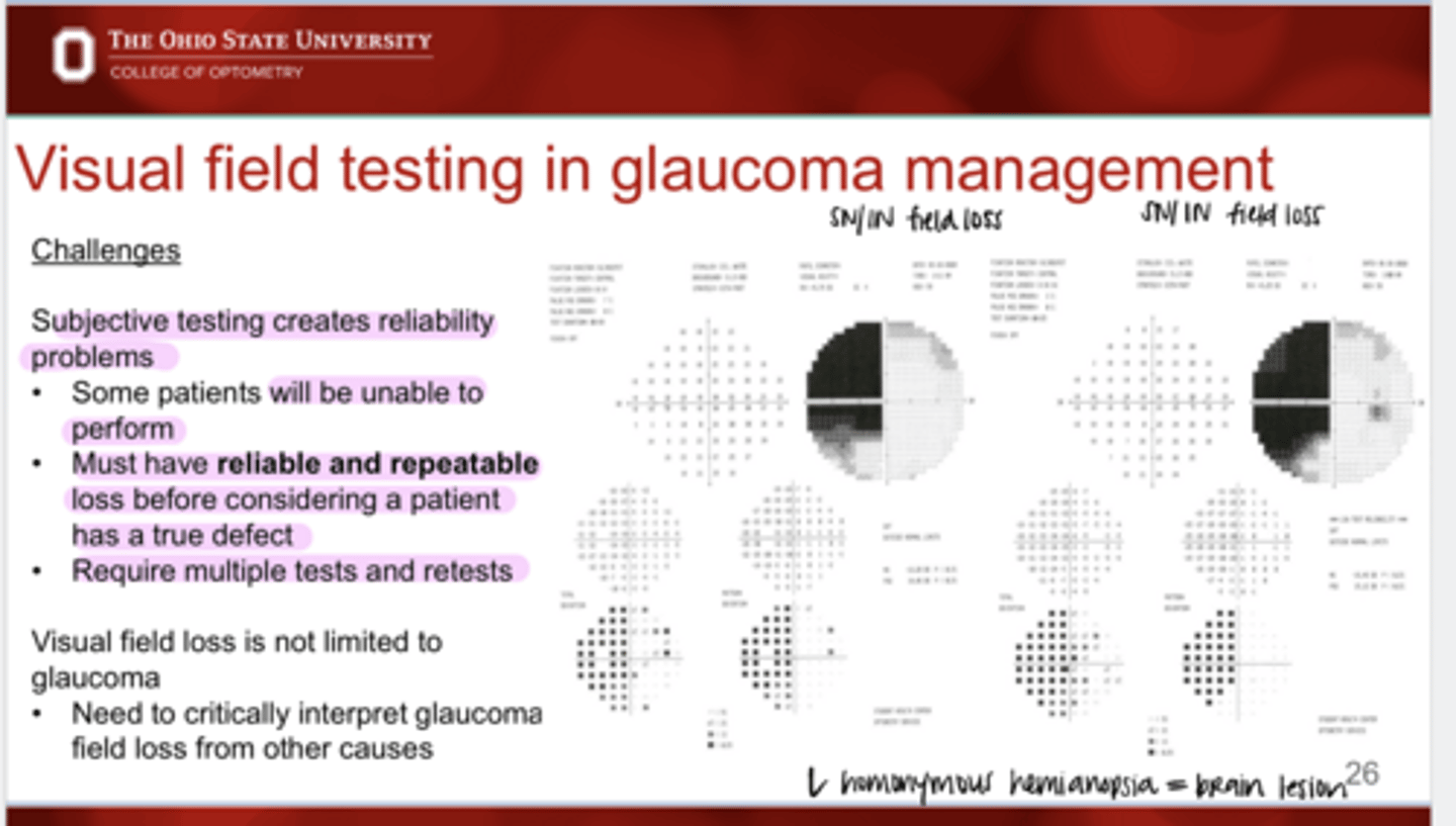
Is visual field loss limited to glaucoma?
No
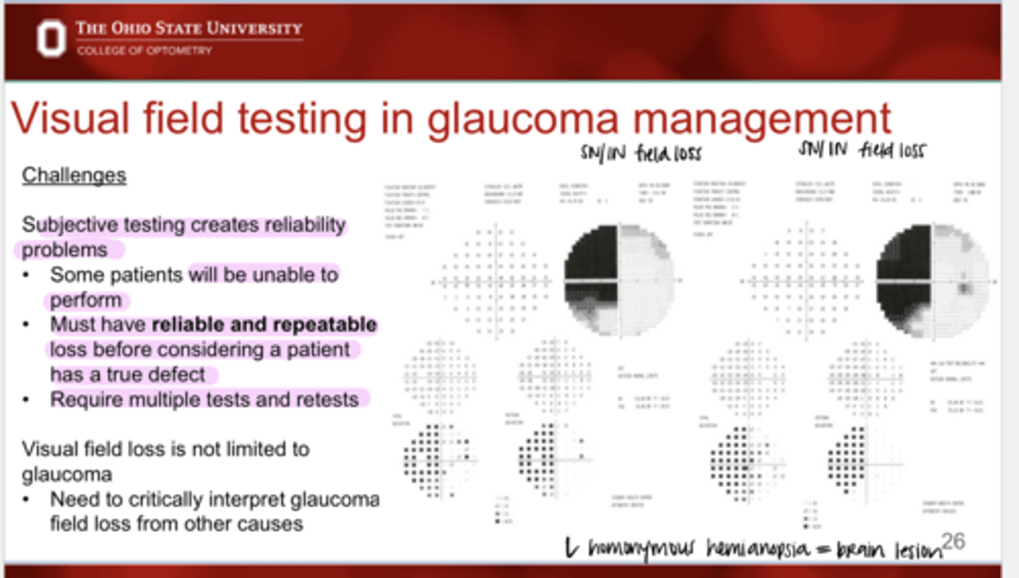
True or False:
You need to critically interpret glaucoma field loss from other causes
true
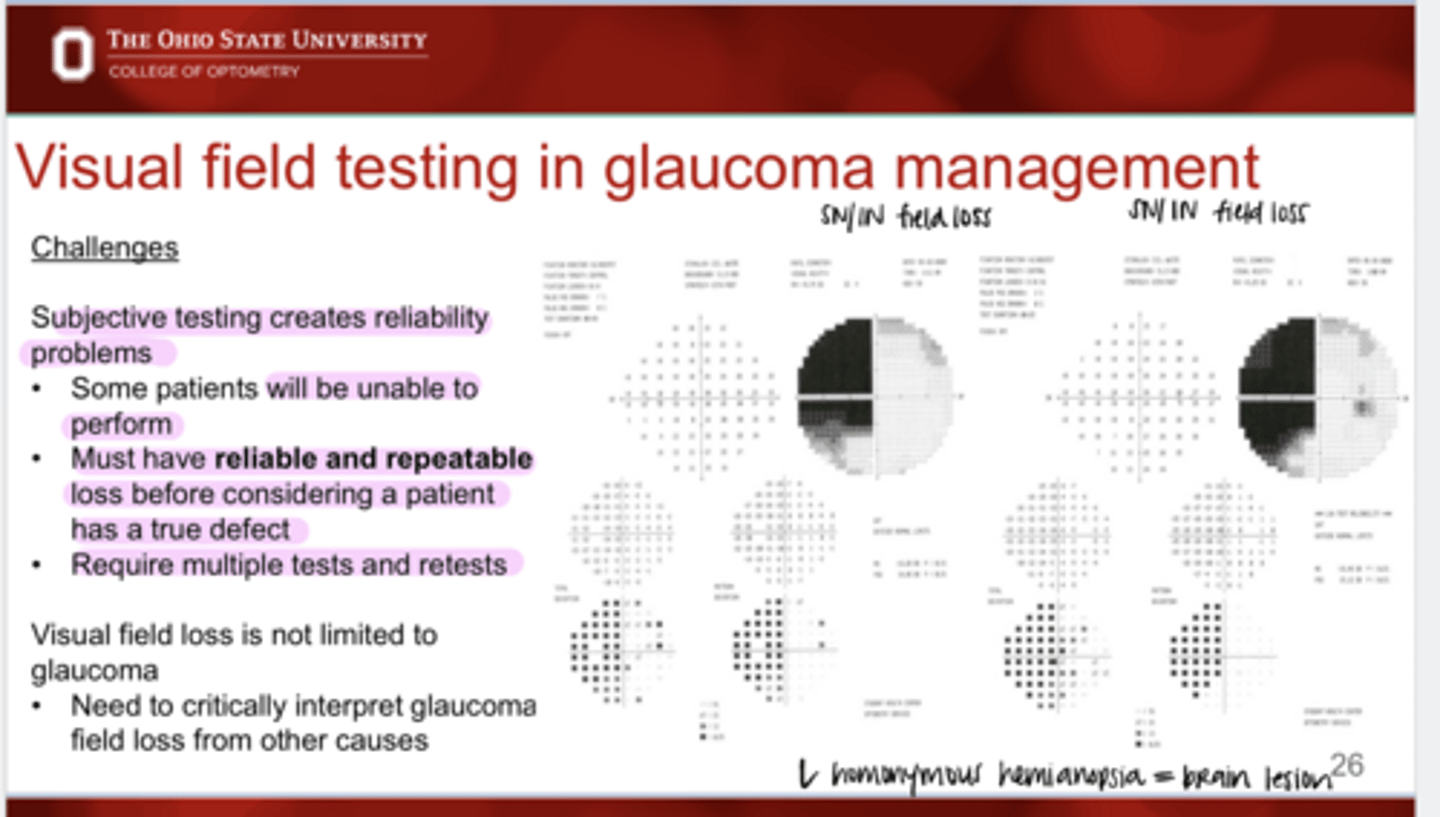
What is shown in this VF?
left homonymous hemianopsia
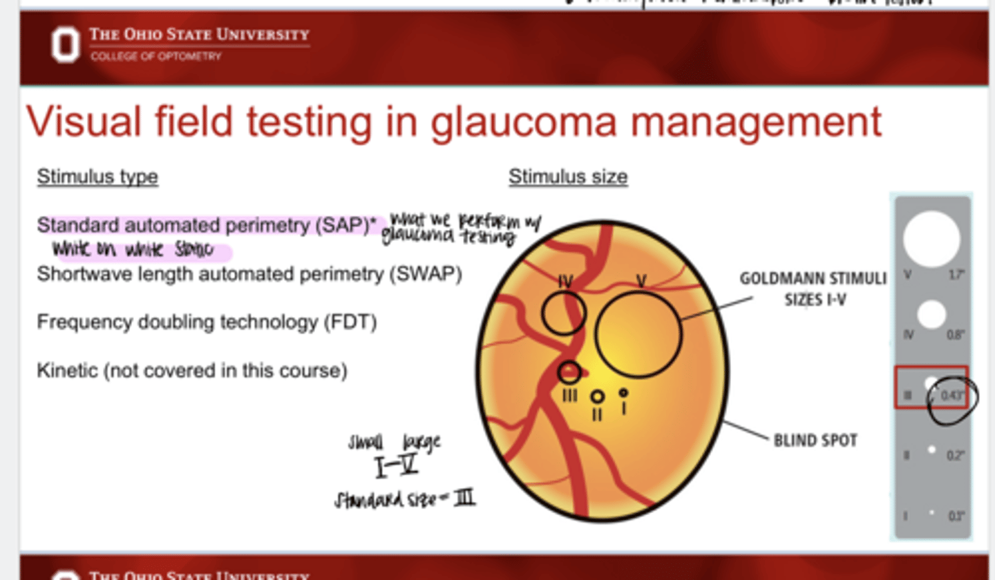
What are the stimulus types that are available in visual field testing?
-Standard automated perimetry (SAP)
-Shortwave length automated perimetry (SWAP)
-Frequency doubling technology (FDT)
-Kinetic
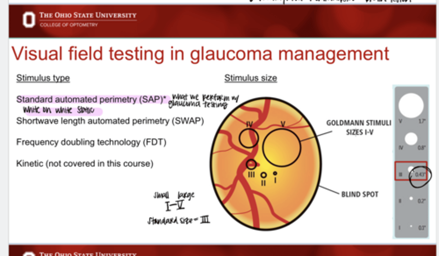
Which testing stimulus type is used in glaucomatous VF testing?
Standard automated perimetry (SAP)
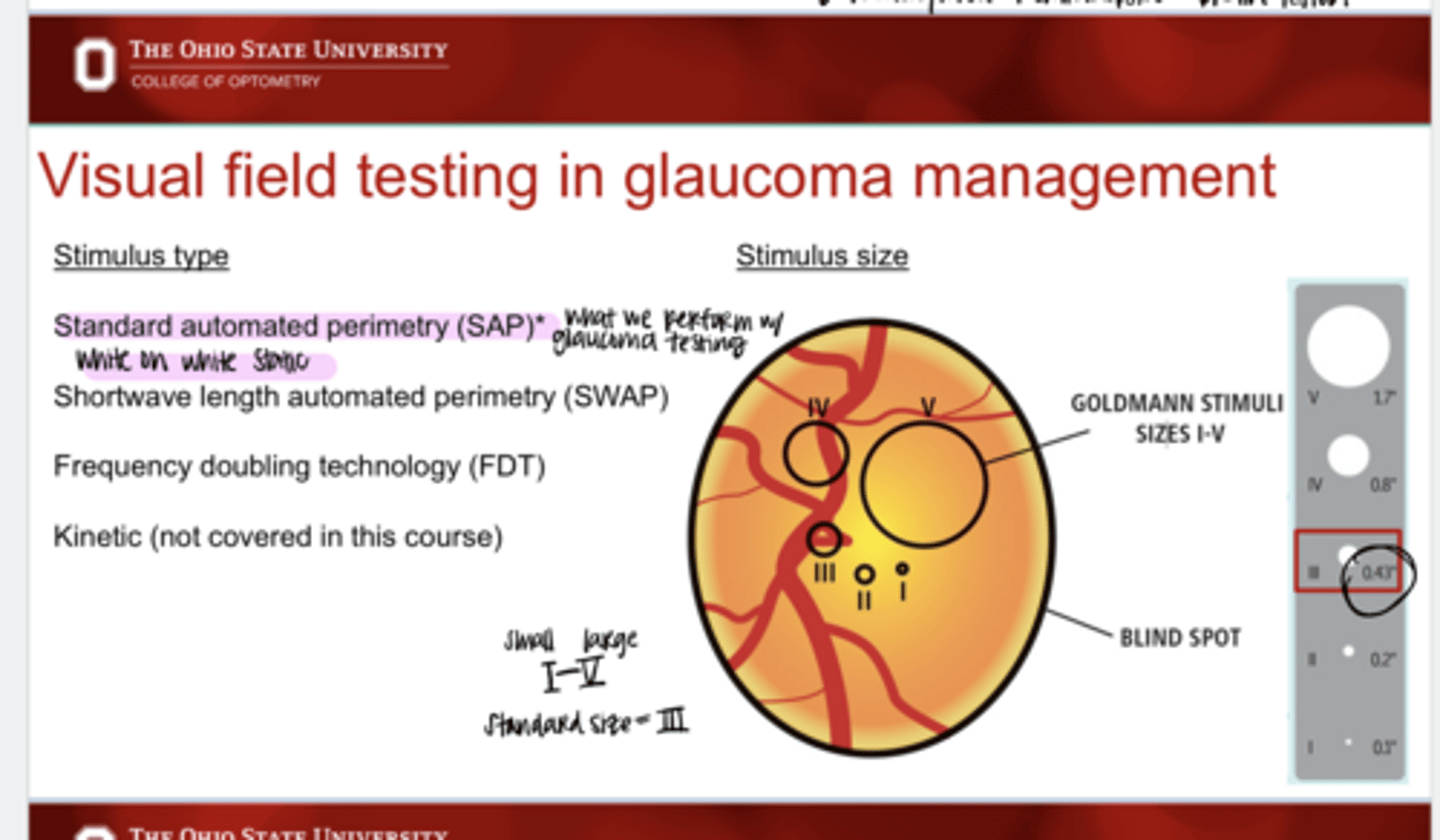
What does a I stimulus size represent?
smallest stimulus
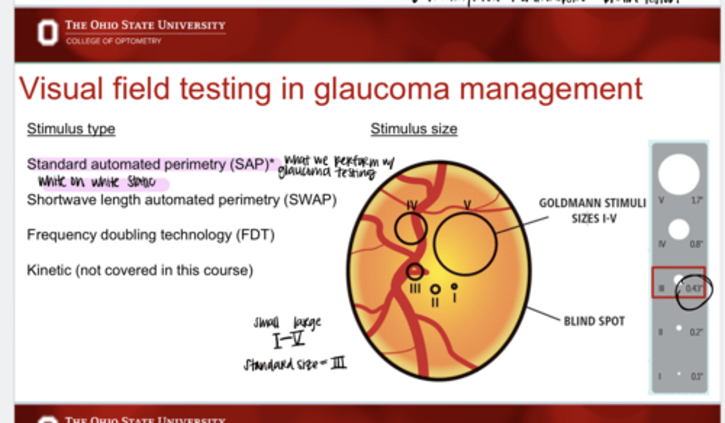
What does a V stimulus size represent?
largest stimulus
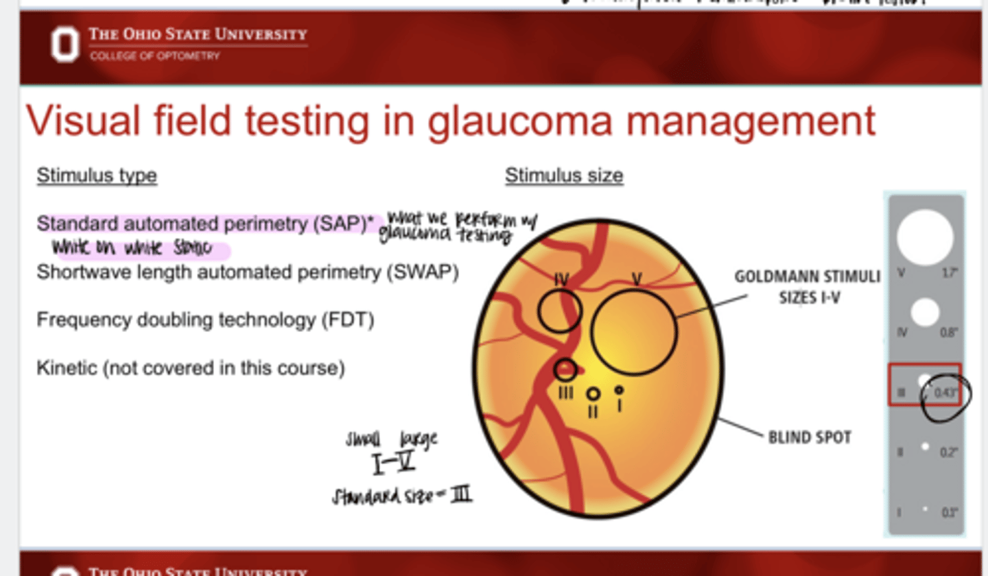
What is the standard stimulus size for VF testing?
III
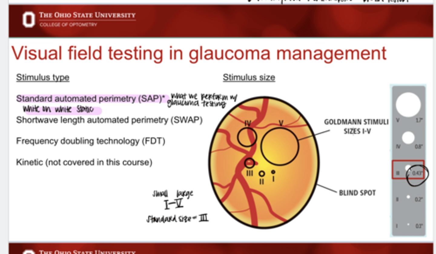
What is the size (in degree) of the Goldmann Stimulus size III?
0.43º
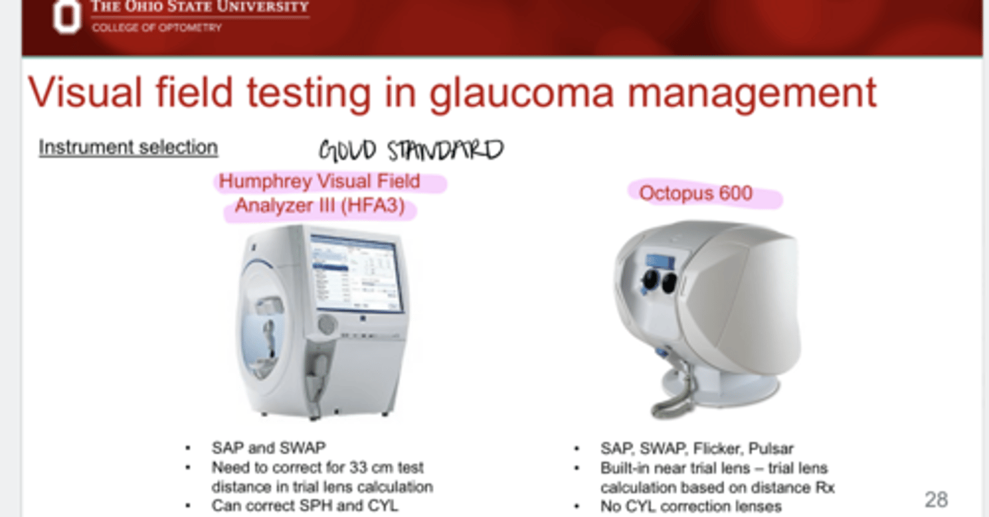
What VF instrument is the gold standard in glaucoma management?
HFA3 Humphrey Field Analyzer
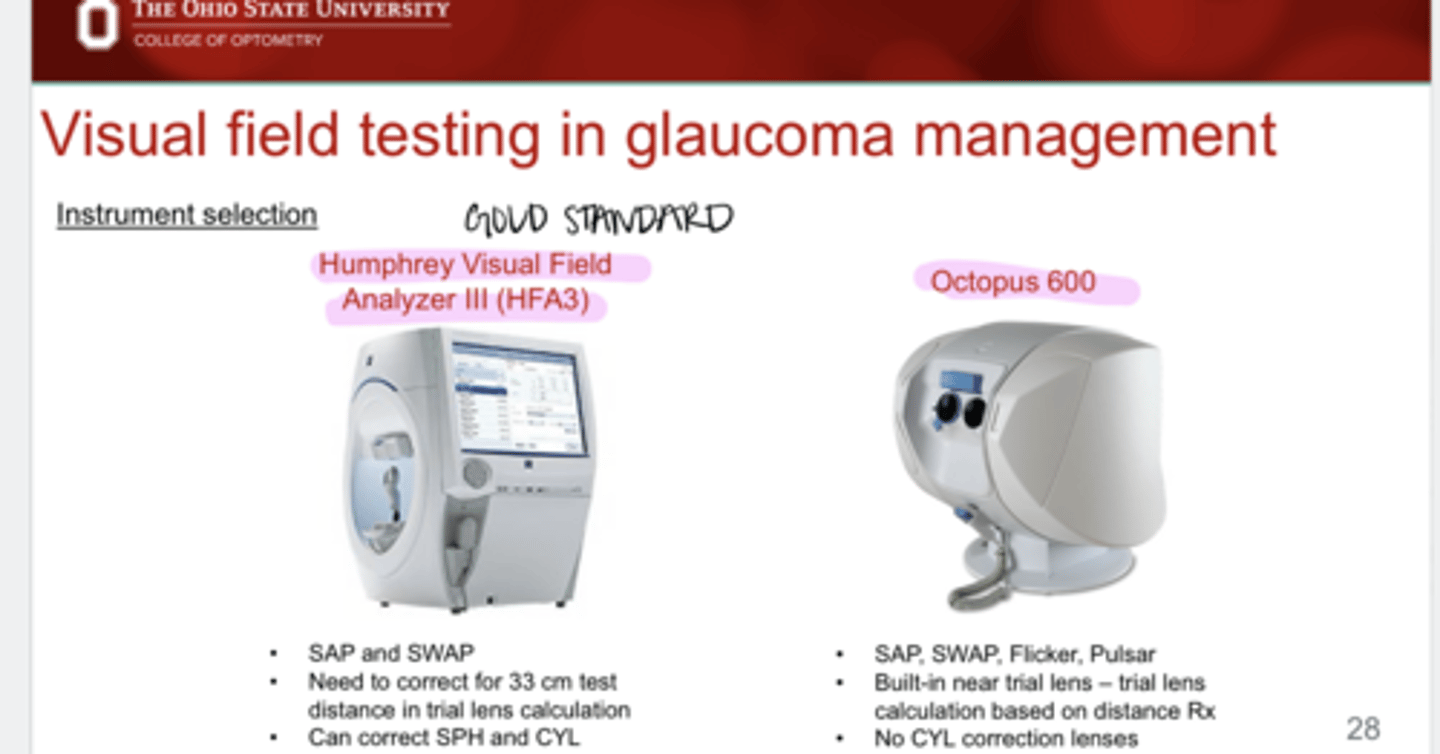
What test strategies are available on the HFA3?
SAP and SWAP
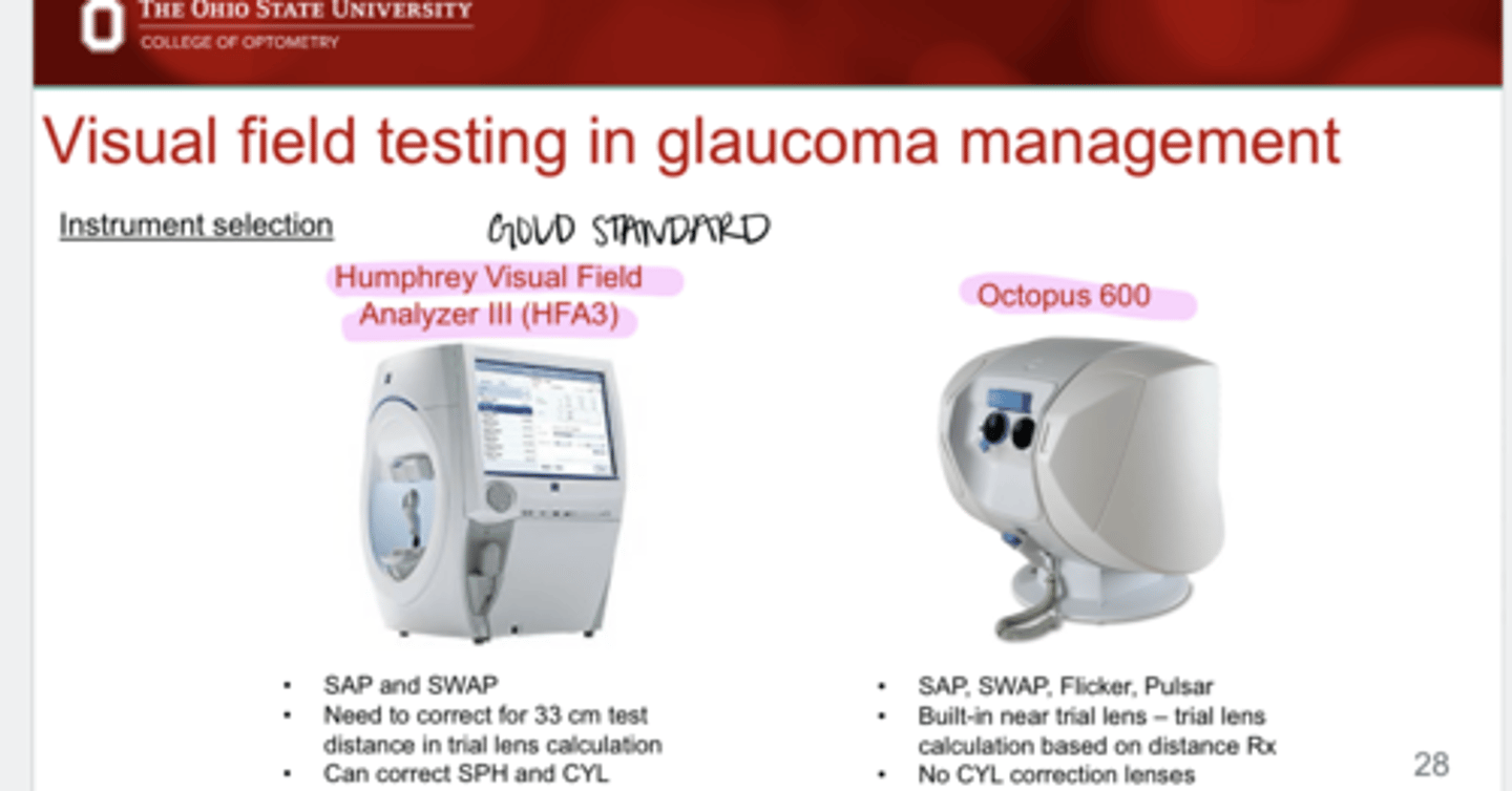
With a HFA3, what needs to be corrected?
Need to correct for the 33cm testing distance
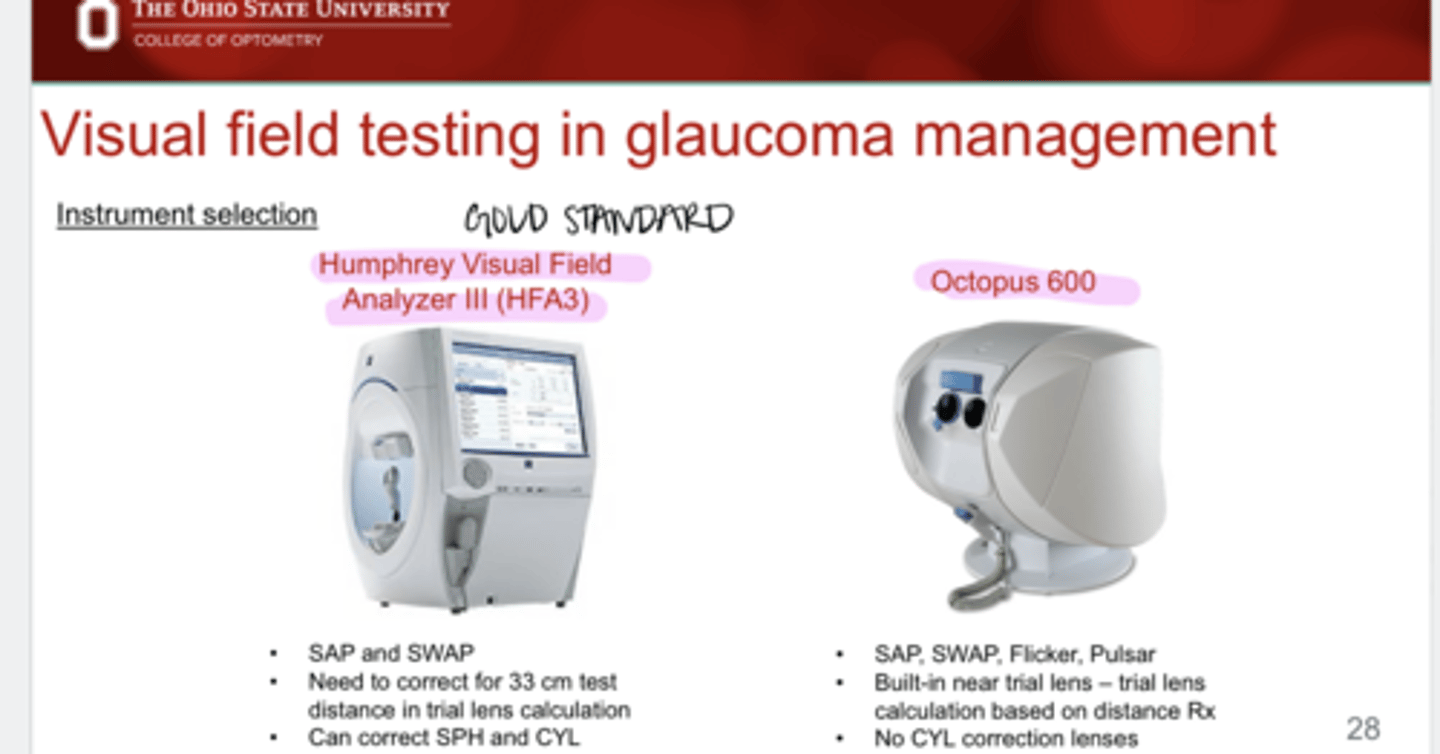
HFA3 can correct for (sph/cyl)
both of these
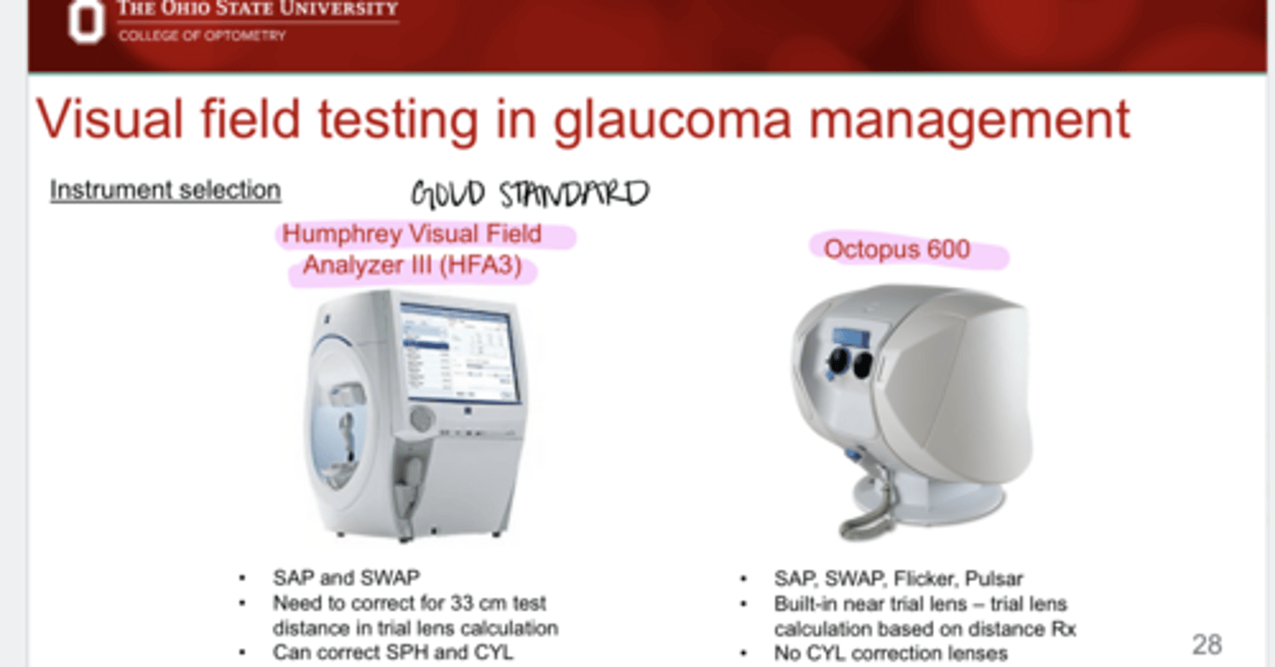
What test strategies are available on the Octopus 600?
SAP, SWAP, Flicker, Pulsar
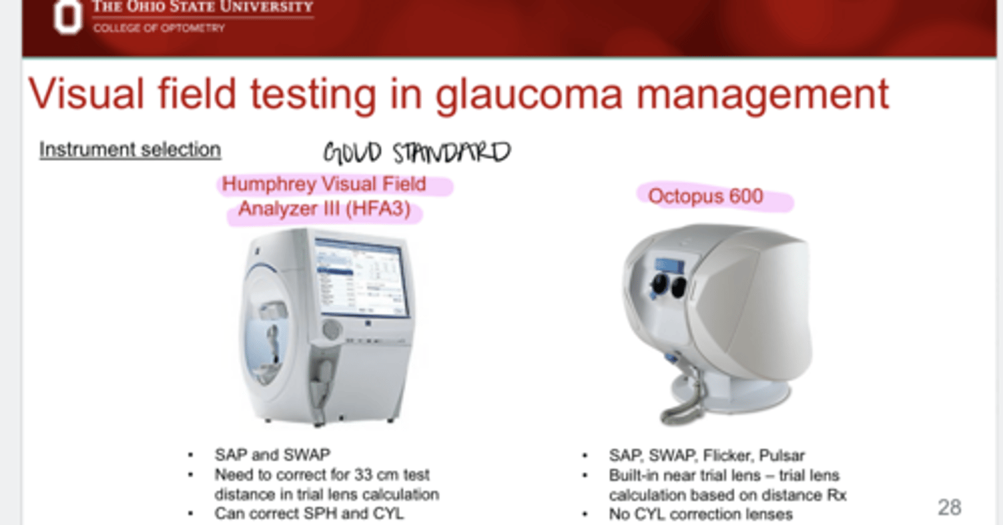
Advantages of the Octopus 600?
Built in near trials lens does not need to be corrected for
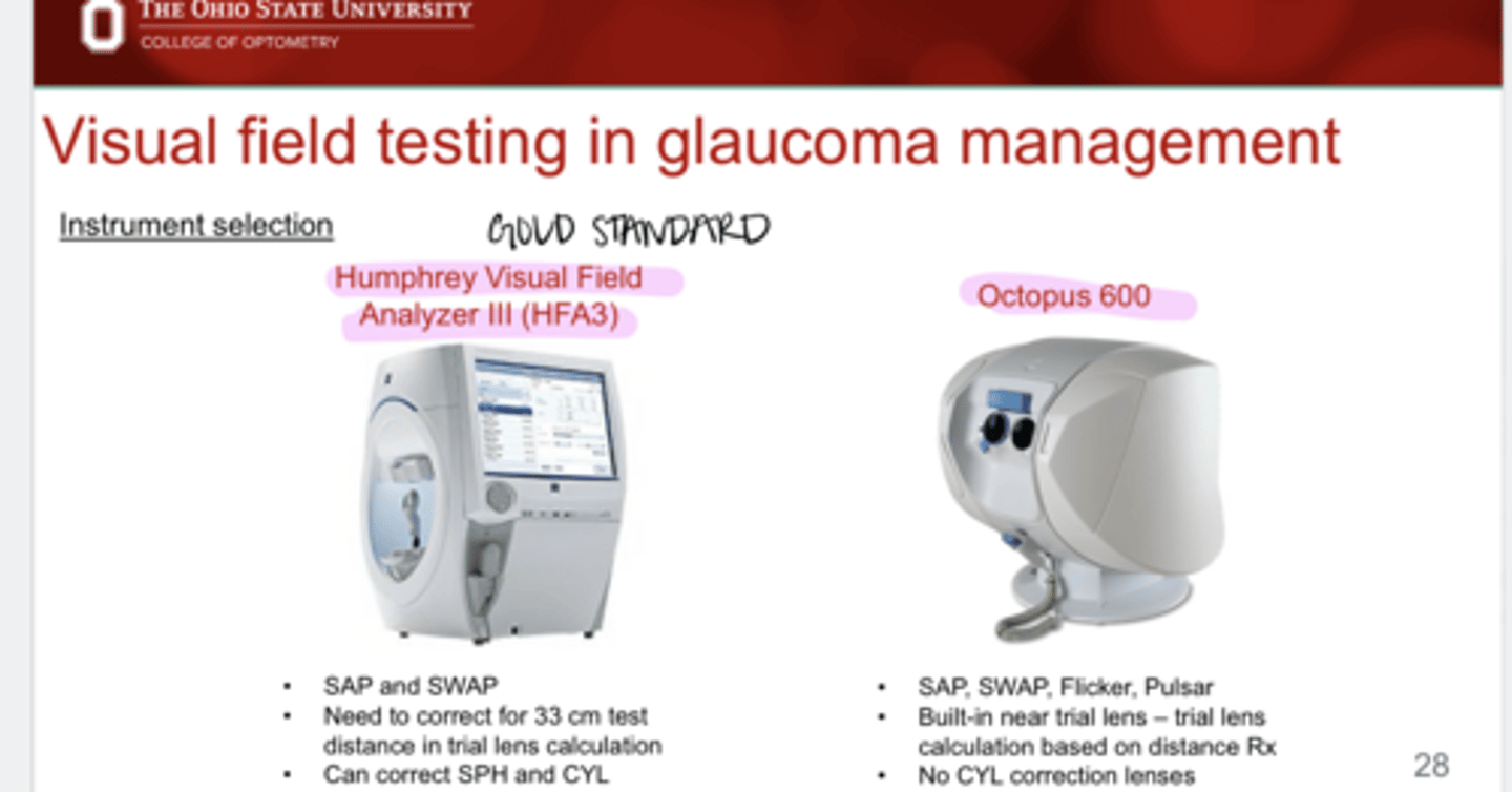
Octopus 600 can correct for (sph/cyl)
sphere
Trial Lens Selection for HFA
-2.00DS distance RX
+1.00
Trial Lens Selection for HFA
+0.75-0.50x089
+3.50DS
Trial Lens Selection for HFA
-6.00-1.00x158
-3.50DS
Trial Lens Selection for HFA
+2.00-1.75x037
+5.00-1.75x037
Over ____D of cyl, you should not use SE anymore in the trial lens calculation of VF
-1.25
Trial Lens Selection for Octopus 600
-2.00DS
-2.00DS
Trial Lens Selection for Octopus 600
+0.75-0.50x089
+0.50DS
Trial Lens Selection for Octopus 600
-6.00-1.00x158
-6.00/-7.00 DS
Trial Lens Selection for Octopus 600
+2.00-1.75x037
Habitual correction -- cannot correct for cyl with Octopus VF
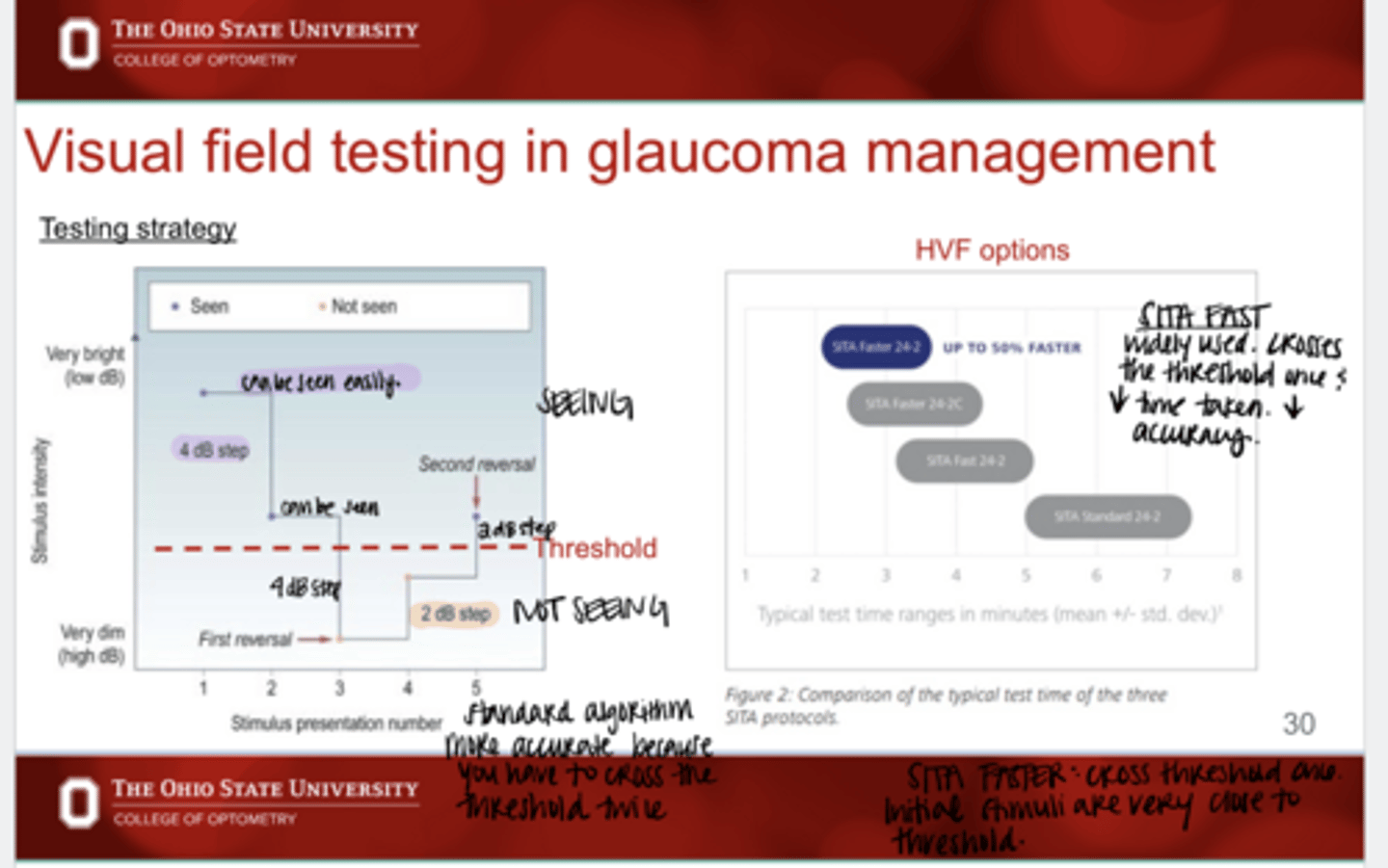
Testing Strategy for SITA STANDARD Testing Strategy
-Present stimulus way above threshold (seen)
-Present another stimulus 4dB lower (seen)
-Present another stimulus 4dB lower (not seen)
-Present another stimulus 2dB higher (not seen)
-Present another stimulus 2dB higher (seen)
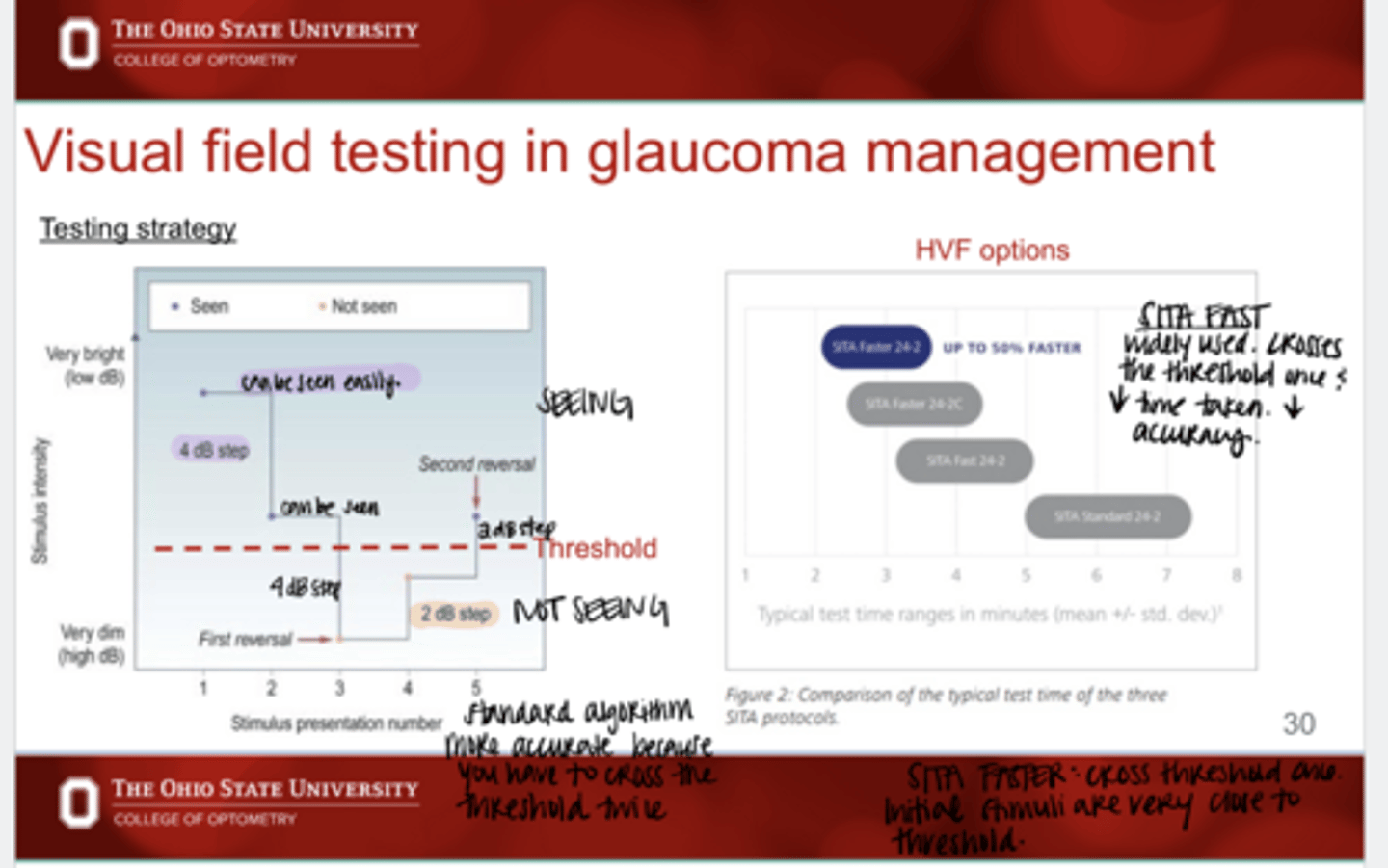
In standard testing strategy, how many times will the threshold line be crossed?
2
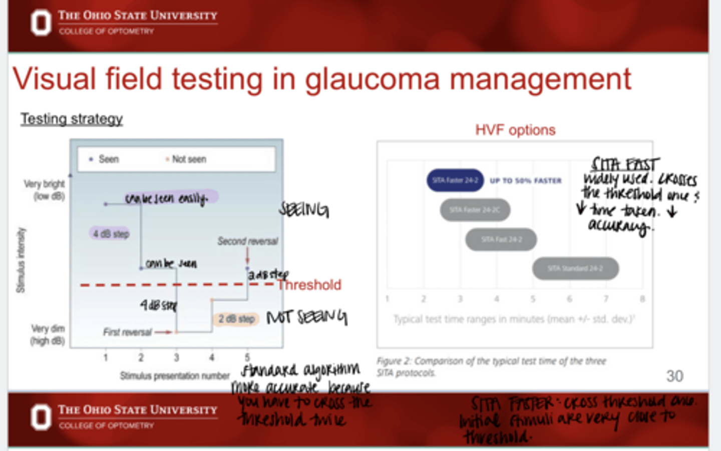
What are the Testing Strategy options for HFA that are faster than SITA standard?
-SITA FAST
-SITA FASTER
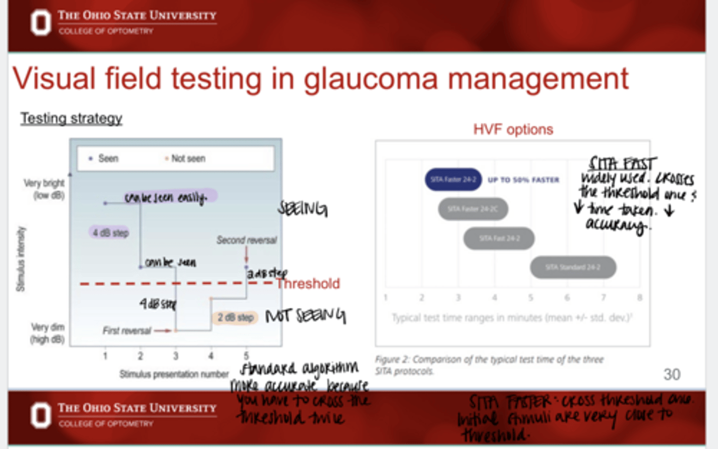
What are the advantages of SITA FAST and FASTER?
less time taken
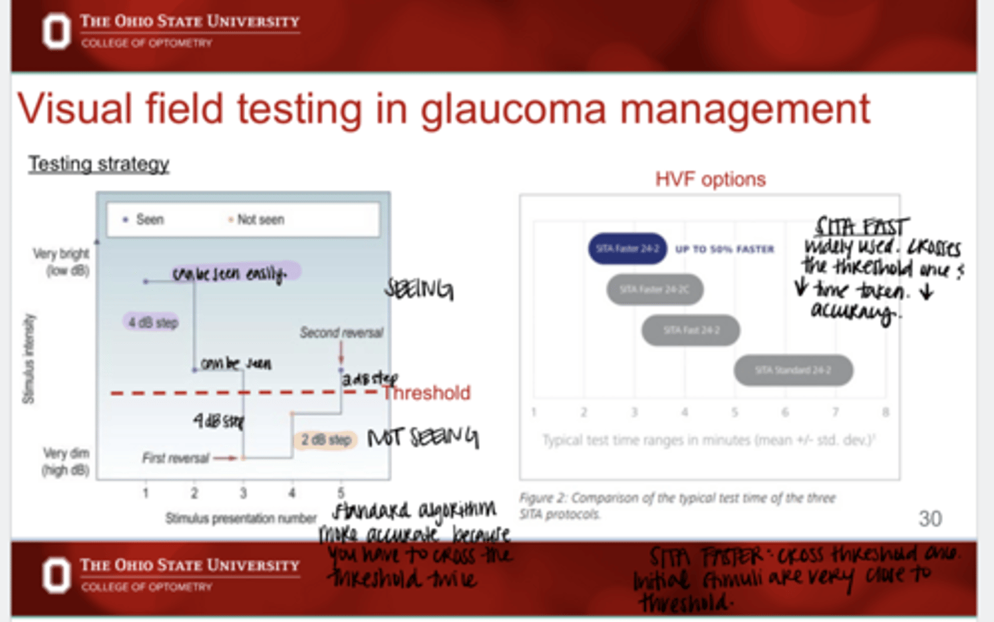
What are the disadvantages of SITA FAST and FASTER?
less accuracy
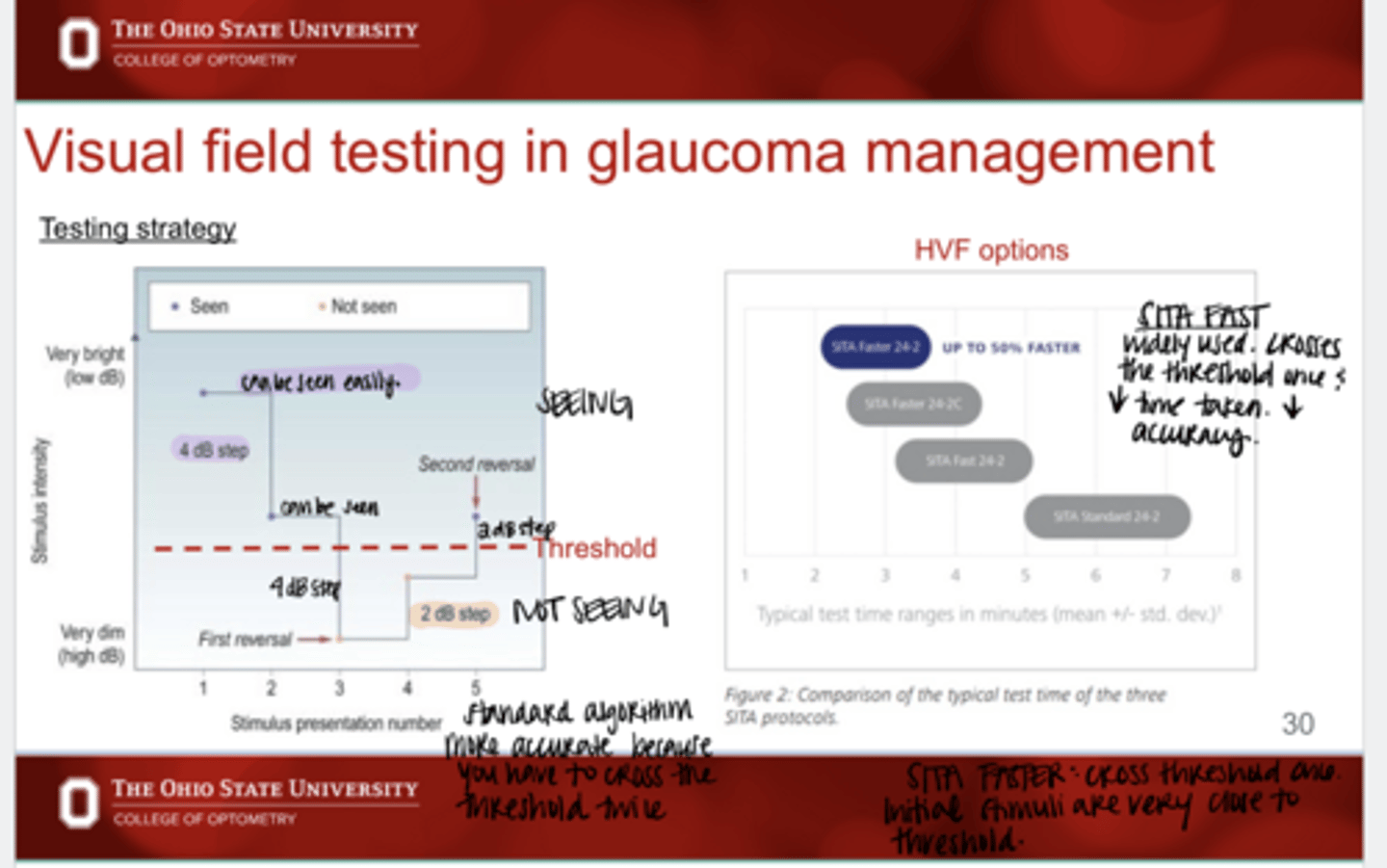
With SITA FAST and FASTER, how many times will the threshold line be crossed?
1
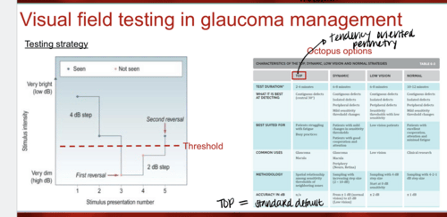
What is the standard default testing strategy on Octopus 600?
TOP
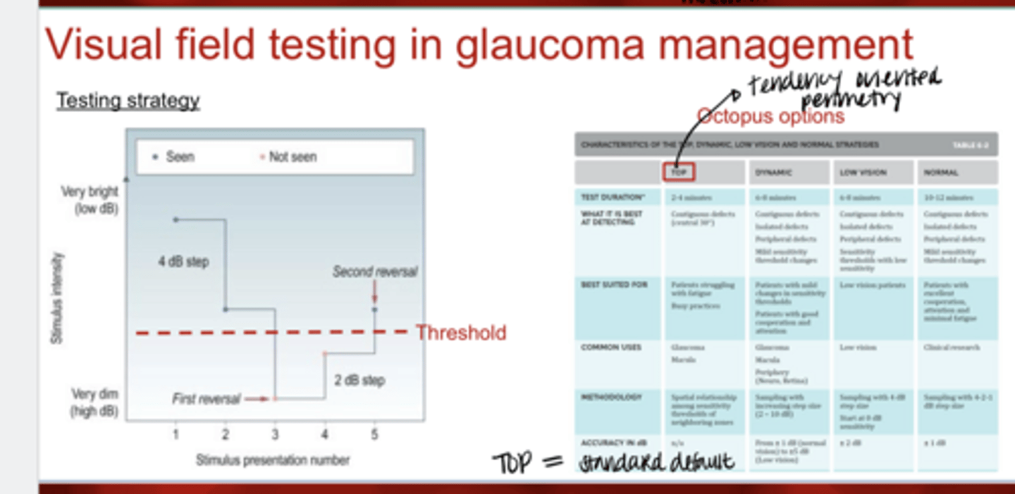
What are the common uses for the TOP VF strategy?
-glaucoma
-macula
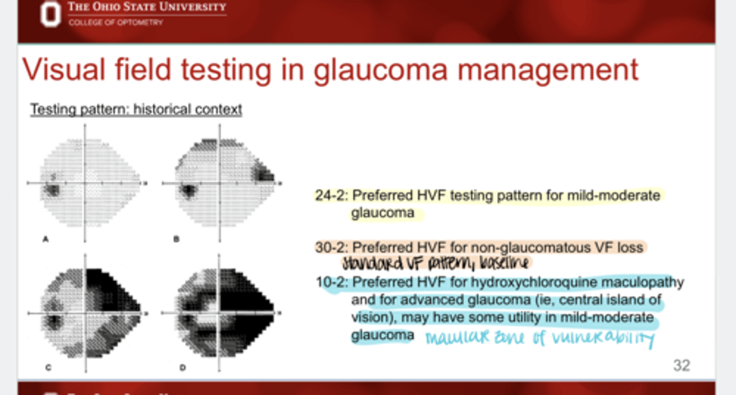
What are 24-2 VF testing patterns used for?
Preferred HVF testing pattern for mild-moderate glaucoma
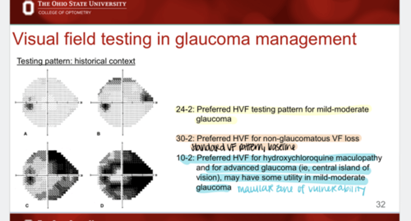
What are 30-2 VF testing patterns used for?
Preferred HVF for non-glaucomatous VF loss
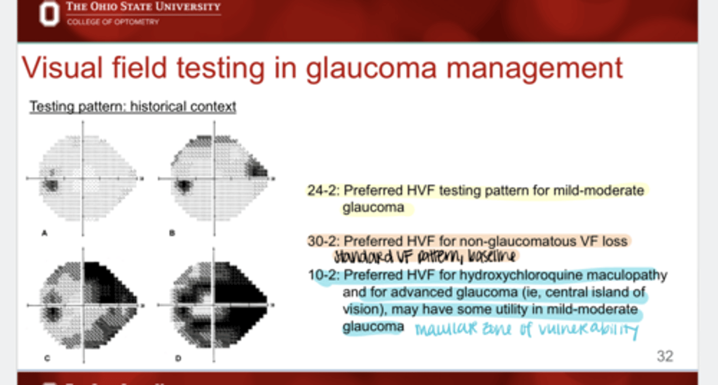
What are 10-2 VF testing patterns used for?
Preferred HVF for hydroxychloroquine maculopathy and for advanced glaucoma (central island of vision); may have some utility in mild-moderate glaucoma
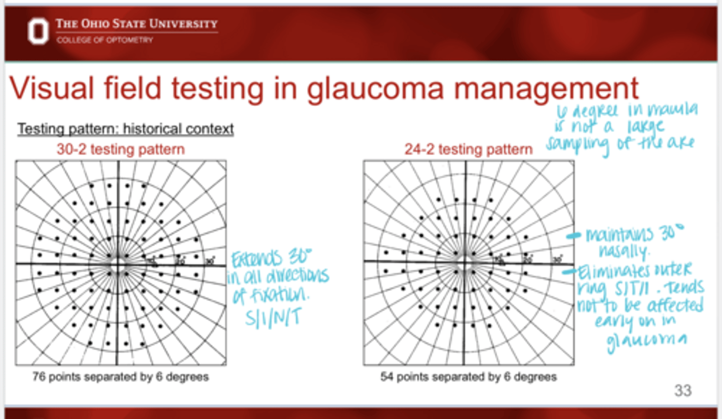
30-2 VF Pattern v 24-2 VF Pattern (Pic)
30-2 VF Pattern v 24-2 VF Pattern (Pic)
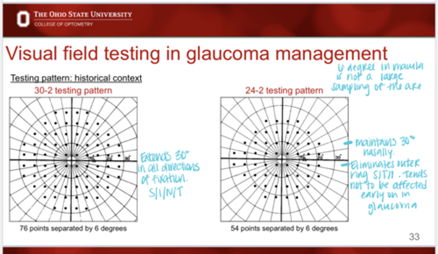
Extend of Testing with 30-2 VF
Extends 30º in all directions of fixation
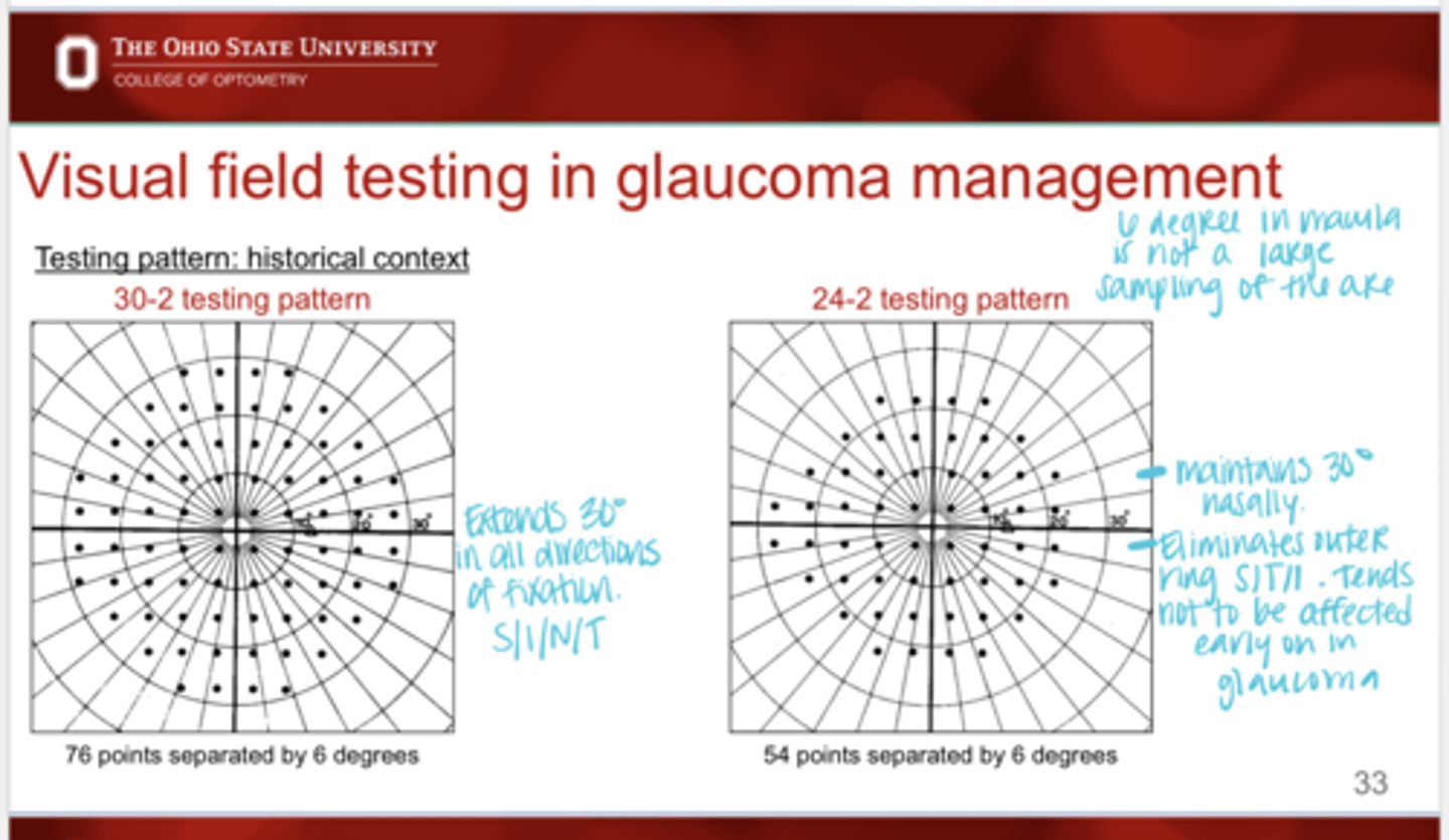
Extend of Testing with 24-2 VF
-Maintains 30º nasally
-Eliminates outer ring of points S/T/I which tends not to be affected early on in glaucoma
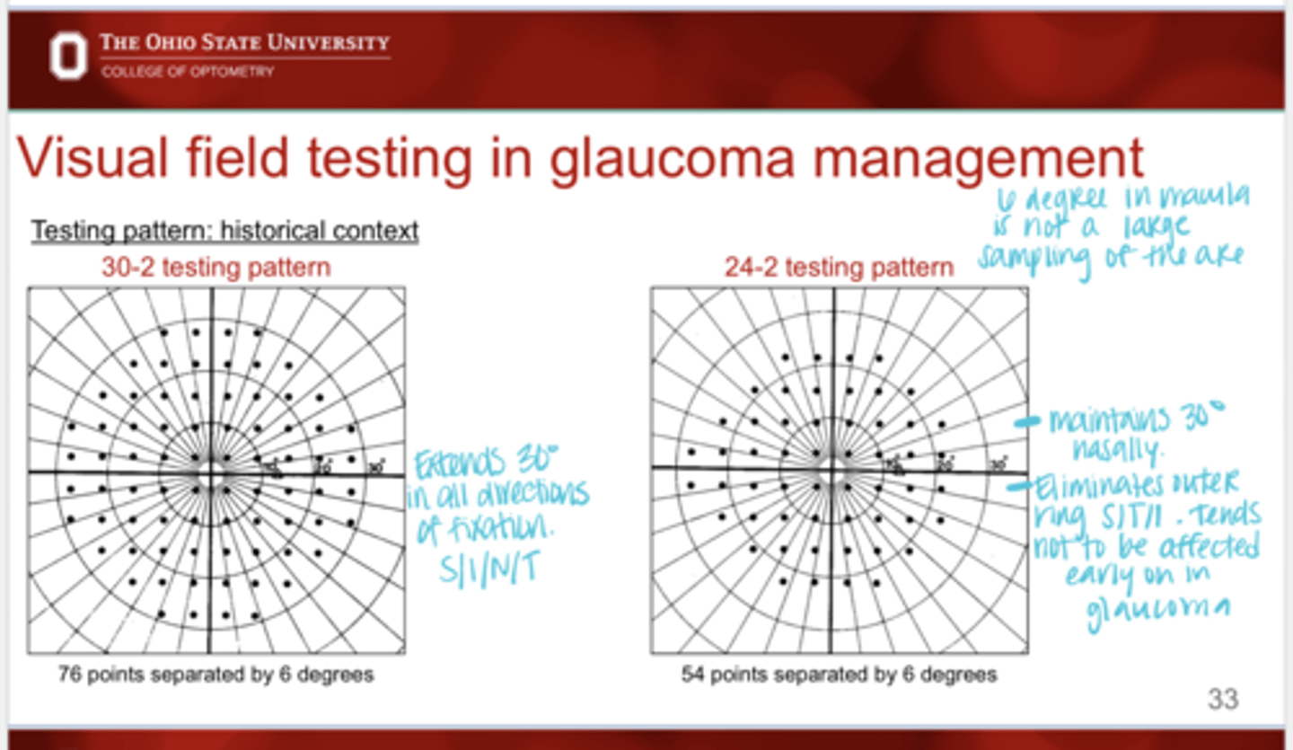
How many points are tested with 30-2 and seperation of the points?
76 points tested, 6 degrees apart
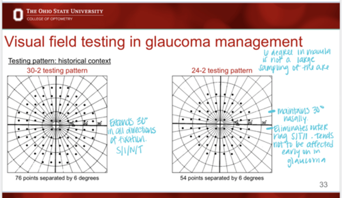
How many points are tested with 24-2 and separation of the points?
54 points tested, 6 degrees apart
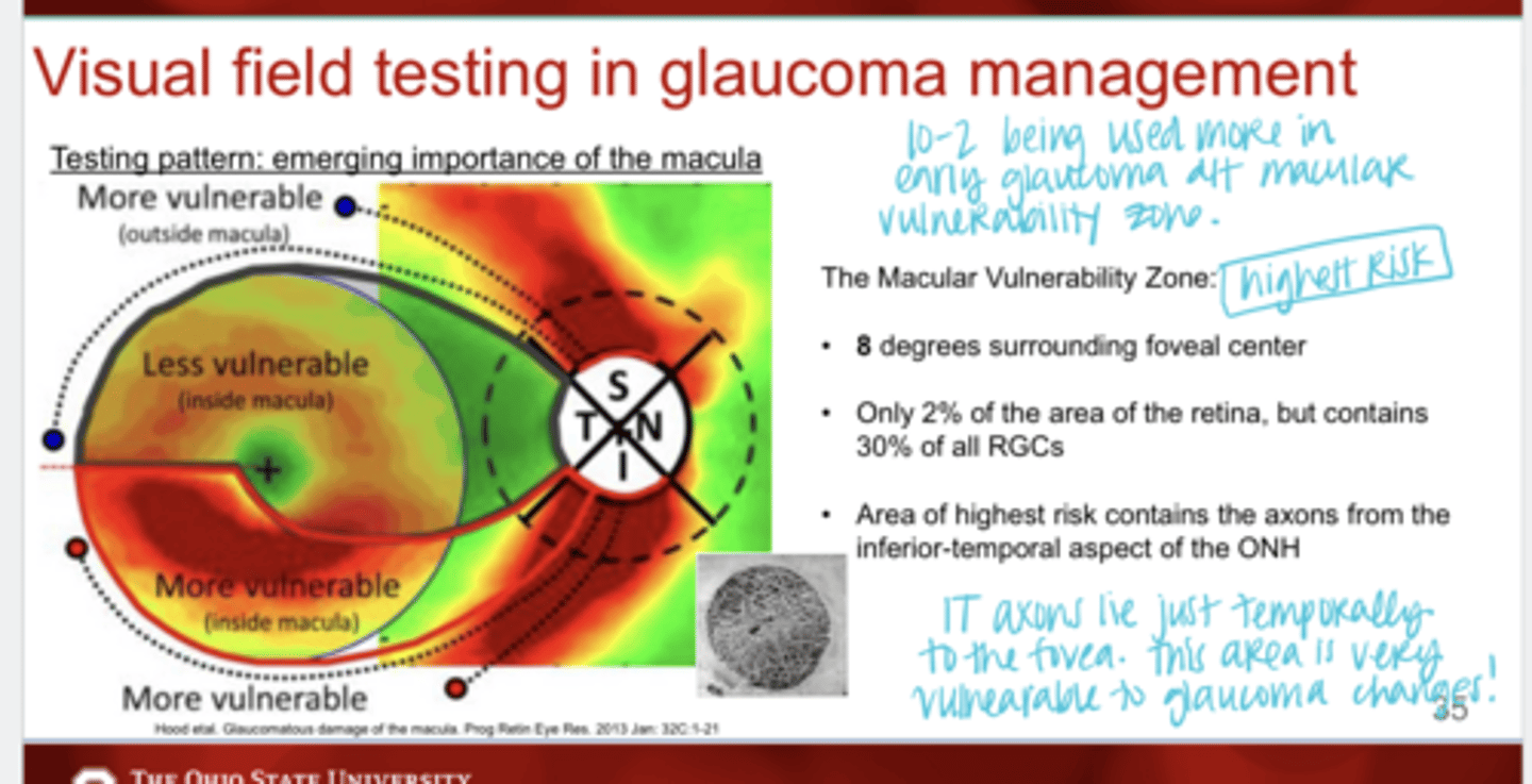
Where is the macular vulnerability zone?
8 degrees surrounding the foveal center
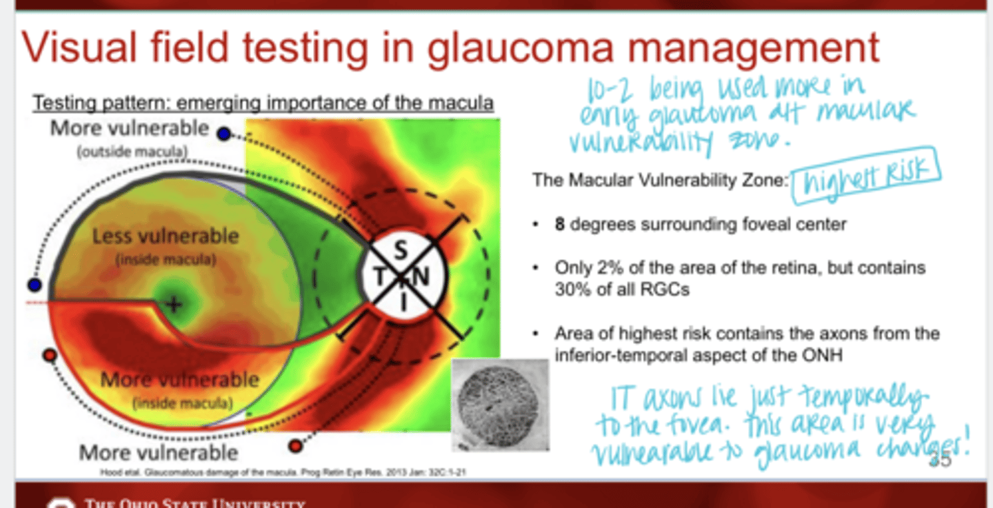
The macular vulnerability zone is only ___% of the retina but contains _____% of all RGCs
2; 30
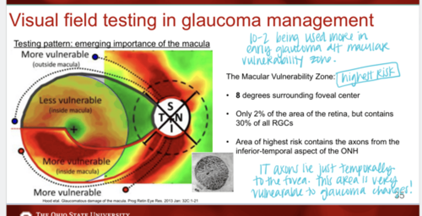
The macular vulnerability zone contains the axons from the ______ aspect of the ONH
inferior-temporal
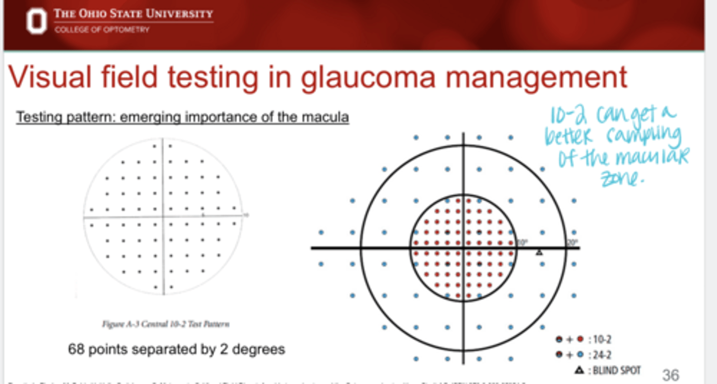
How many points are tested with 10-2 and separation of the points?
68 points separated by 2 degrees
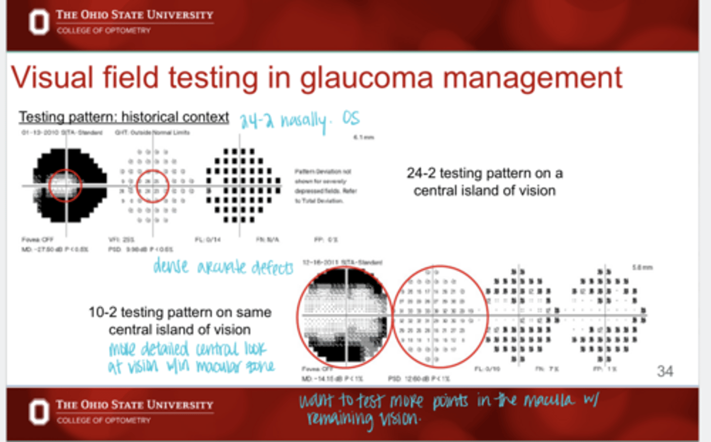
What is the advantage to 10-2 testing?
Can get a better sampling of the macular zone that may/may not be detected by 24-2
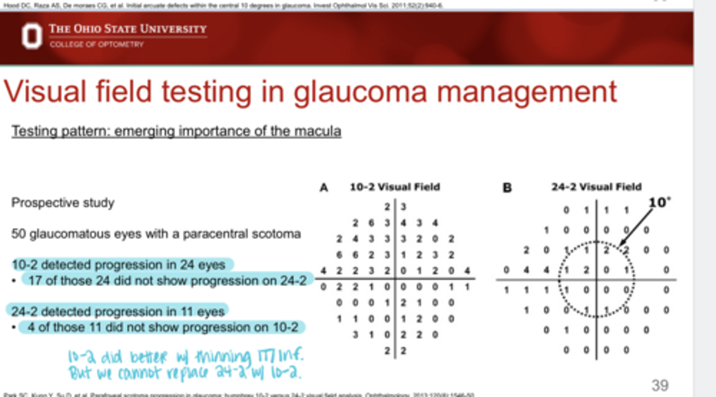
Can we replace 24-2 with 10-2 to detect glaucomatous damage?
No -- 4/11 times it does not detect progression that 24-2 will detect
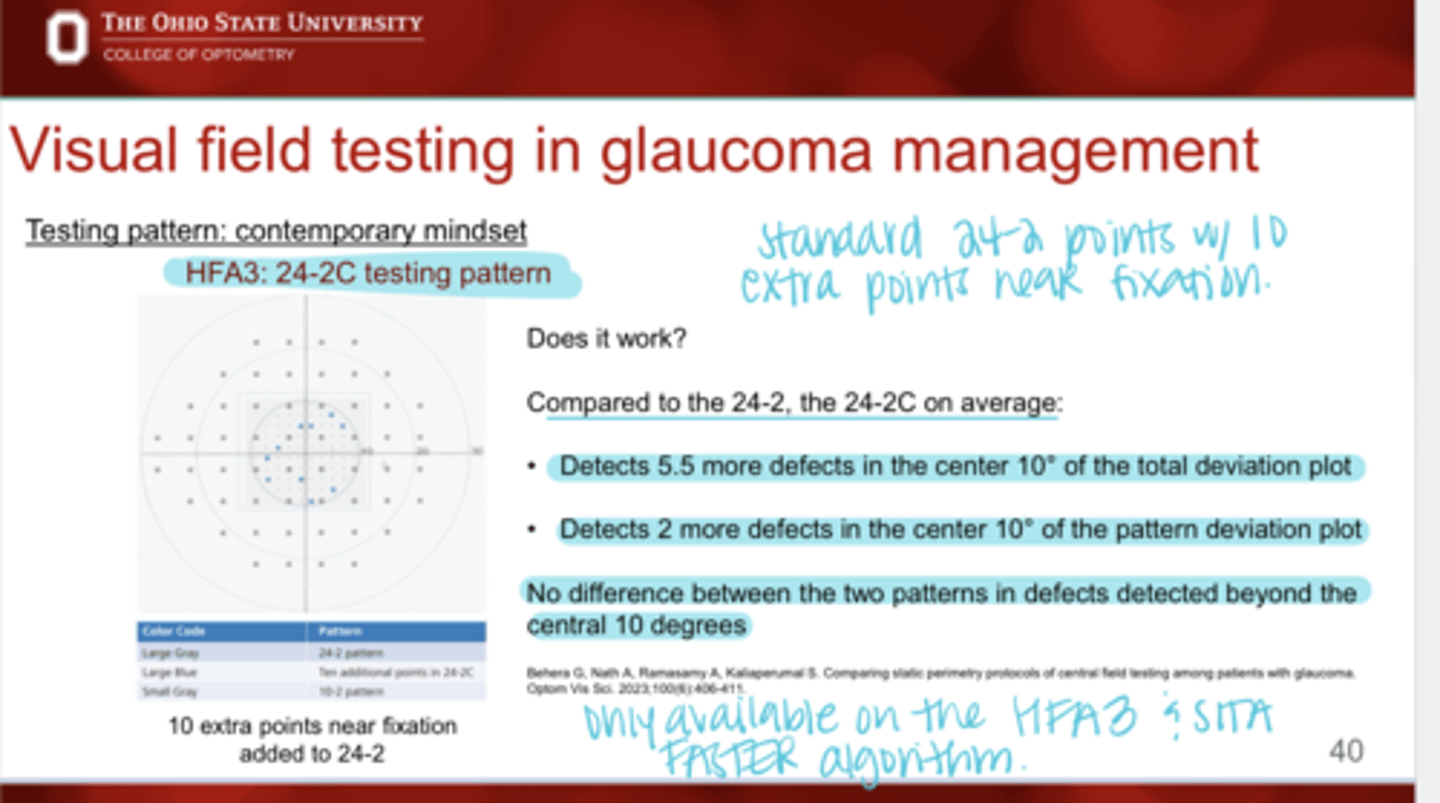
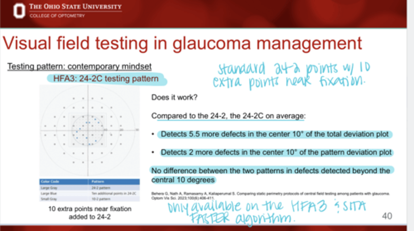
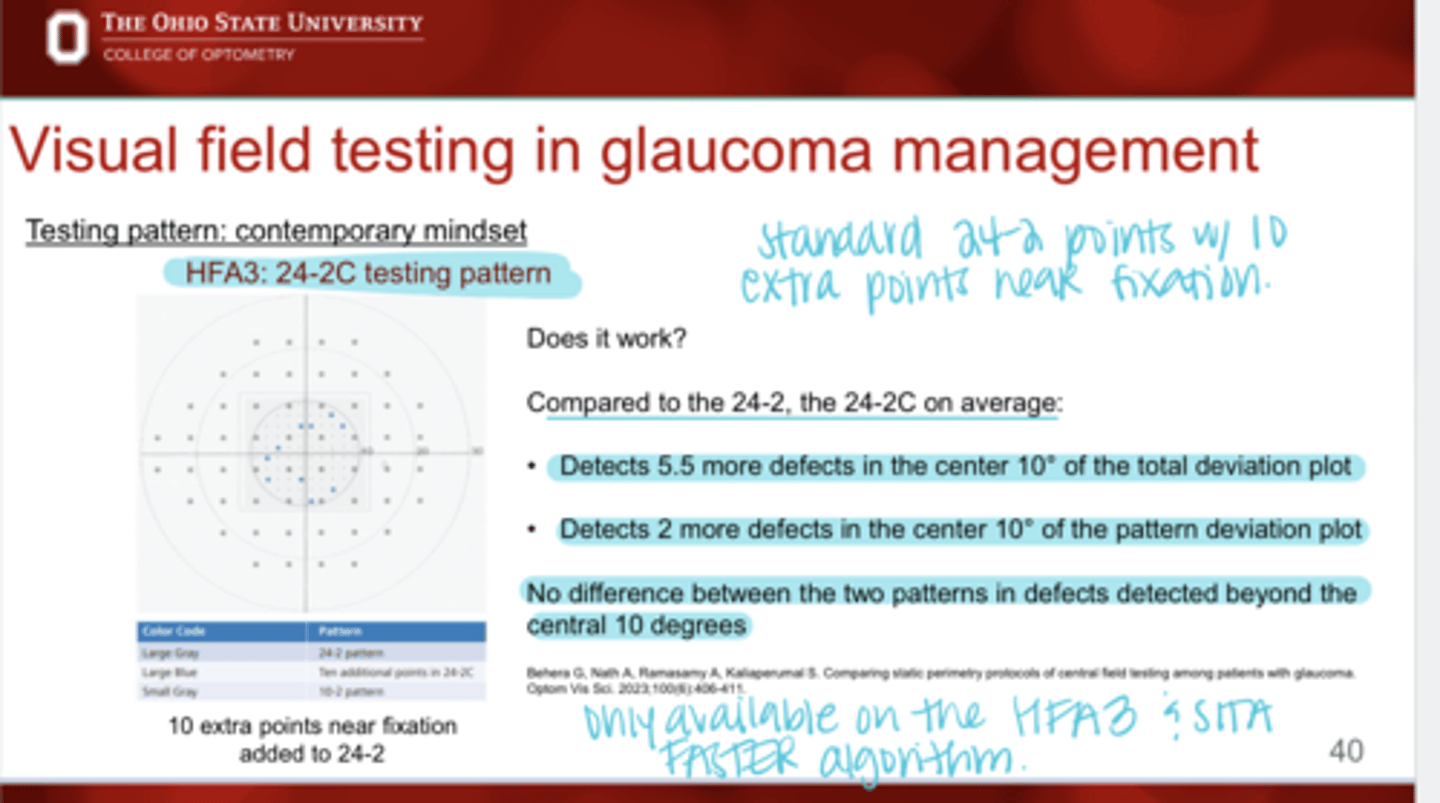
In order to test more points in the macular region, what did the makers of Humphrey do?
Testing Pattern 24-2C with 10 extra points near fixation
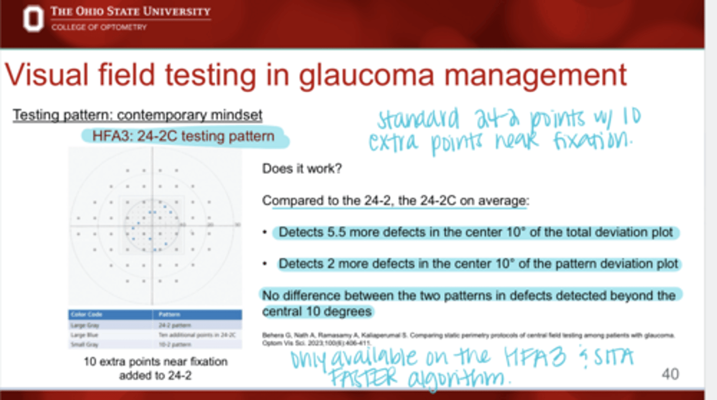
Compared to the 24-2, the 24-2C will detect ______ more defects in the center 10º of total deviation plot
5.5
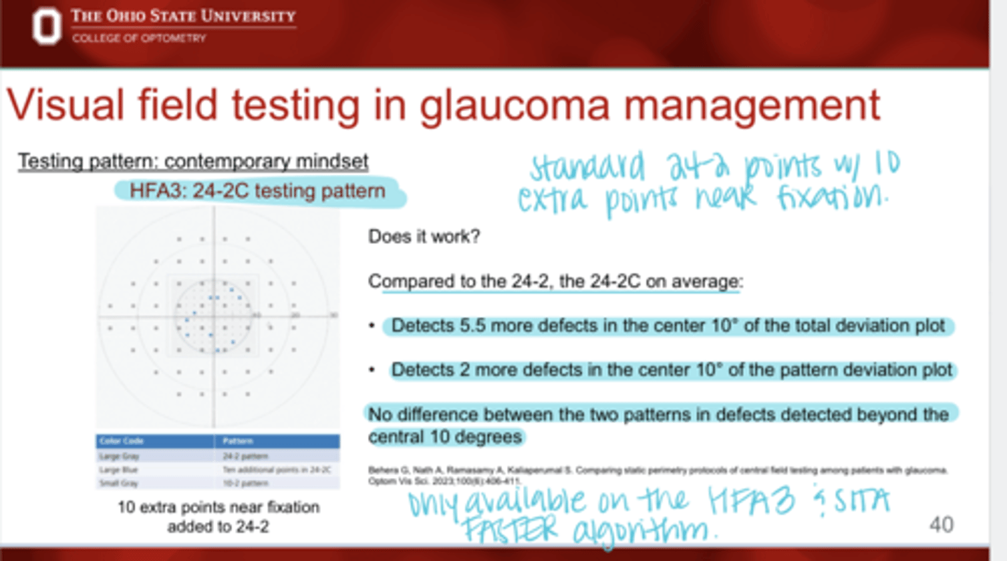
Compared to the 24-2, the 24-2C will detect ______ more defects in the center 10º of pattern deviation plot
2
Compared to the 24-2, is there any difference between the number of defects detected beyond the central 10º with 24-2C?
No
24-2C is only available how?
-with HFA3
-with SITA FASTER strategy
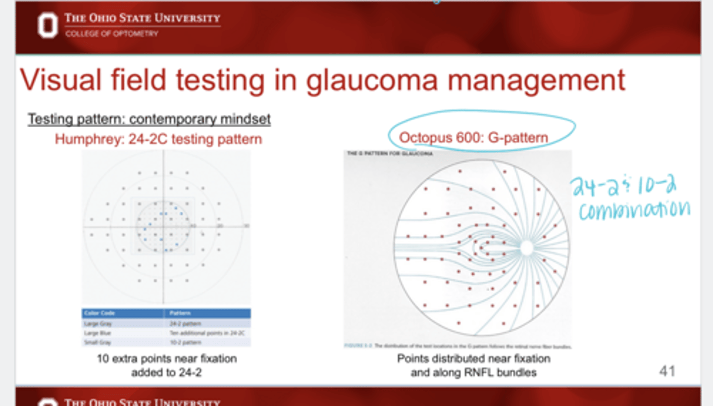
Octopus 600 comparison to a 24-2
G pattern
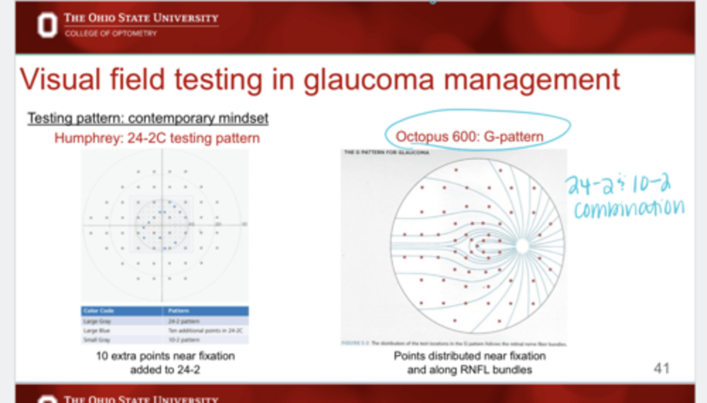
What is different with G pattern (Octopus) compared to HFA 24-2C?
With G pattern, the points are distributed near fixation and along the RNFL bundles
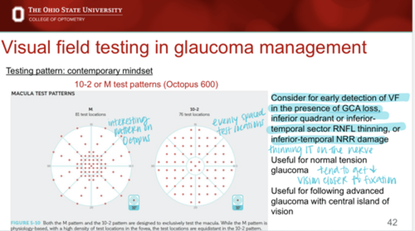
10-2 or M testing patterns should be considered when in glaucoma management?
-For early detection of VF in the presence of GCA loss, inf quadrant RNFL thinning, IT sector RNFL thinning, or IT NRR damage
-Useful in normal tension glaucoma
-For following advanced glaucoma with central island of vision
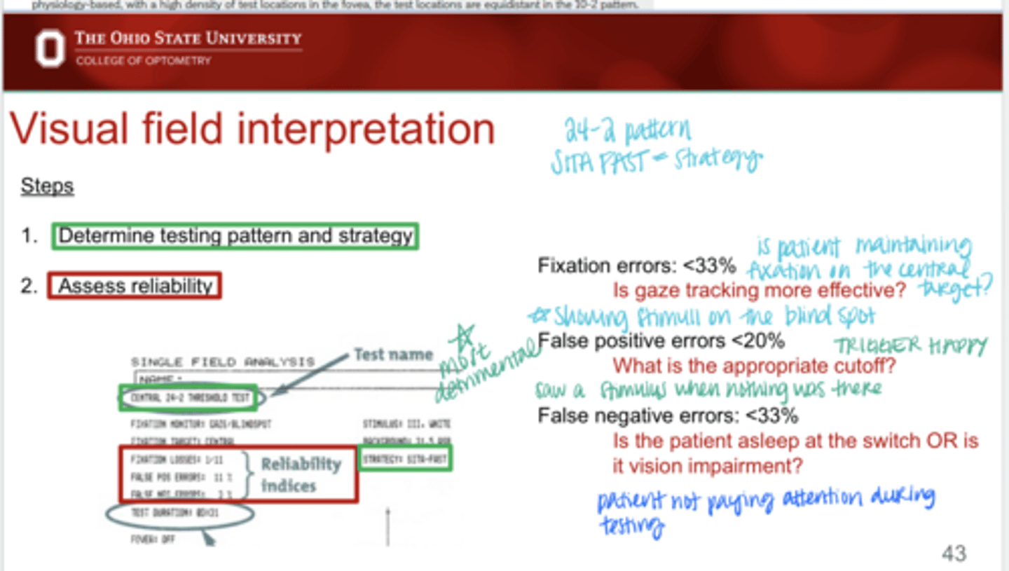
When doing VF testing, you want fixation errors to be under _____%
33
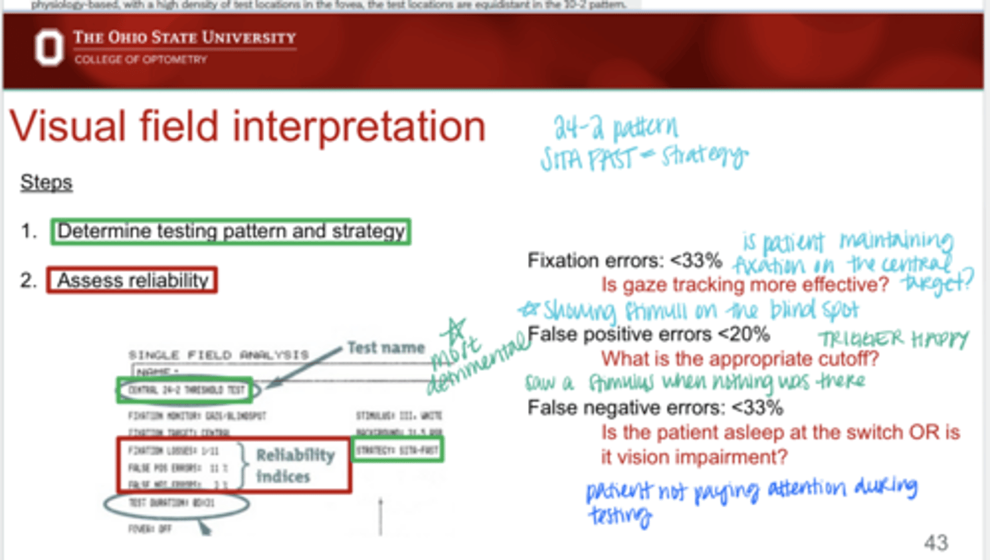
What are fixation errors on VF testing?
occur when your eyes wander or shift during a visual field test off of the central target
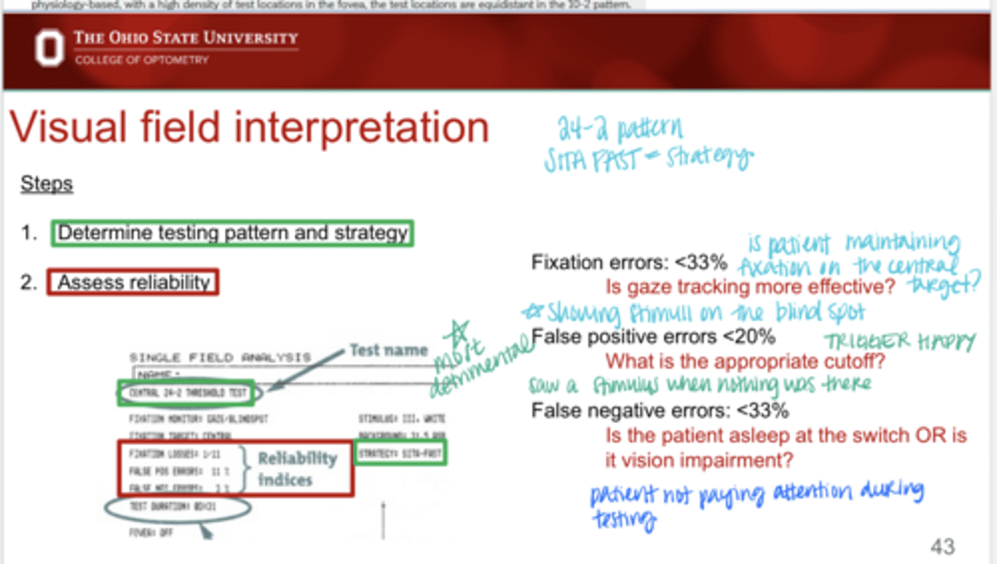
When doing VF testing, you want false positive errors to be under _____%
20
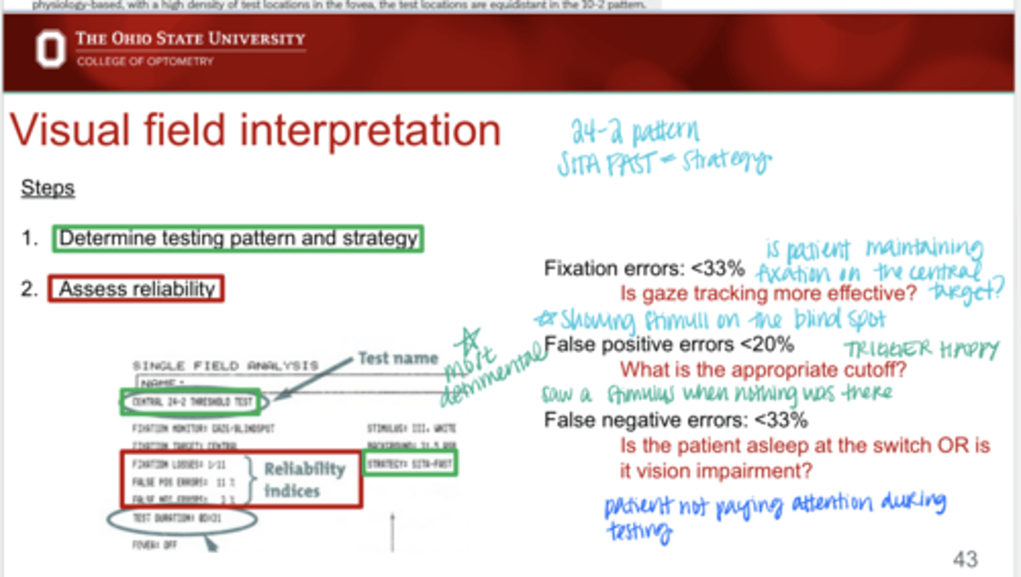
What do false positives during VF testing indicate?
-trigger happy patients
-saw a stimulus (clicked) when no lights were present
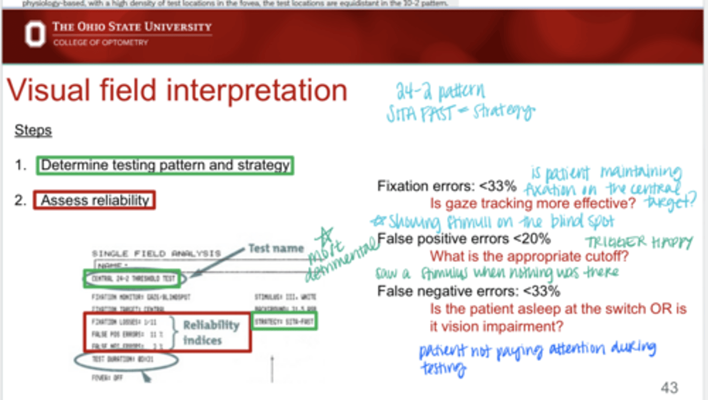
Most detrimental value to a VF measurement?
high false positive value
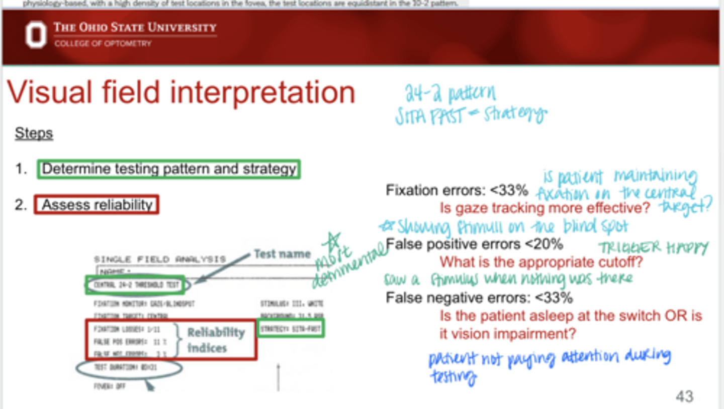
When doing VF testing, you want false negative errors to be under _____%
33
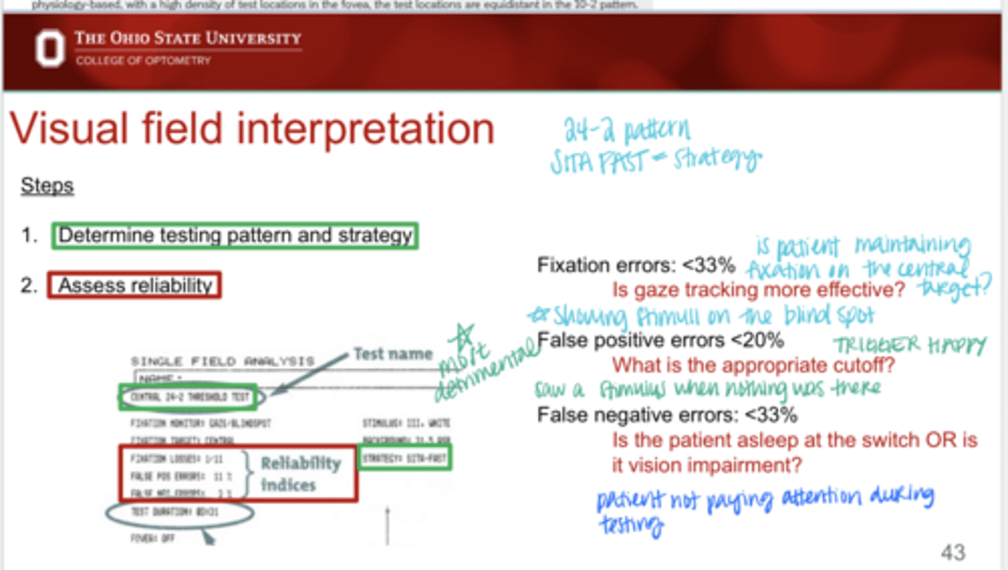
What do false negatives indicate during VF testing indicate?
-Patient not paying attention during testing
-Severe vision impairment
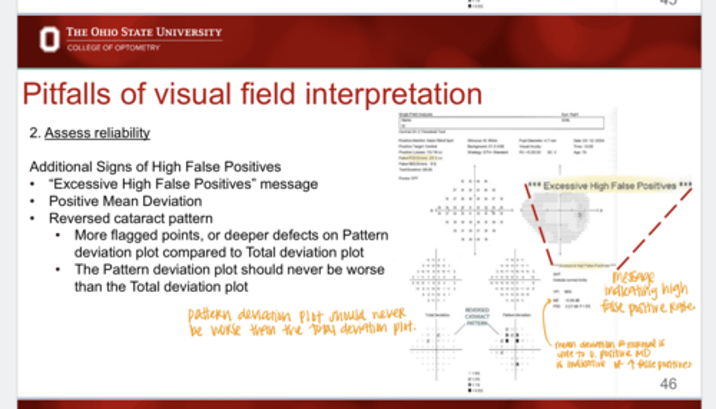
Signs of High False Positives
-"Excessive High False Positives" message on the printout
-Positive mean deviation
-reversed cataract pattern
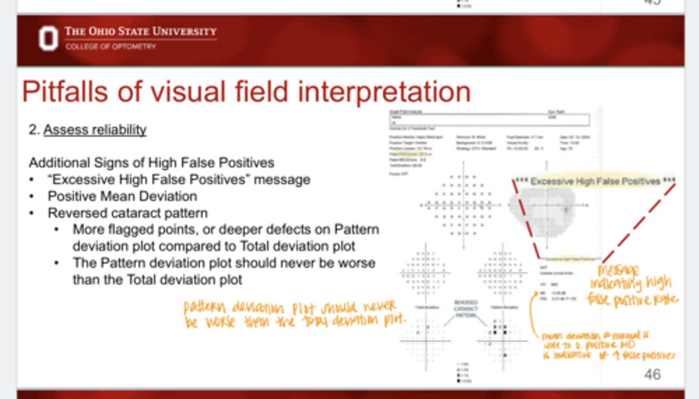
What is a reversed cataract pattern that could indicate high false positives during the VF test?
More flagged points or deeper defects on Pattern deviation plot compared to the total deviation plot
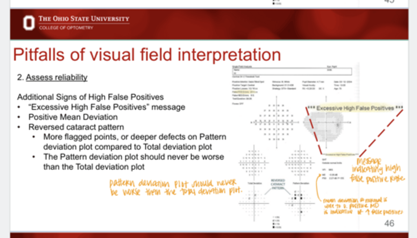
Should the pattern deviation plot ever look worse than the total deviation plot?
no
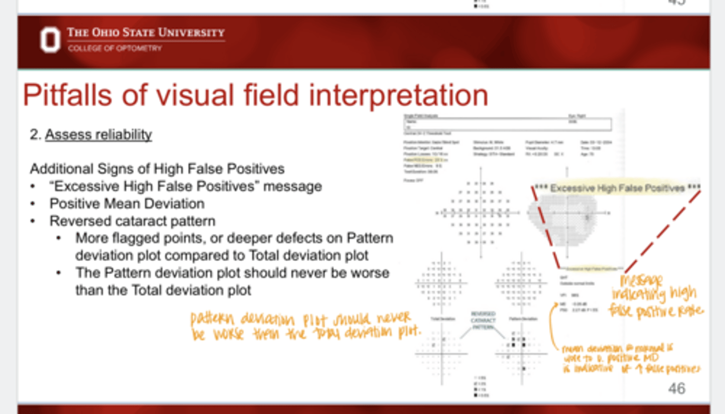
A normal mean deviation is close to _____
0
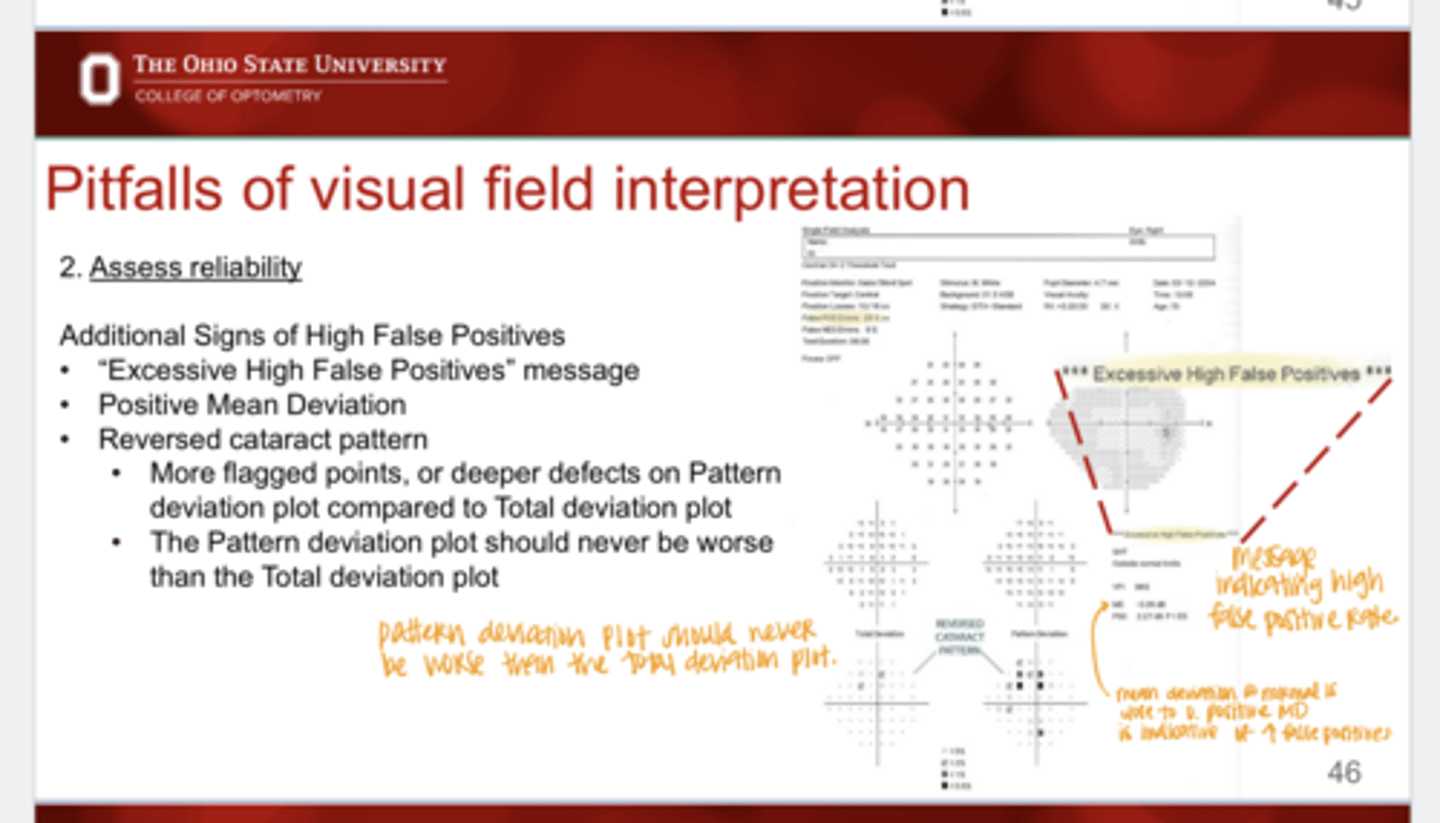
A (negative/positive) MD is indicative of high false positives
positive
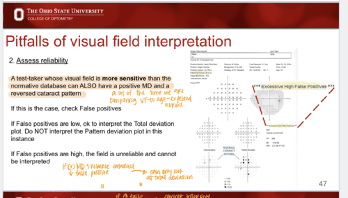
Besides with high false positives, when else could a positive MD occur w/ a reverse cataract pattern?
When a test taker has a VF more sensitive than the normative database
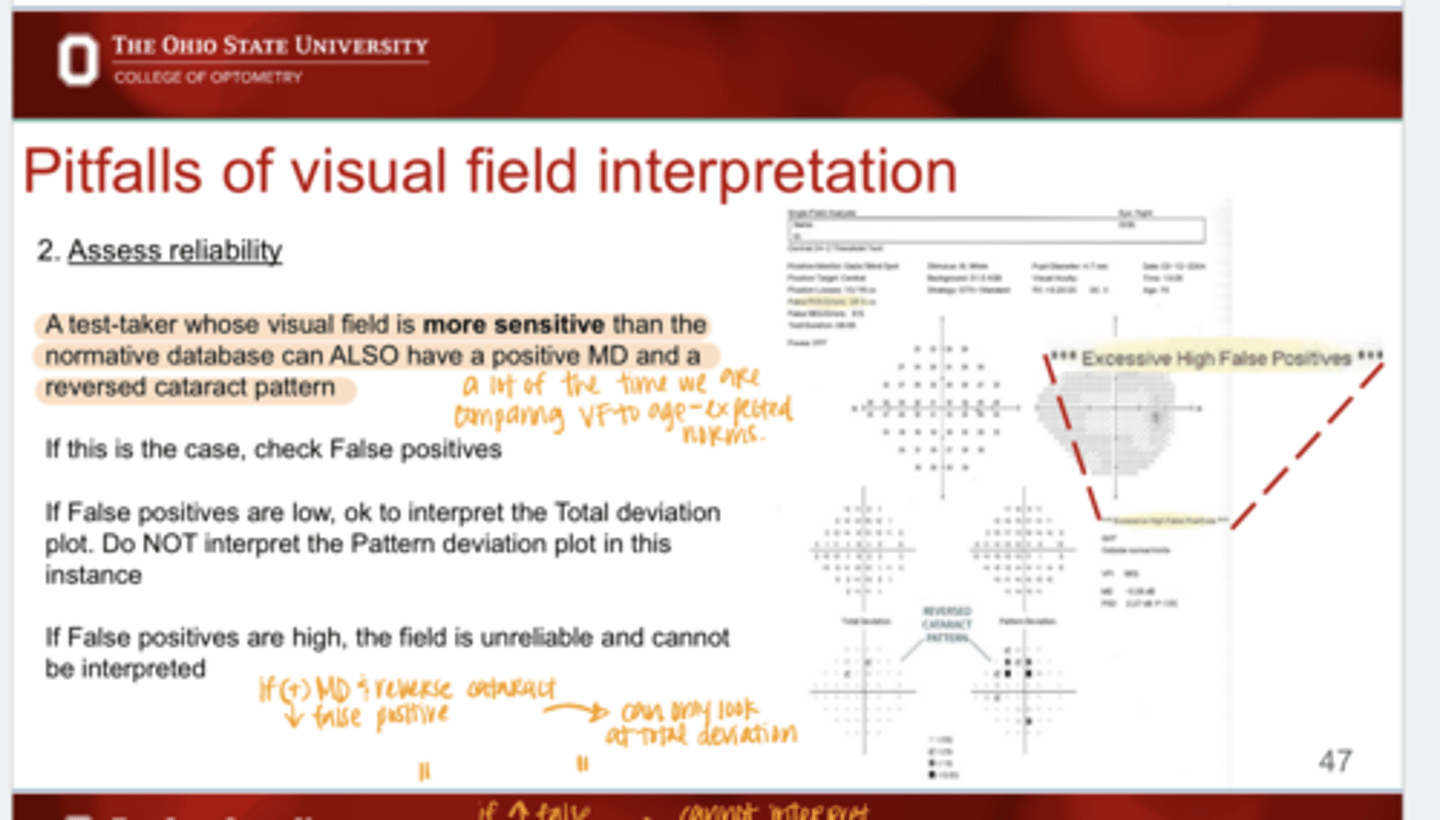
If false positives are low, positive MD present, and reverse cataract pattern, what should you do with the VF?
OK to interpret. Use the total deviation plot & NOT the pattern deviation plot in this instance
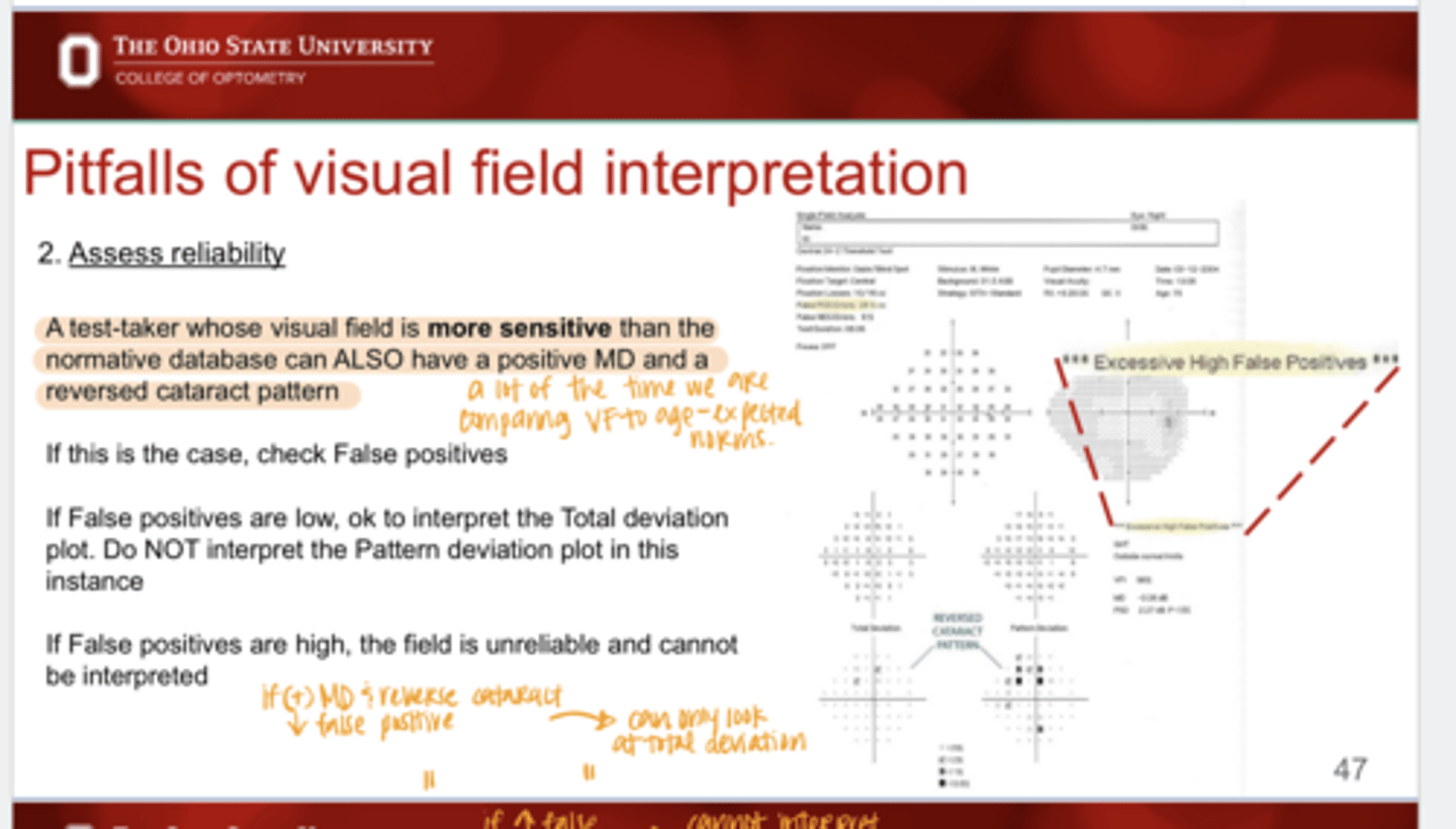
If false positives are high, positive MD present, and reverse cataract pattern, what should you do with the VF?
The field is unreliable and cannot be interpreted
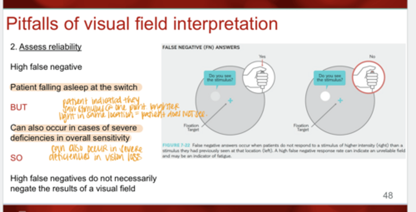
When do false negatives occur?
-Patient falling asleep
-Can occur in causes of severe deficiencies in overall sensitivity
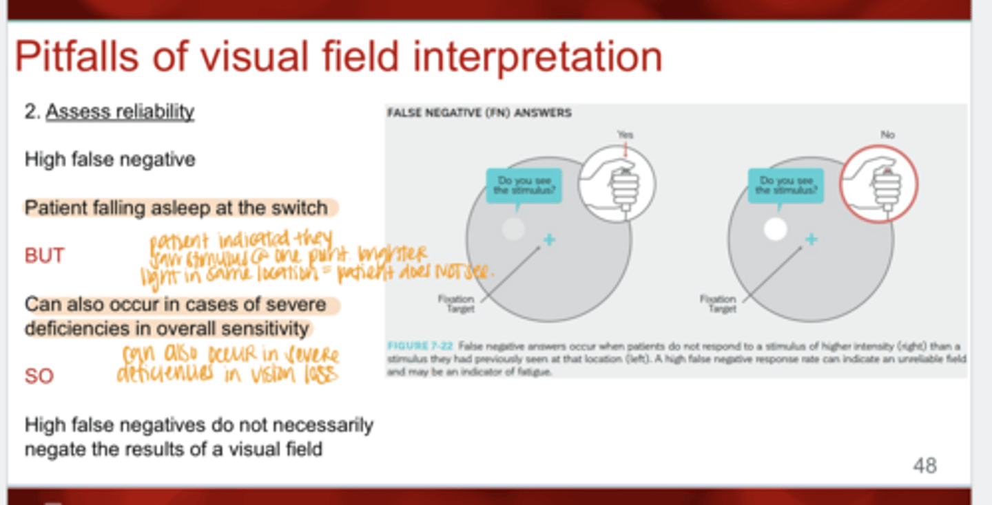
Do high false negatives necessarily negate the results of the visual field?
No
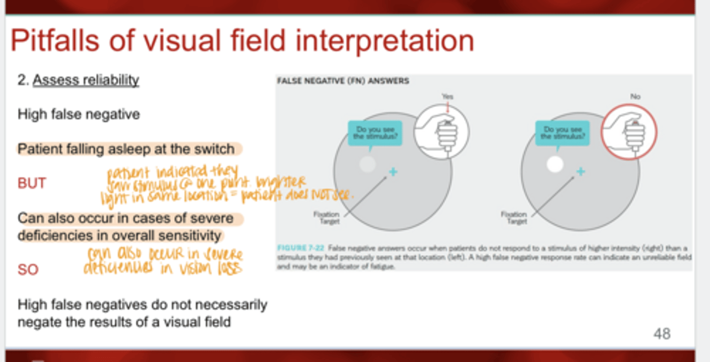
When do false negatives occur in VF testing?
occur when patients do not respond to a stimulus of higher intensity than a stimulus they had previously seen at that location
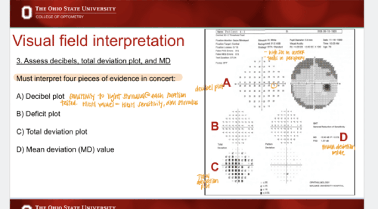
When interpreting VF, you will need to assess what 4 pieces of evidence?
1) Decibel plot
2) Deficit plot
3) Total deviation plot
4) Mean Deviation plot (MD)
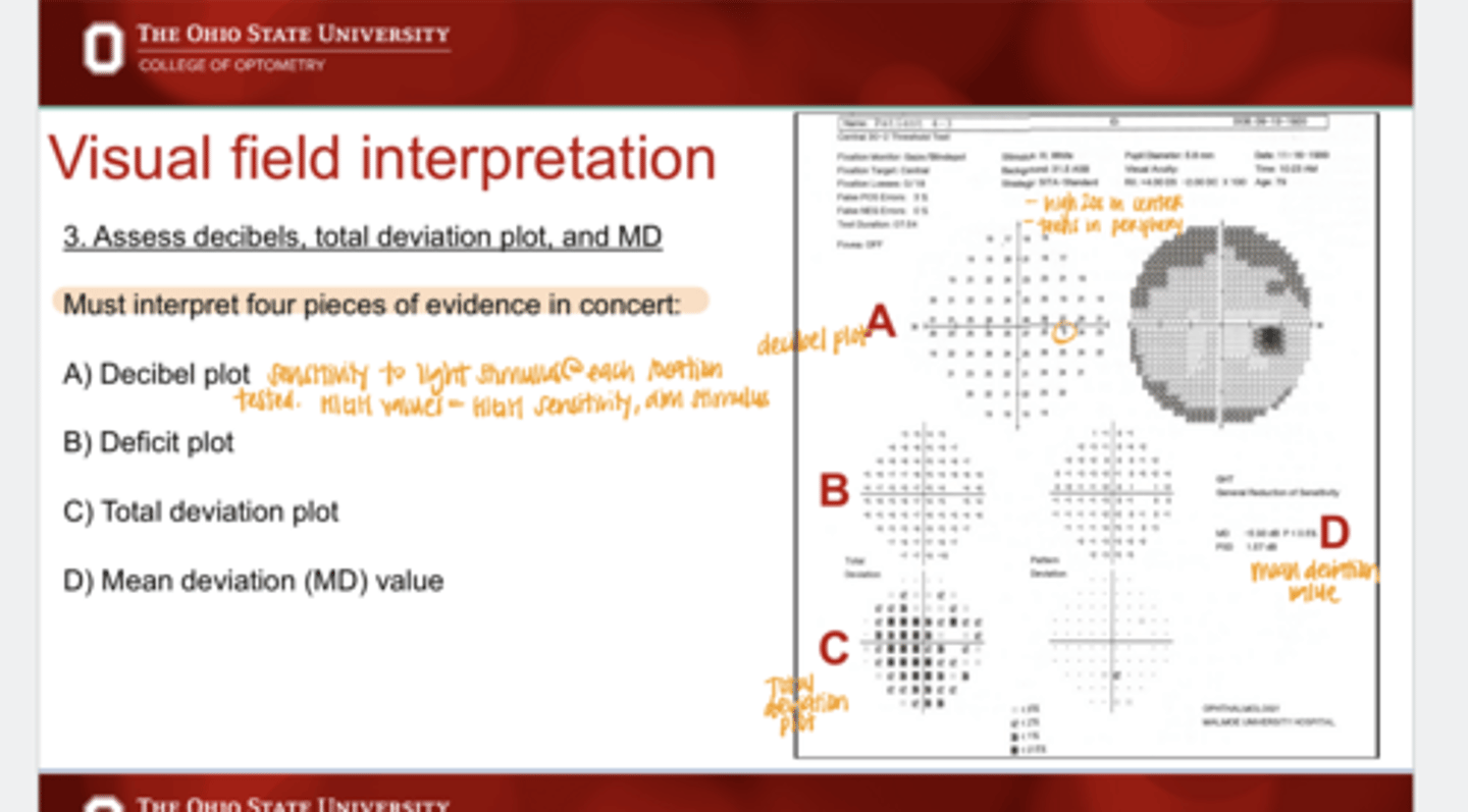
What does a decibel plot show?
maps the retina's sensitivity to light with decibel levels at each retinal location that was tested.
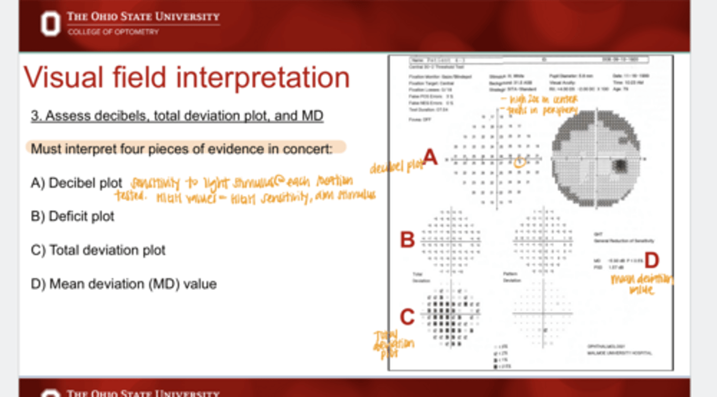
High decibel values on the decibel plot indicate what?
higher sensitivity, dimmer stimulus seen
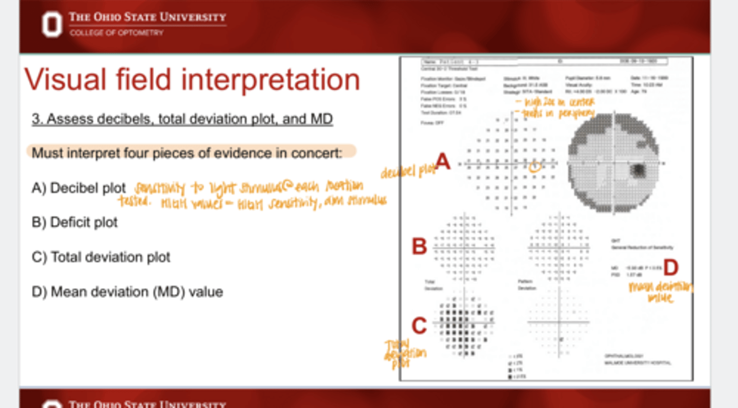
Low decibel values on the decibel plot indicate what?
lower sensitivity, needed brighter stimulus
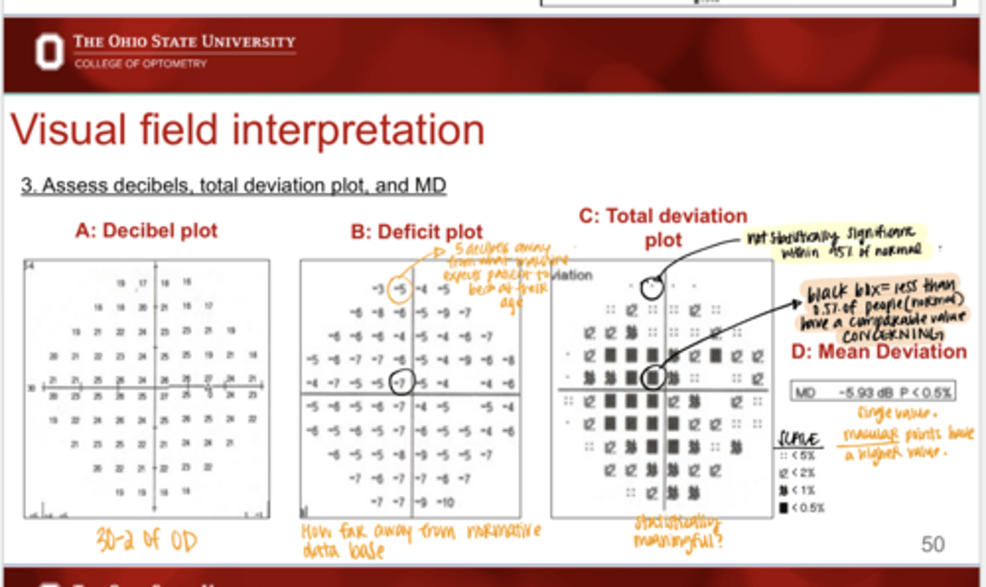
You want values in the high ______ in the center of the decibel plot
20s
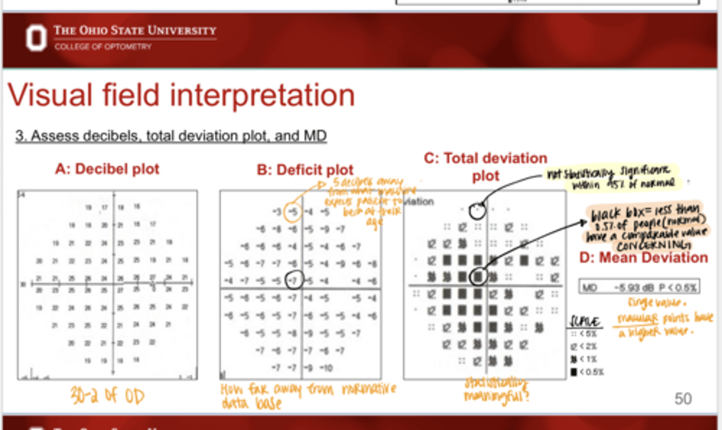
Values in the ____ are normal peripheral values on the decibel plot
teens
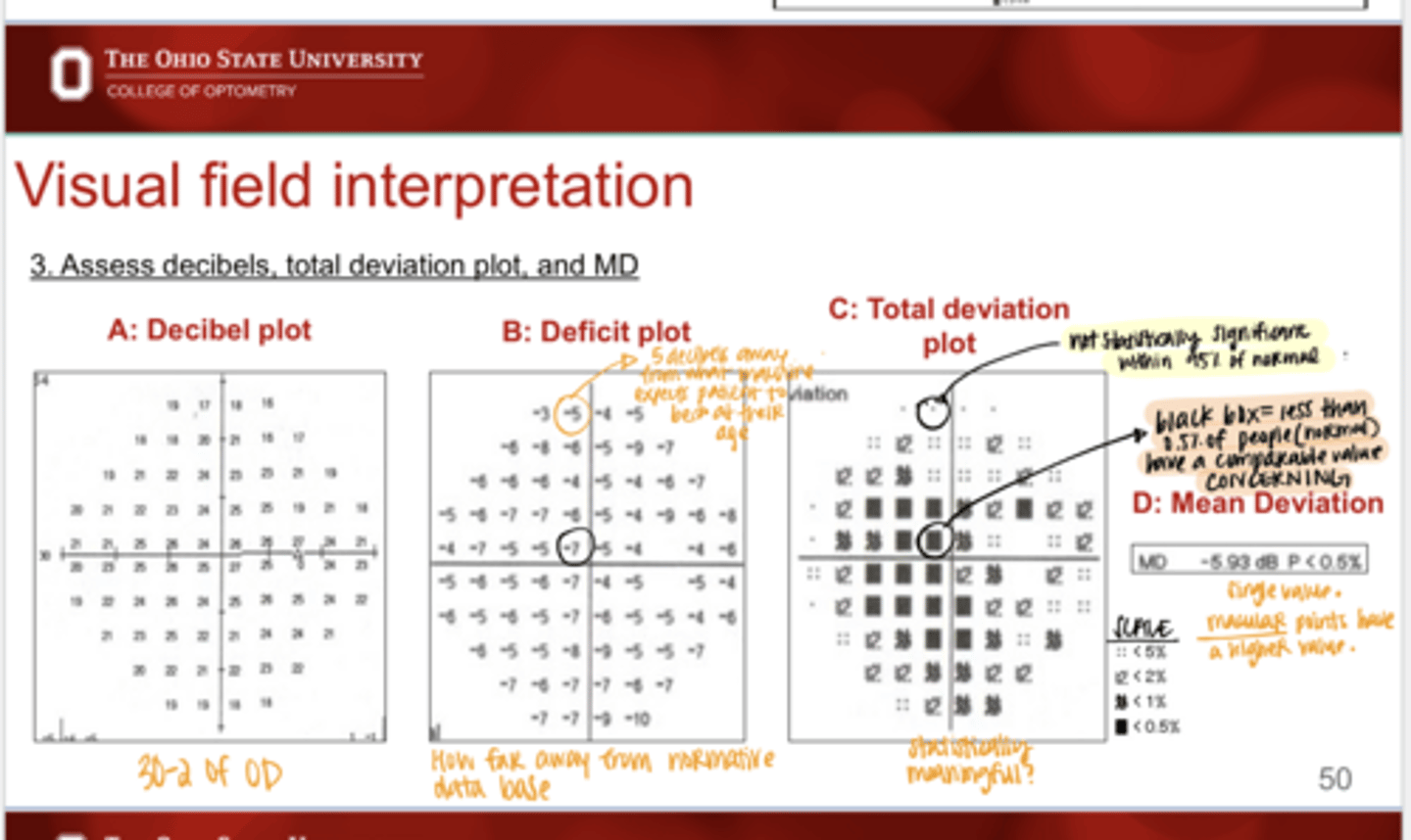
What is the deficit plot?
Shows how far away from normative data base each decibel level is at all points tested
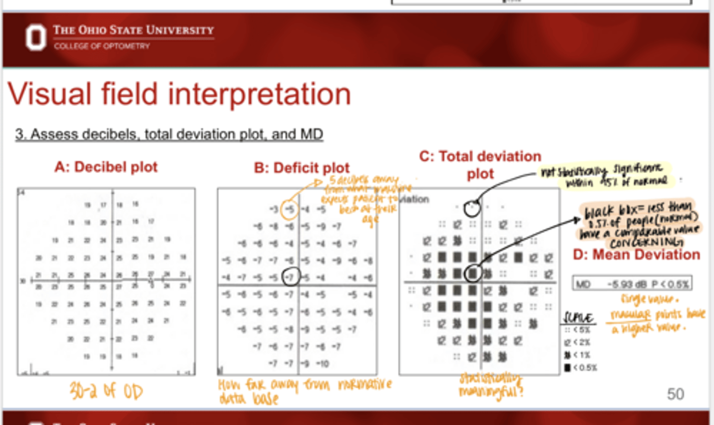
What does the total deviation plot show in VF testing?
Are the deviations from normal statistically significant
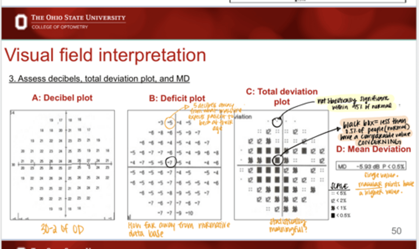
What is the mean deviation in VF testing?
a key global index in visual field testing that calculates the average difference between a patient's visual sensitivity and age-matched normal values; one number value
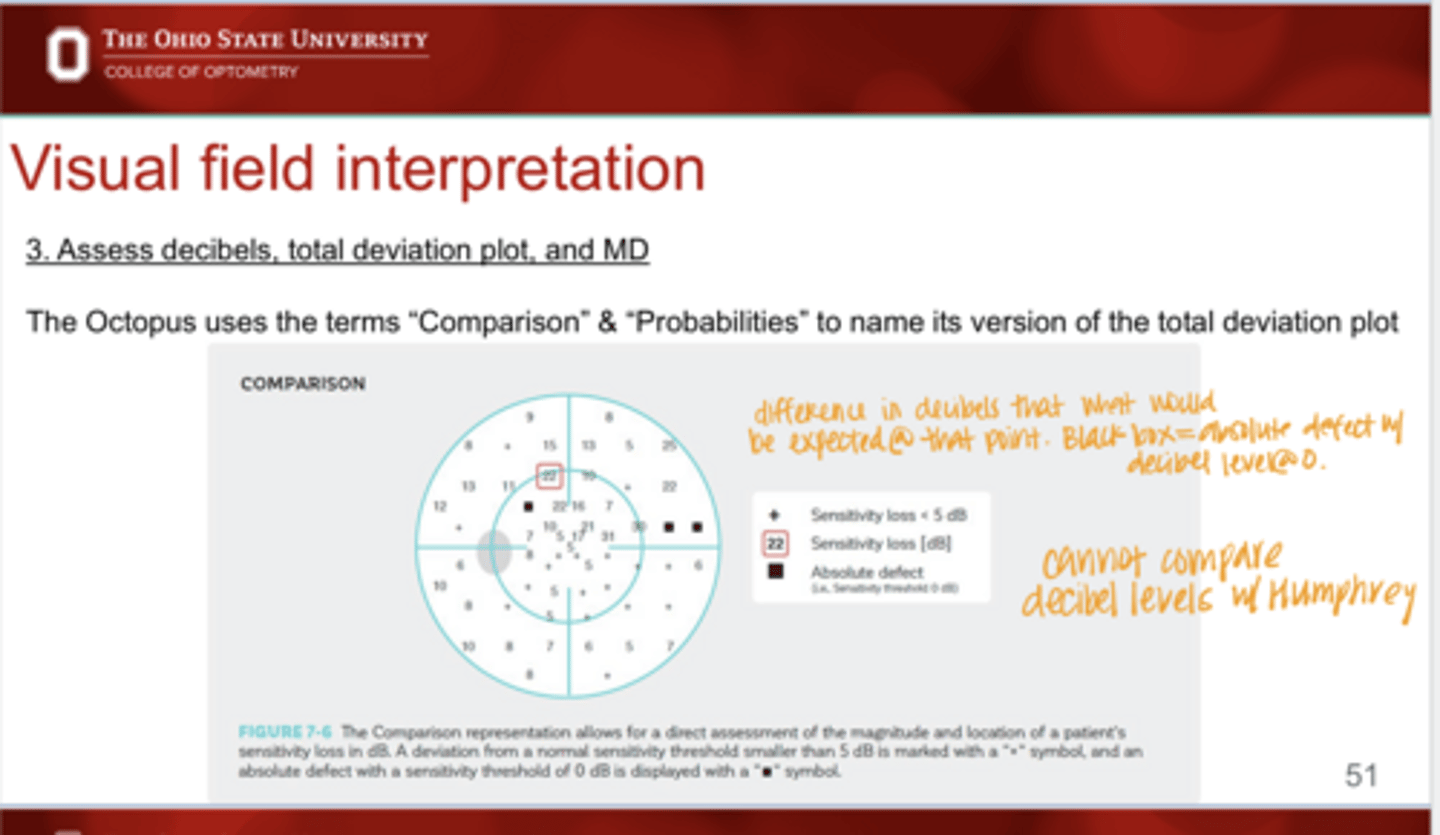
What term does the Octopus 600 use to name its version of the total deviation plot?
Comparison & Probabilities
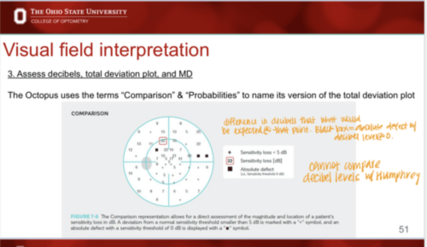
Are you able to compare decibel values between Humphrey and Octopus 600?
No