Abbott EP Product Portfolio Equipment
 Call Kai
 Learn
Learn Practice Test
 Spaced Repetition
 Match
Match Flashcards
 Knowt Play
1/41

There's no tags or description
Looks like no tags are added yet.
Name | Mastery | Learn | Test | Matching | Spaced | Call with Kai | Chat |
|---|
No analytics yet
Send a link to your students to track their progress
42 Terms
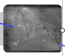
RAO
Right Anterior Oblique
Spine is on the left
CS catheter pointed away
long axis view of HRA, HIS, RVA
Posterior to Anterior
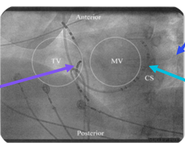
LAO
Left Anterior Oblique
HIS points directly at us
Spine is on the right
CS catheter forms a smile
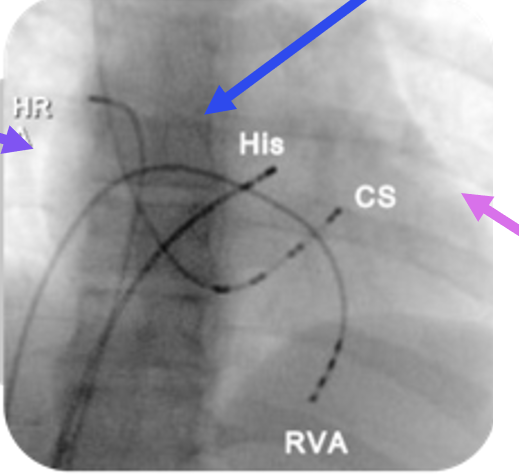
AP
Anteriorposterior
RA forms a left cardiac border
spine is in the middle
LVOT forms right cardiac border
Fluoroscopy (FLURO)
RAO, LAO, AP
radiation flows from the X ray tube to the image intensifier
structures that resist the flow of radiation (higher density) have increased opacity
bones are whiter than soft tissue
ALARA (As low as reasonably achievable)
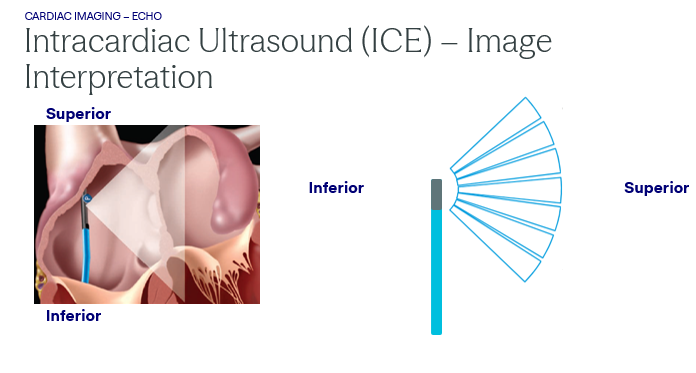
Intracardiac Ultrasound (ICE)
Probe maneuvered within the heart to:
guide catheters and visualize blood flow
assist in transseptal complication
Benefits: no radiation required
limitations:
clarity is dependent on transducer type and distance of structure to probe
requires groin access
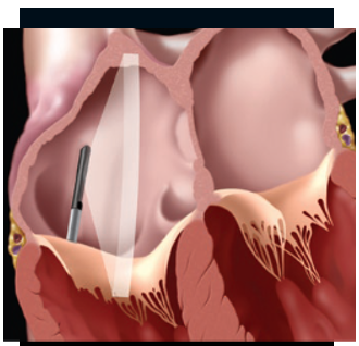
ICE Home View
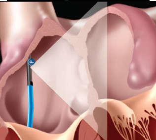
ICE Transseptal view
Echocardiography (ECHO)
How it works:
transducer: houses piezoelectric crystals
crystals vibrate when stimulated by an electrical current
crystal produce mechanical ultrasound waves (high frequency sound pressure waves)
ultrasound waves echo back to crystals
crystals convert waves - electrical current with varying brightness based on density
Examples:
ICE
TEE
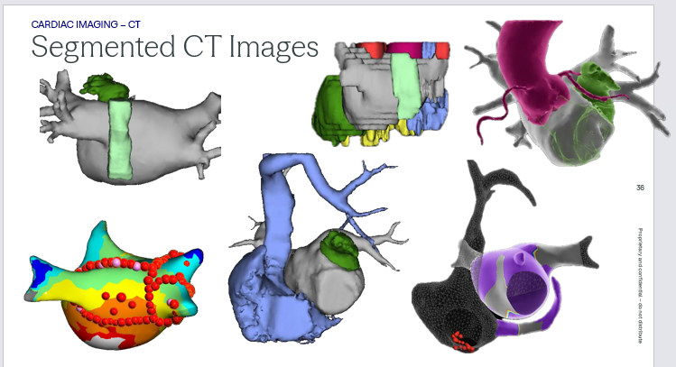
Computed Tomography (CT)
cross sectional slices are digitally stacked together to form a 3D image of the patient’s heart
benefits: provides detailed patient anatomy prior to the case/ catheter placement
limitations: relies on radiation and contrast
static image of a dynamic structure
uses gating to image at one portion of the cardiac cycle
slice thickness and spacing determine image resolution
Scanning Planes
Coronal: divides structure from front to back
Transverse: divides the structure into upper and lower portions perpendicular to the long axis
Sagittal: divides structure vertically through the long axis creating right and left
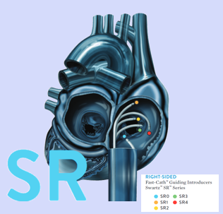
SR (Swartz series right sided)
fixed sheath
Swartz right sided
typical RA AFL
Accessory pathways and focal tachycardias located along the TVA
Series: (the number determines the curve so 4 has the largest curve)
SR0
SR1
SR2
SR3
SR4
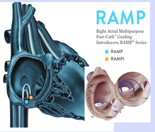
RAMP (right sided)
fixed sheaths
Right Atrial multi-purpose
CTI dependent flutter
orients catheter tip perpendicular against the tissue
Series
RAMP
RAMP1
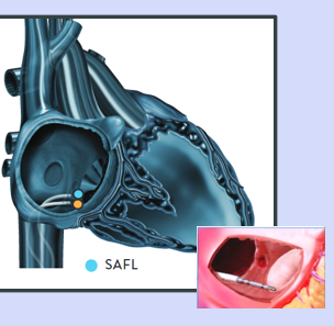
SAFL (right sided)
fixed sheath
S-shaped/ atrial flutter
provides excellent stability on the CTI
Orients catheter tip flat against the tissue
S- shape shaft stabilizes sheath in IVC
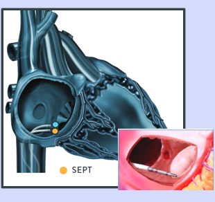
SEPT (right sided)
septal
helps place ablation catheter on the septal CTI
Orients catheter tip flat against the tissue
S-shape shaft stabilizes sheath in the IVC
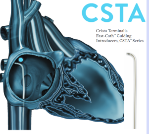
CSTA (right sided)
crista terminalis
useful for mapping and ablation in the high right atrium
guides to the crista terminalis and RA free wall
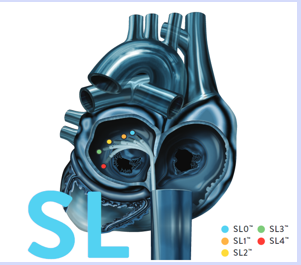
SL (left sided)
Swartz Left sided
designed to contour the left atrium
braided (for stability across septum and atraumatic tip)
81 cm SL accommodates 89cm length Abbott BRK transseptal needles
Series:
SL0
SL1
SL2
SL3
SL4
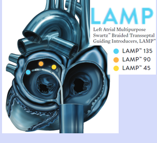
LAMP (Left sided)
left atrial multi-purpose
multiple reach angles provide options for gaining access to the pulmonary veins and mitral isthmus areas
braided (for stability across septum and atraumatic tip)
series
LAMP 135
LAMP 90
LAMP 45
Agilis NxT Steerable Introducer
8.5 F
Inner Diameter - 8.5F
Outer Diameter - 11.5F
Qualities:
auto lock steering
offset venting holes
atraumatic tip
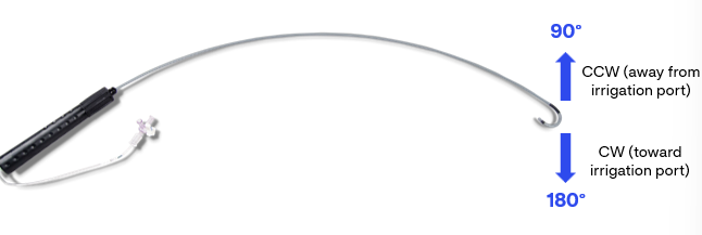
Agilis Curl Options
curl options:
small: 61cm
medium: 71cm
large: 82cm
Deflection:
bi-directional: 180 and 90 degree deflection
no alignment knotch on handle
Why use Agilis Nxt 8.5F?
Efficacy:
higher acute ablation success rates
higher rate of complete pulmonary vein isolations
fewer acute PV reconnections
higher rate of bidirectional MI block and decreased need for CS ablation
Contact force:
higher average contact force in AF ablation
shorter AF procedures with CF catheters in severe LA enlargements
fewer acute left PV segment reconnections
Time:
shorter procedure time in AF and flutter ablations
shorter fluoro time
shorter RF energy time
Reoccurrence:
fewer chronic PV reconnections
lower rate of AF re ablation
lower rate of cardioversion after AF ablations
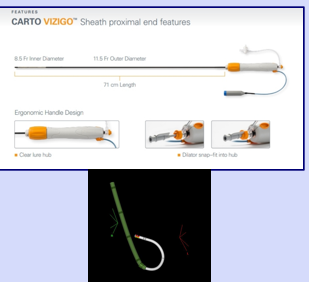
Competitor for Agilis?
Carto Vizigo:
8.5F/ 11.5F outer diameter
71cm useable length
braided sheath
small, medium, and large curve
rotating collar handle, deflects the tip clockwise 180 and counterclockwise 180
advantages: has electrodes on the outside of the sheath to allow for visualization on the Carto System
Agilis NxT steerable introducer Dual Reach 13F
inner diameter: 13F
outer diameter: 17.5F
atraumatic tip: reducing the stepdown when crossing the septum
6 offset venting holes: 4 distal and 2 that are more proximal
material transition: 6 inches from deflectable shaft to create a more pliable tip
bidirectional rotating collar
3-war stopcock: for air or blood aspiration, fluid infusion, blood sampling
braided shaft
Curl options for Agilis Steerable Dual Reach
small: 18.4
medium: 24.8
large: 45.1mm
total length: 91cm
useable length: 71cm
fill volume: 15cc
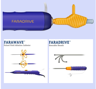
Who is the competitor for the Agilis Dual Reach 13F
Boston Scientific Faradrive
paired with farapulse and versacross
inner 13F, outer 16.8F
useable length: 74cm
overall length: 91cm
radio opaque tip marker
bidirectional steering
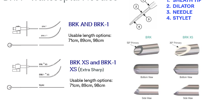
Transseptal Needles
BRK and BRK-1
usable length options: 71cm, 89cm, 98cm
BRK XS and BRK-1 (extra sharp)
usable length options: 71cm, 89cm, 98cm
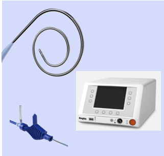
Competitor to BRK transseptal needles?
Boston Scientific VersaCross
access product includes:
VersaCross RF wire ( J-tip or Pigtail)
RF connector cable
Versacross transseptal sheath (8,5F)
Dilator
0.035 guidewire
clinical advantage: less manual force needed
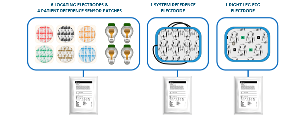
Ensite X surface electrode kit
6 locating electrodes and 4 patient reference sensor patches
1 system reference electrode
1 right leg ECG electrode
System Reference Electrode
what does it do?
serves as our systems electrical reference for impedance and ECG measurements
Placement
the electrode is places on the patient’s abdomen
should be the first and last electrode to be connected/ disconnected
this is sometimes put on the patients back because of the belly button interference (no air should be under the patch)
Right leg ECG Electrode
what does it do?
serves as the right left 12 lead ECG electrode during a procedure
cancels out powerline noise that may be picked up by the system reference
placement?
RL ECG placed on the patients right left
sometimes moved into inner thigh so physician doesnt constantly bump it
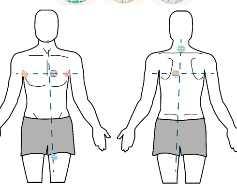
What are the locating electrodes?
6 electrodes impart low intensity currents generated by the amplifier
supports the acquisition of impedance signals used for locating EP catheters
the locating electrodes work in patch pairs
placement:
left (red)
right (orange)
front (black)
neck (back of neck sometimes on traps) (green)
back (brown)
Lleg (blue)
Patient Reference Sensors (PRS) Patches
patches are used to secure PRS sensors on the patient
usage: patches are single use and included in the kit
PRS sensors are reusable and part of the system hardware
Placement
front: 1 patch holds the anterior PRS (PRS-A) which detects metal distortion
Back: 3 patches hold posterior PRS sensors (PRS - P2, P3) forming a triangle
the center of the triangle defines the magnetic coordinate systems zero points
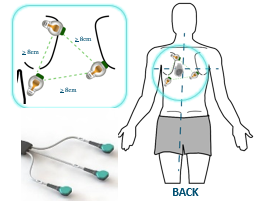
PRS Placement
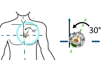
green side up
the anterior patch guidelines:
must not be inclined more than 45 degrees relative to the patient table
Diagnostic EP Catheters
solely for diagnostic purpose and are not designed for RF energy delivery
catheters are named by their number and electrodes
catheters are measured (F size) by their outer diameter
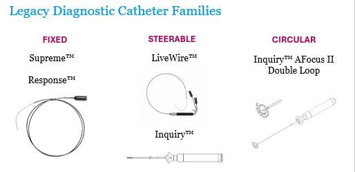
What are the diagnostic catheter families?
Fixed:
supreme
response
Steerable:
LiveWire
Inquiry
Circular:
Inquiry AFocus II double loop
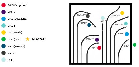
Supreme and Response Catheter Curves
JSN
JSN-1
CRD
CRD-1
CRD-2
CSL
DAO
DAO-1
STR
fixed diagnostic cath
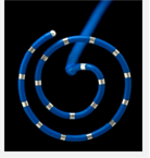
Inquiry AFocus II Double Loop
shaft/handle:
7F shaft
push/pull plunger handle
unidirectional 180-degree deflection
loop:
fixed loop catheter
20mm, 4F loop
20 electrodes with equidistant 4mm electrode spacing
not indicated for retrograde access or ventricular use
What is the PA interval and how long should it be?
the PA interval is a measure from onset of the p wave on surface ECG to rapid depolarization of A wave on HIS catheter
35-45 msec
the time it takes for signals to travel from the SA node to AV node
What is the A-H interval and how long should it be?
measure from A signal to onset of H deflection on HIS catheter
time for electrical signals to travel through AV node
70-80 msec
Normal signal sequence in normal sinus rhythm?
HRA- HIS - CS - H deflection - RVA - HIS V and CSV
Lead II can be used to locate the “surface p wave”
What is the normal PS interval?
120 - 200 msec
from the start of atrial depolarization to the start of ventricular depolarization
What is the normal QRS interval?
< 120 msec
time for ventricular depolarization
What is the normal HV interval?
35 - 55 msec
earliest deflection of HIS to the onset of earliest ventricular signal on any channel
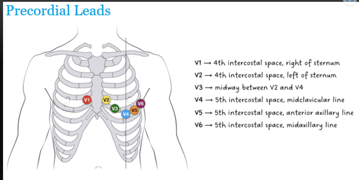
Where is each precordial lead located?
V1 - 4th intercostal space, right of the sternum
V2 - 4th intercostal space, left of the sternum
V3 - midway between V2 and V3
V4 - 5th intercostal space, midclavicular line
V5 - 5th intercostal space, anterior axillary line
V6 - 5th intercostal space, midaxillary space