The Fetal Head and Brain
 Call Kai
 Learn
Learn Practice Test
 Spaced Repetition
 Match
Match Flashcards
 Knowt Play
1/120

There's no tags or description
Looks like no tags are added yet.
Name | Mastery | Learn | Test | Matching | Spaced | Call with Kai | Chat |
|---|
No analytics yet
Send a link to your students to track their progress
121 Terms
The neural plate is the
Structure that will form into the central nervous system (developed by 4.5 wks)
The neural tube becomes the
Spine and brain
The brain is divided into 3 vesicles called the
Prosencephalon (forebrain)
Mesencephalon (midbrain)
Rhombencephalon (hindbrain)
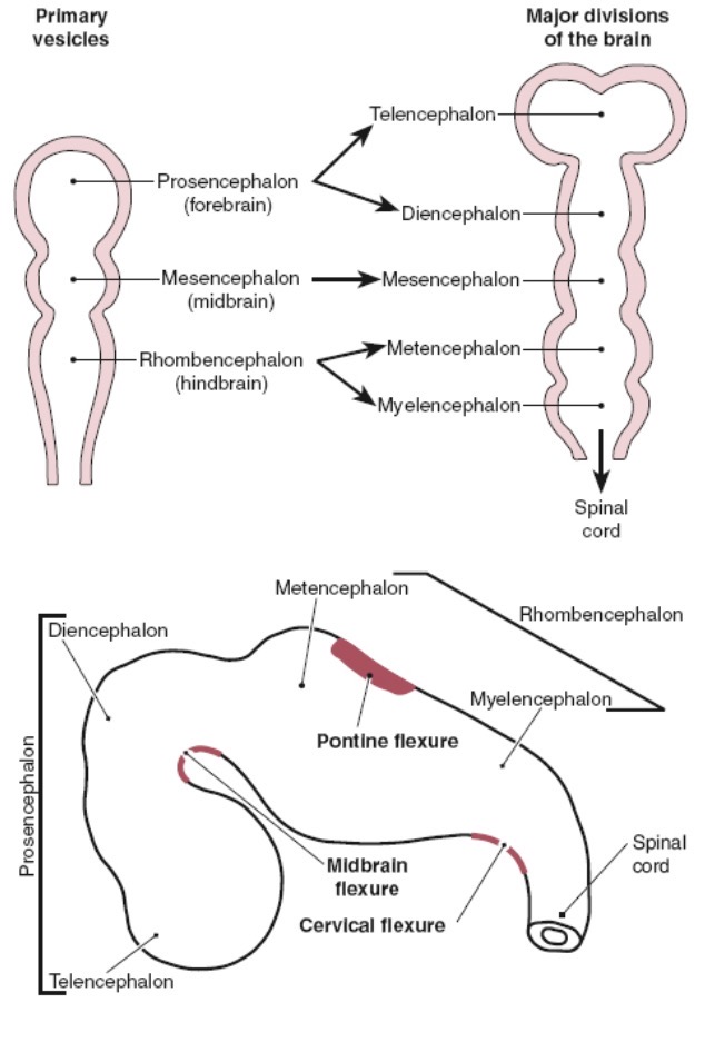
The prosencephalon will mature into
Telencephalon and diencephalon
The telencephalon will form the
Cerebral hemispheres, lateral ventricles, and third ventricle
The diencephalon will become the
Thalamus and hypothalamus
The mesenchephalon will become the
Midbrain, superior parts of brainstem
The rhombencephalon will become the
4th ventricle and medulla oblongata and cerebellum
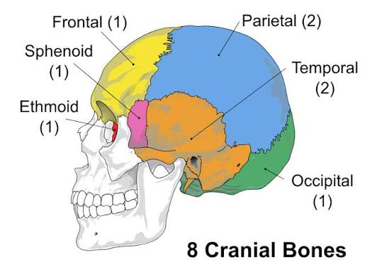
Cranial bones
Fetal cranial sutures (connective tissue that lies btw cranial bones)
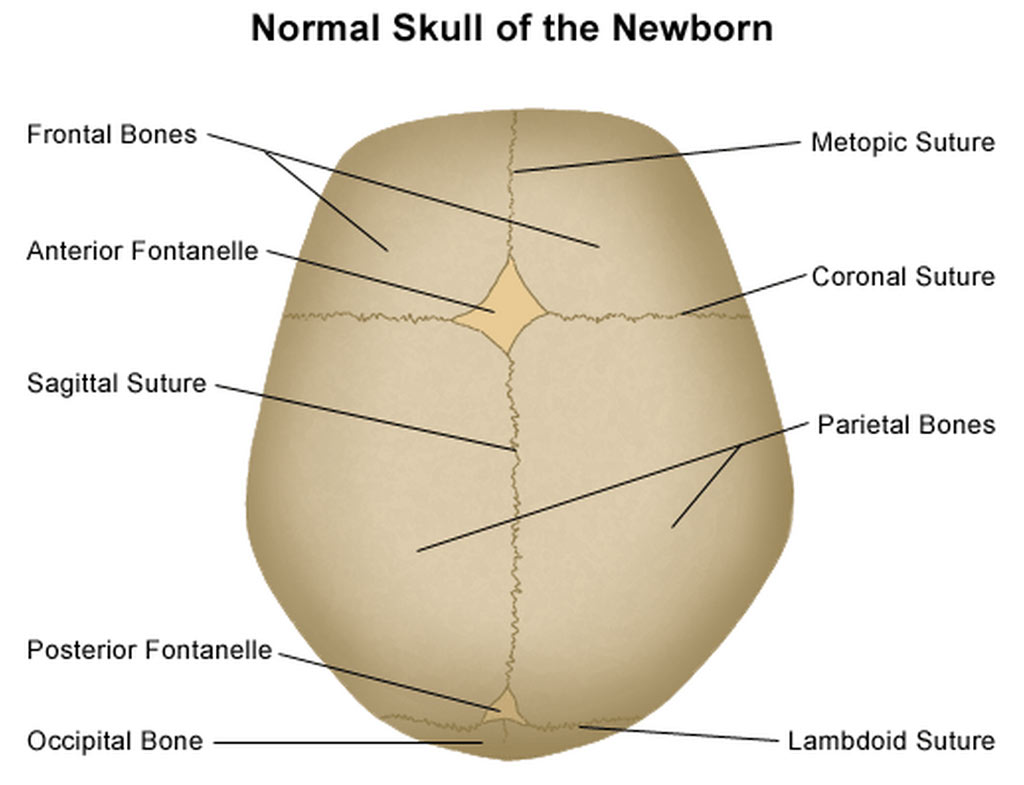
Premature fusion of sutures is termed
Craniosynostosis
Fontanelles/ soft spots
The opening in the base of cranium through which the spinal cord travels is the
Foramen magnum
Meninges of the brain
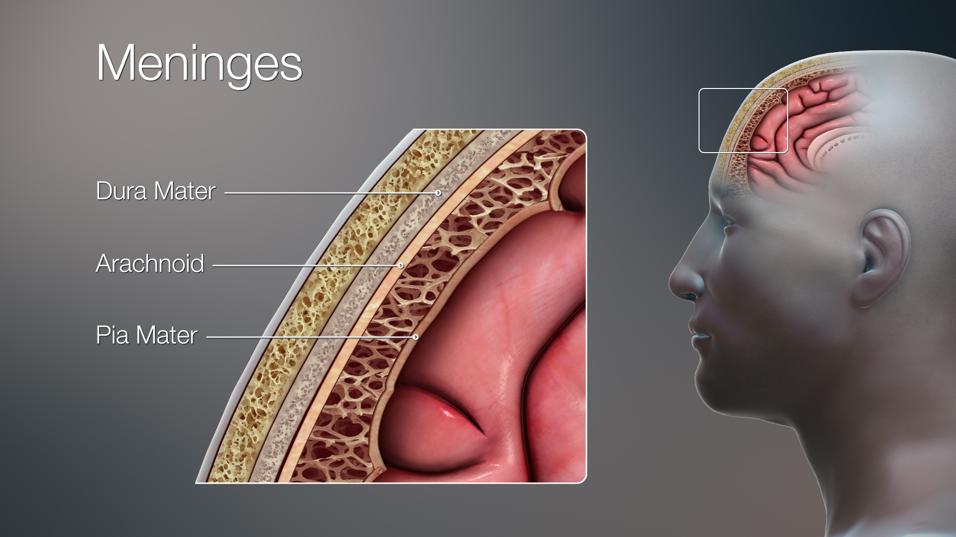
The brain can be divided into 2 parts:
Cerebrum and cerebellum
The cerebrum is the largest part of the brain and the normal cerebrum contains multiple:
Sulc: grooves within brain
Gyri: folds in cerebral cortex (wrinkly outer layer)
The cerebrum has 4 main cerebral lobes
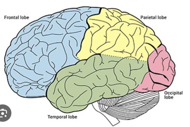
The cerebrum is divided into a right and left hemisphere by
Interhemispheric fissure
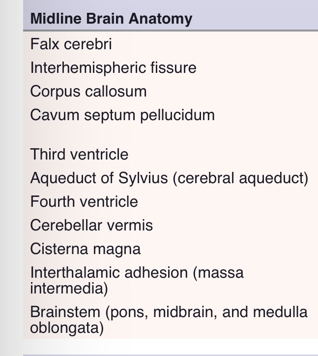
Midline brain anatomy
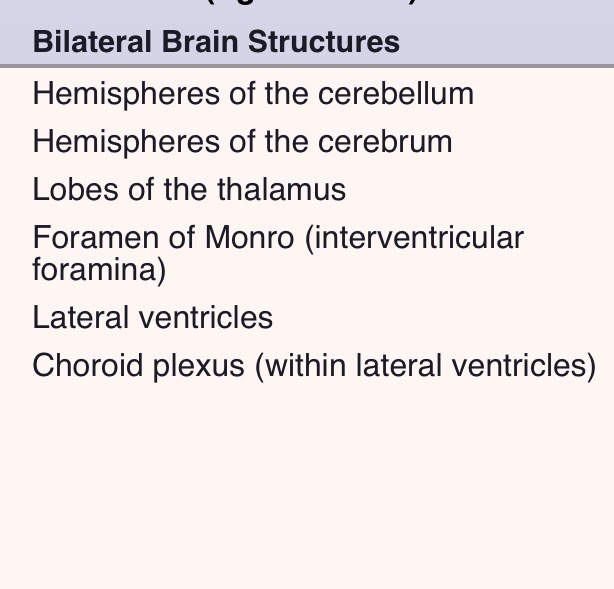
Bilateral brain structures
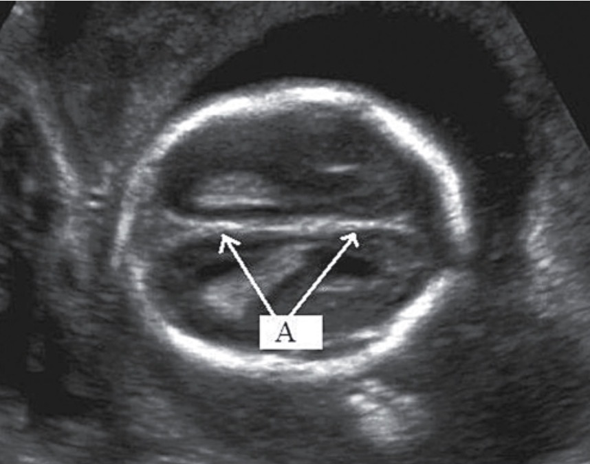
What is the falx cerebri
A double fold of dura mater, located in the interhemispheric fissure and seperates the cerebral hemispheres
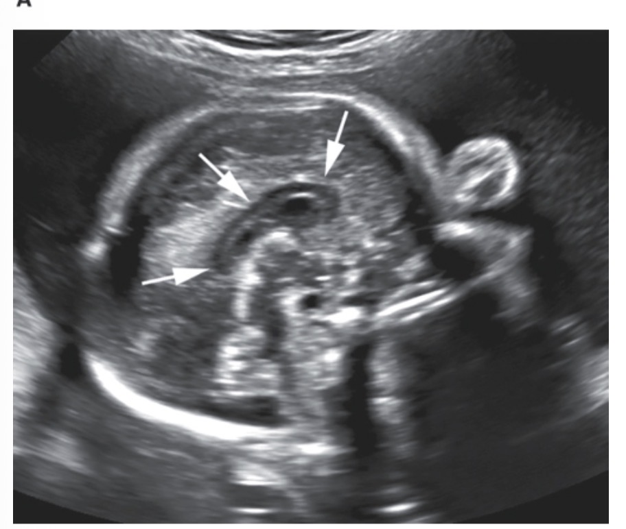
What is the corpus callosum
Connects the cerebral hemispheres and allows communication between them
The corpus callosum (developed btw 18-20wks) develops form anterior to posterior and has 4 parts termed the
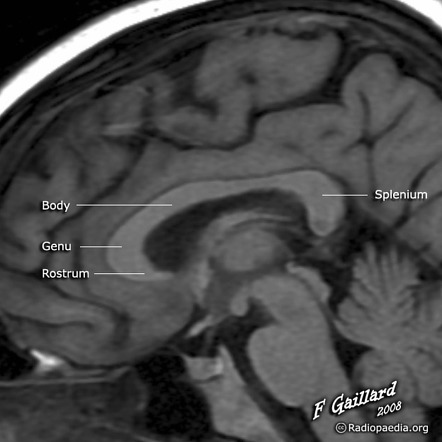
The absence of the corpus callosum is referred to as
Agenesis of the corpus callosum
Anterior portion of brain between frontal horns of the lateral ventricles
Cavum septum pellucidum CSP
Sonographically, the Cavum Septum Pellucidum (CSP) will present
“Box shaped” structure in axial/transverse plane
Should be seen between 18 and 37 weeks
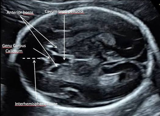
The CSP does NOT communicate with the ? And its absence is associated with?
Ventricular system.
Multiple cerebral malformations including agenesis of corpus callosum
The two lobes of thalamus are located and connected by?
Located on Both sides of third ventricle and conencted by massa intermedia/ interthalamic adhesion
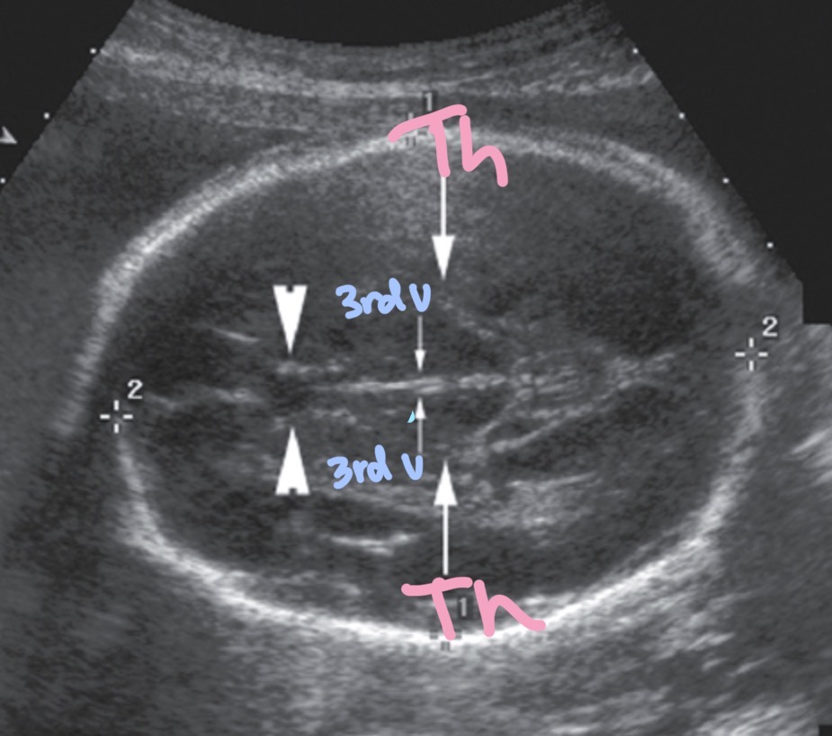
The paired lateral ventricles are located?
Both sides ot the falx celebri
Within the atria of both lateral ventricles lies the choroid plexus, which is
Mass of cells responsible for production of cerebrospinal fluid ( CSF )
Cerebal ventricle system
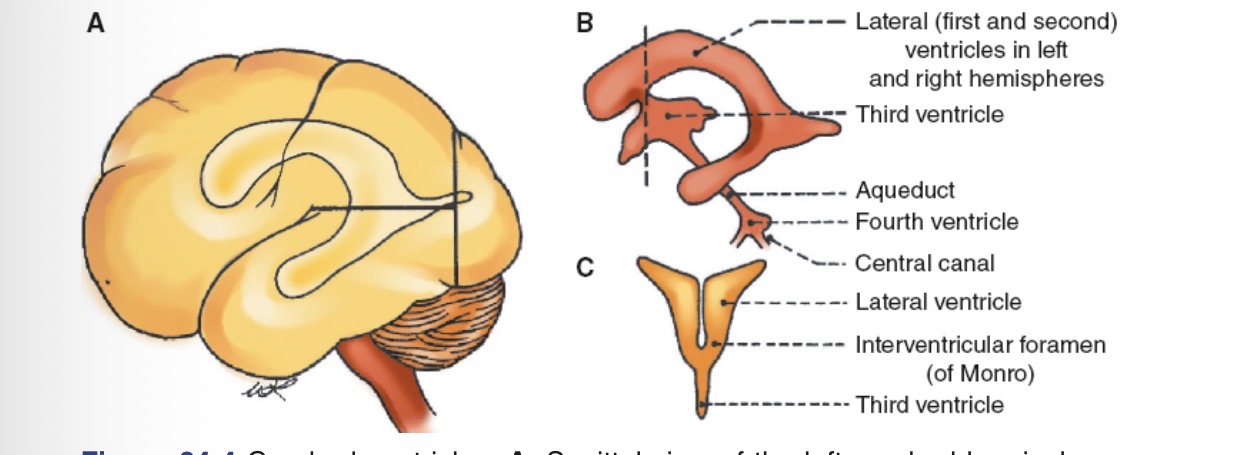
Lateral ventricle measurement
Measured in axial plane at level of atrium. Should not measure more than 10mm (ventriculomegaly)
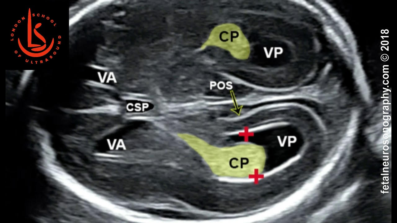
The lateral ventricles communicate with the third ventircle at the ?
Foramen of monro/ interventricular foramina
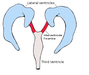
The third ventricle connects to the 4th ventricle inferiorly by means of a long tubelike structure via the?
Aqueduct of sylvius or cerebral aqueduct.
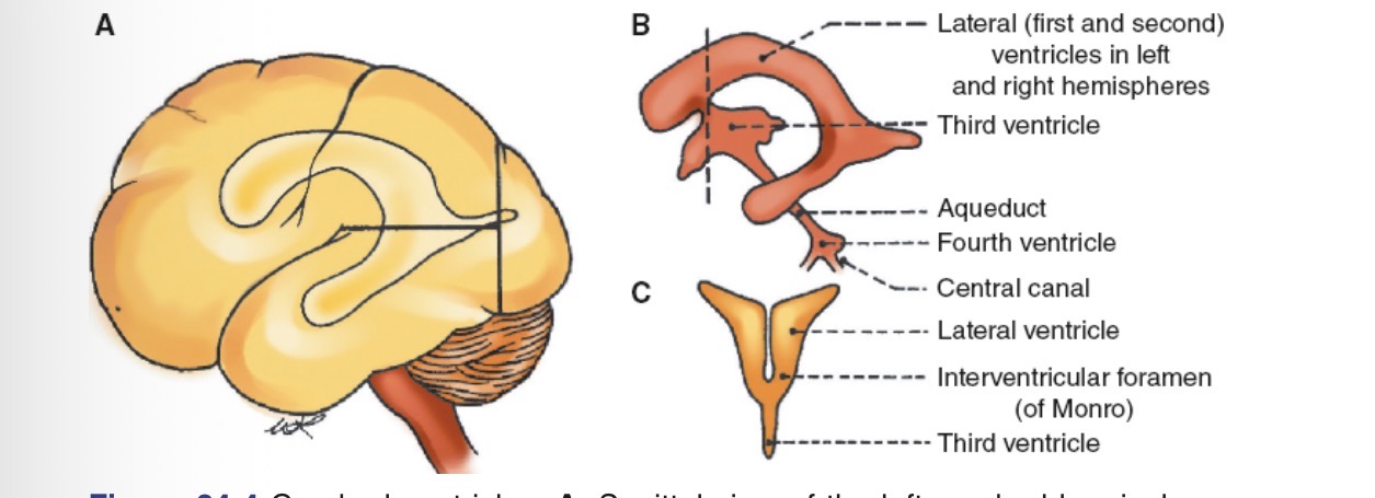
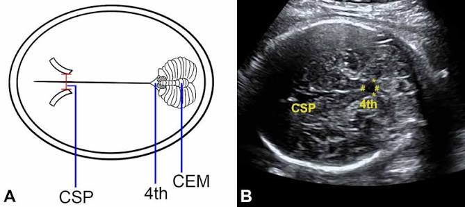
The 4th ventricle is located
Anterior to cerebellum
The foramina of luschka of the 4th ventricle allows CSF from 4th ventricle to travel to
Subarachnoid space
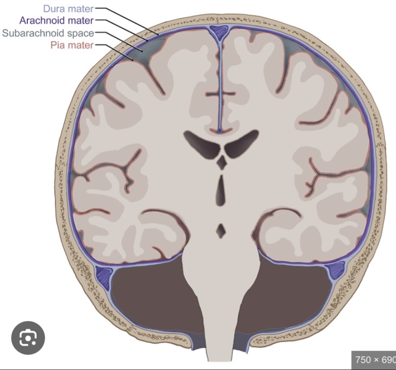
The median aperture of 4th ventricle aka the foramen of magendie allows CSF to travel to
Cisterna magna and subarachnoid space
What is the cisterna magna?
Located in the posterior fossa of cranium, it is the largest cistern in the head
The cisterna magna is sonographically located
Posterior to cerebellum between the cerebral vermis and inner surface of occipital bone
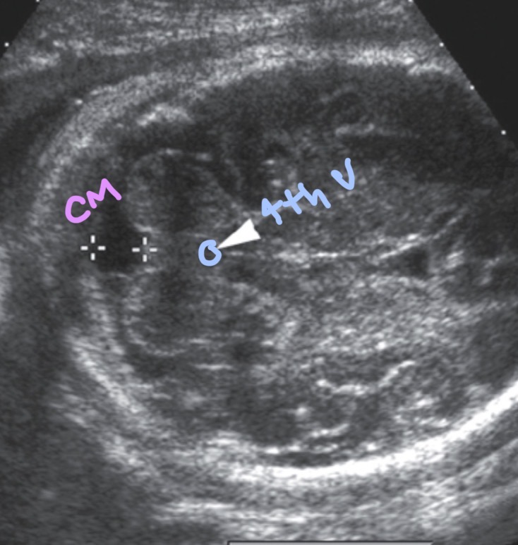
The depth of the cisterna magna should not measure
More than 10 mm or less than 2 mm in trans cerebellar plane
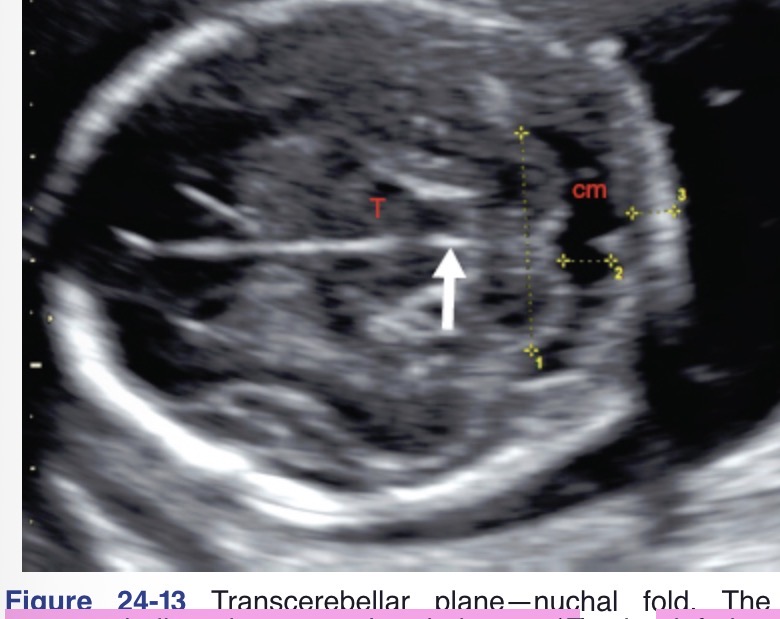
CM meas of more than 10mm is consistent with
Mega cisterna magna and dandy-walker complex
CM meas of less than 2mm is worrisome for
Arnold chiari 11 malformation
The cerebellum is located in the posterior fossa of the cranium and consists of two hemispheres: right and left. These two hemispheres are joined by the
Cerebellar vermis
The cerebellum appears
Normal cerebellum is dumbbell or figure 8 shaped
2 hemispheres should be symmetric although hypoplasia of one hemisphere may occur, causing one to appear smaller than normal
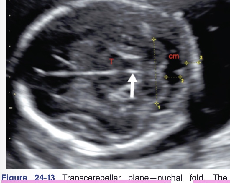
The transcerebellar diameter (TCD)
TCD= gestational week of fetus
Measured at transverse plane at level of cisterna magna and thalamus
What is the cephalic index?
Tool for head shape indication calculated by
CI = BPD/OFD x 100
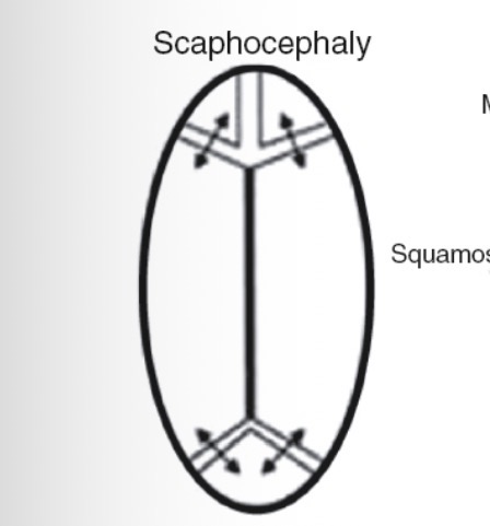
A CI of less than 75 denotes a
Dolichocephy/spachocephaly (elongated and narrow)
A CI of more than 85 denotes
Brachycephaly (short and round)
The normal-to-medium sized skull is termed
Mesocephalic
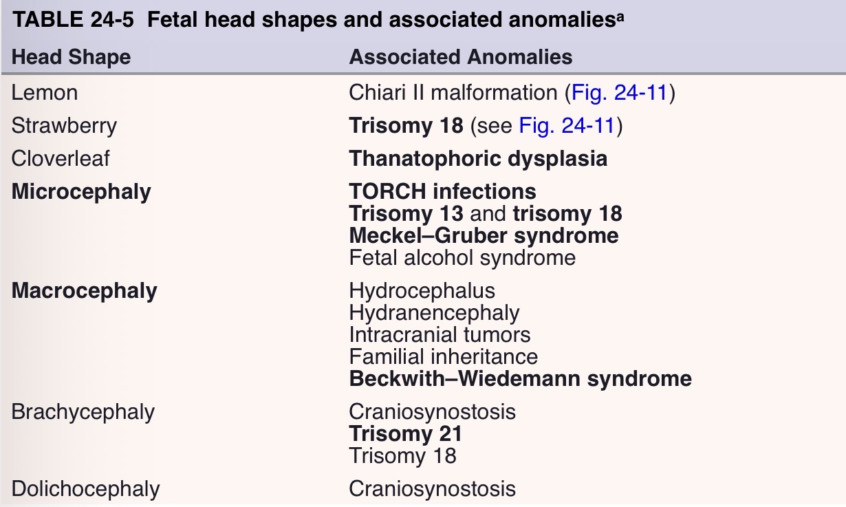
Fetal head shapes and associated anomalies
Lemon: chiari 2 malformation
Strawberry: trisomy 18
Cloverleaf: thanotophoric dysplasia
Microcephaly: torch infection, trisomy 13 or 18, meckel-gruber syndrome
Macrocephaly: hydro or hydranecephaly, beckwith- wiedeeman syndrome
Brachycephaly: trisomy 21
Dolichocephaly: craniosynostosis
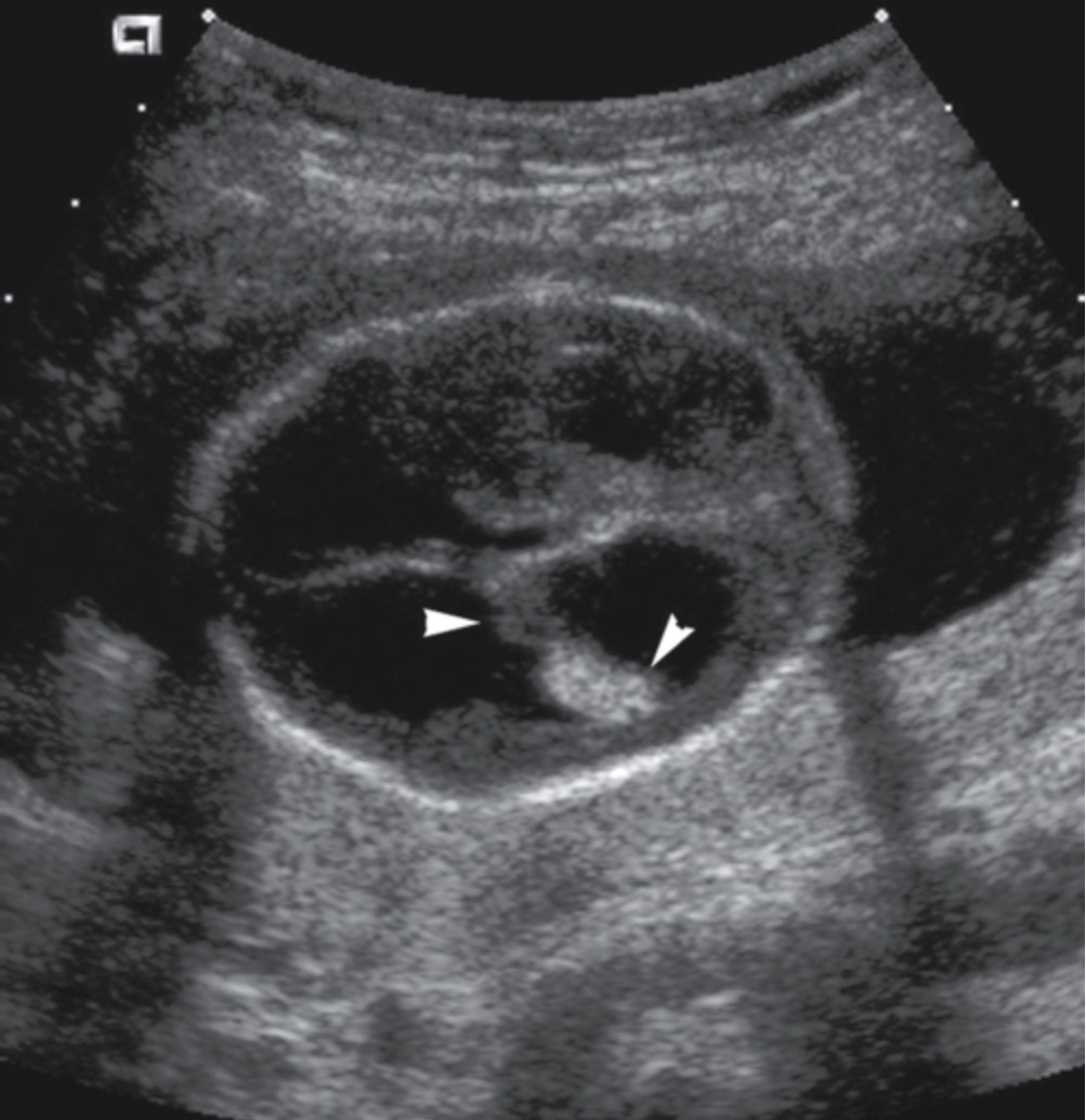
Ventriculomegaly is the most common cranial abnormality and occurs when atrial diameter of lateral ventricle measures?
More than 10 mm
What are the sonographic findings of vetriculomegaly?
Atrium of LV meas >10mm
Atrial meas >15mm is considered moderate to marked ventriculomegaly
Dangling choroid sign
Dilation of any part of ventricular system
How does hydrocephalus differ from ventriculomegaly
Hydrocephalus is the dilation of ventricular system secondary to an obstruction
What are the 2 types of hydrocephalus?
Communicating hydrocephalus (obstruction outside of ventricular system)
Noncommunicating hydrocephalus (obstruction is located within ventricular system)
What is the most common cause of hydrocephalus in utero
Aqueductal stenosis
what is aqueductal stenosis?
when the cerebral aqueduct/aqueduct of sylvius is narrowed, thus preventing the flow of CSF from the third to the fourth ventricle.
sonographic findings of aqueductal stenosis
Atrium of the lateral ventricle measures greater than 10 mm
Atrial measurement greater than 15 mm is considered moderate to marked ventriculomegaly
Dangling choroid sign
Dilatation of the lateral ventricles and the third ventricle;
the fourth ventricle remains normal
what is hydranencephaly and what happens with this condition
fatal condition in which the entire cerebrum is replaced by a large sac containing CSF;
what are possible causes of hydranencephaly
bilateral occlusion of the internal carotid arteries with subsequent destruction of the cerebral hemispheres.
TORCH infections (cytomegalovirus or toxoplasmosis)
Sonographic Findings of Hydranencephaly
Fluid-filled cranium
Absent or partial absence of the falx cerebri
Maintained brainstem, basal ganglia, and, perhaps, the thalamus
No identifiable cerebral cortex
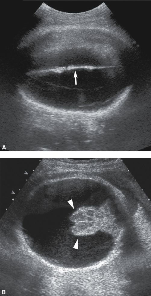
Hydranencephaly can be difficult to differentiate with the sonographic findings of severe ventriculomegaly and alobar holoprosencephaly. It is important to note that with both severe ventriculomegaly and holoprosencephaly,
there will typically be a rim of cerebral tissue maintained, whereas with hydranencephaly, there is no cerebral mantle present
what is Holoprosencephaly
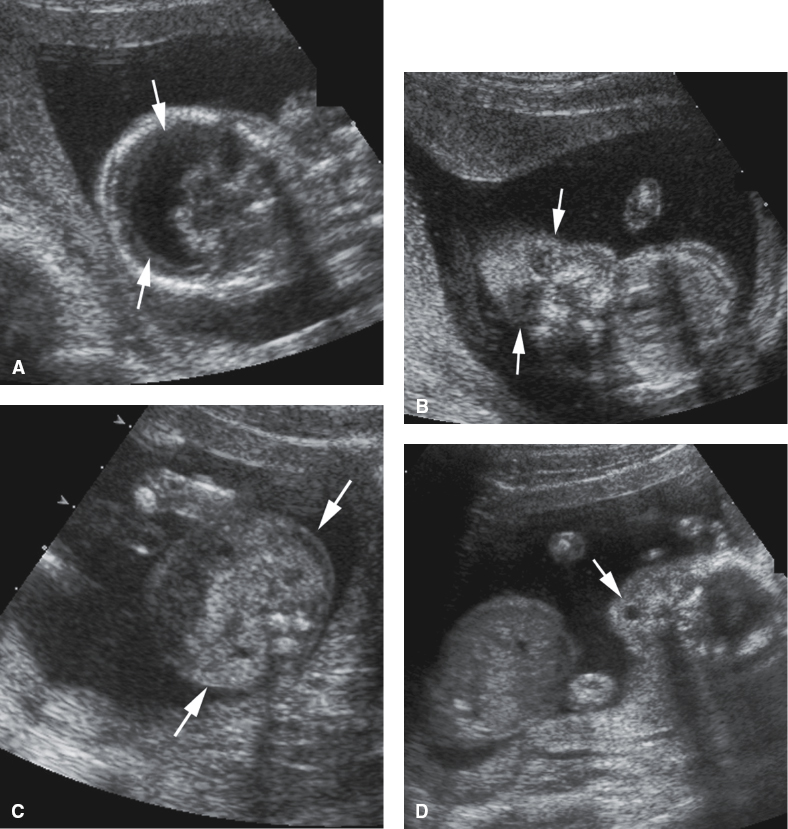
a group of brain abnormalities consisting of varying degrees of lateral ventricle fusion, absence of the midline structures, and associated facial anomalies
what are the three main types of holopresencephaly
alobar (cortex can resemble “pancake” “cup” or “ball”) this is the most severe form
semilobar
lobar
Alobar holoprosencephaly is diagnosed when there is absence of the
corpus callosum
CSP
third ventricle
interhemispheric fissure
falx cerebri
There will also be evidence of a horseshoe-shaped monoventricle
and the lobes of the thalamus may be fused and echogenic in appearance
Facial anomalies associated with alobar holoprosencephaly
Cyclopia (fusion resulting in one single eye)
Hypotelorism (close eyes)
Proboscis (false nose)
(Median) cleft lip
Anophthalmia (absence of one or both orbits)
Cebocephaly (closely spaced eyes, one nostril, missing upper lip, irregular head shape)
Infants with lobar holoprosencephaly may experience
extreme mental retardation
in 50% to 70% of fetuses diagnosed with holo-prosencephaly also have
Trisomy 13/ Patau syndrome
Sonographic Findings of Alobar Holoprosencephaly
Horseshoe-shaped monoventricle
Fused echogenic thalami
Absence of the CSP, interhemispheric fissure, falx cerebri, corpus callosum, and third ventricle
Normal cerebellum and brainstem
Facial anomalies (e.g., cyclopia, proboscis, cebocephaly, facial clefts, hypotelorism)
What is dandy-walker complex
spectrum of posterior fossa abnormalities that involve the cystic dilatation of the cisterna magna and fourth ventricle.
what other midbrain anomalies associated with dandy-walker malformation
agenesis of the corpus callosum, ventriculomegaly, holoprosencephaly, and cephaloceles
If the cerebellar vermis is absent and the fourth ventricle is enlarged, then
DWM must be suspected.
what is the difference between mega cisterna magna and DWM
the fourth ventricle is normal with mega cisterna magna and enlarged with DWM
Sonographic Findings of Dandy–Walker Malformation
Enlargement of the cisterna magna greater than 10 mm
Communication of the enlarged cisterna magna with a dilated fourth ventricle
Agenesis or hypoplasia of the cerebellar vermis
splayed lobes
Varying degrees of ventriculomegaly
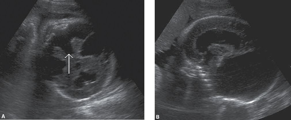
Sonographic Findings of Mega Cisterna Magna
Enlargement of the cisterna magna greater than 10 mm
Normal cerebellum and fourth ventricle
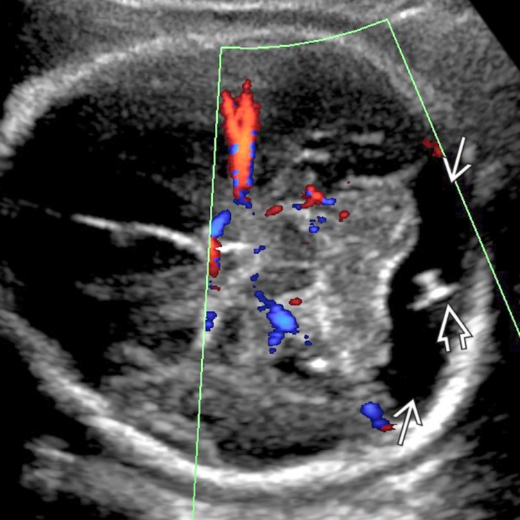
The congenital lack of corpus callosum and CSP is termed
agenesis of the corpus callosum, and CSP. There can be partial or complete absence of the corpus callosum. Most often, if the corpus callosum is absent, the CSP will be absent as well
agenesis of corpus callosum and csp has been linked to as many as 50 to 200 different syndromes and anomalies such as
Apert syndrome (genetic disorder that includes craniosynostosis, midline facial hypoplasia, and syndactyly)
holoprosencephaly
DWM
aqueductal stenosis
trisomy 18
trisomy 8
trisomy 13
With agenesis of the corpus callosum, the sulci tend to have a more
perpendicular or radial arrangement and often appear to have a “spoke-wheel” pattern.
Sonographic Findings of Agenesis of the Corpus Callosum and Cavum Septum Pellucidum
Partial or complete absence of the corpus callosum and absence of the CSP (after 18 weeks)
“Sunburst” sign/ “spoke-wheel” pattern
Colpocephaly—(teardrop-shaped lateral ventricles)
Elevated and dilated third ventricle
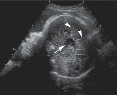
what is Schizencephaly
development of fluid-filled clefts within the cerebrum, may be described as open lip or closed lip, with open lip being more readily identified in utero
Sonographic Findings of Schizencephaly
Fluid-filled clefts within the cerebrum
Agenesis of the CSP and corpus callosum (50% of the time)
Ventriculomegaly
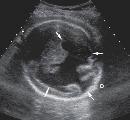
what is porencephaly
a rare condition in which a cyst communicates with the ventricular system. Porencephaly can occur after the fetus has experienced hemorrhage within one or both of the cerebral hemispheres.
porencephaly may be caused by
ischemic events or vascular occlusion within the brain
Arachnoid cysts can be confused with porencephaly. It is important to note that
arachnoid cysts will not communicate with the ventricular system.
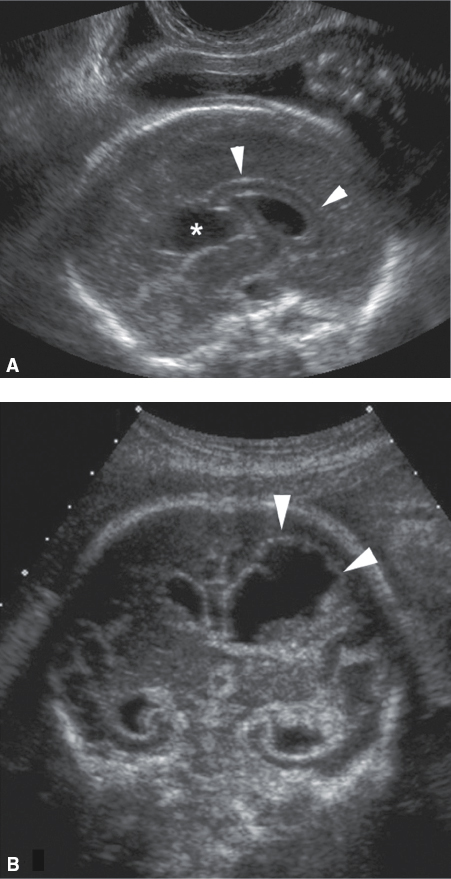
Sonographic Findings of Porencephaly
Cystic mass that communicates with the lateral ventricle
Most often unilateral
what is lissencephaly
“smooth brain.” It is a condition in which there are no gyri within the cerebral cortex. Agyria, and the absence of sulci within the brain, is not typically diagnosed until the third trimester or postnatally and almost always carries a poor prognosis.
Sonographic Findings of Lissencephaly
Lack of sulci and gyri within the cerebrum
what are Choroid plexus cysts
cysts located within the choroid plexus of the lateral ventricles. These are frequently encountered during a routine sonographic examination and typically regress by the end of the third trimester, although there is an association with trisomy 18
Sonographic Appearance of a Choroid Plexus Cyst
Anechoic, round, smooth-walled cyst located within the choroid plexus of the lateral ventricle
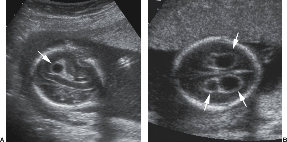
Among the list of neural tube defects are
cephaloceles, various spinal dysraphisms, anencephaly, and spina bifida;
with Anencephaly and spina bifida the most common
Screening for neural tube defects is achieved by a combination of
sonography, amniocentesis, and/or maternal serum screening
Alpha-fetoprotein (AFP) is initially produced by the
yolk sac, fetal gastrointestinal tract, and the fetal liver. AFP exits the fetus through an opening in the neural tube if one is present
what test is especially helpful for detecting neural tube defects?
maternal serum alpha-fetoprotein (MSAFP)
increased levels of AFP may not always indicate that a neural tube defect is present. Elevated levels of AFP are also found with
omphalocele, gastroschisis, multiple gestations, fetal demise, and incorrect gestational dating.
what is acrania
one of the most common neural tube defects. Acrania is defined as the absence of the cranial vault above the bony orbits. It can be further divided into two main subtypes depending on the amount of cerebral tissue present, anencephaly and exencephaly.
Clinical Findings of Acrania/Anencephaly
Elevated MSAFP
Sonographic Findings of Acrania/Anencephaly
Absent cranial vault
Some cerebral tissue may be present
“Froglike” facies or bulging eyes
Possible fetal movement and active fetal heart
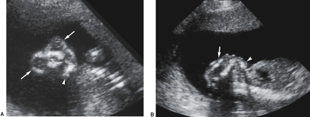
what is Arnold–Chiari II or Chiari II malformation
group of cranial abnormalities associated with the neural tube defect spina bifida
Spina bifida may result in a mass that protrudes from the spine. This mass can be referred to as a
meningocele or myelomeningocele, depending on its contents
The most common location of spina bifida is within the
distal lumbosacral region
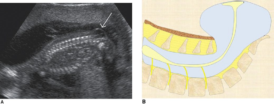
A posterior fossa abnormality such as Chiari II malformation should be suspected if the
cisterna magna is not visualized.
The lateral ventricles will also be distorted in shape.
Colpocephaly (teardrop shaped lateral ventricles)