PSY 351 exam 1
 Call Kai
 Learn
Learn Practice Test
 Spaced Repetition
 Match
Match Flashcards
 Knowt Play
1/251

There's no tags or description
Looks like no tags are added yet.
Name | Mastery | Learn | Test | Matching | Spaced | Call with Kai | Chat |
|---|
No analytics yet
Send a link to your students to track their progress
252 Terms
Neurons/nerve cells
specialized cells that receive inputs, integrate them and distribute info to other neurons
80-90 billion in brain
Glial cells
same number of them as neurons
provide various support functions
directly participate in info processing
Synapses
tiny gaps between neurons where info is transmitted between them
Principal divisions of neuron
input zone
integration zone
conduction zone
output zone
Input zone
dendrites of neurons receive info via synapses from other neurons
Dendritic spines
small projections from surface of dendrite that add more space for synapses
Integration zone
cell body/soma of neuron integrates info received to determine whether to send signal
Conduction zone
axon/nerve fiber carries neuron’s own electrical signals away from cell body
axon may split into multiple branches — axon collaterals
Output zone
axon terminals transmit neuron’s signals across synapses to other cells
Axon terminals
transmit the neuron’s signal across synapses to other cells
Motor neurons
trigger movements
large with long axons reaching out to synapse on muscles, causing muscular contractions
Sensory neurons
convey info from sense organs to brain
take on different shapes depending on which sense the neuron is signalling
longer axon
Interneurons
this is what most of the neurons in the brain are
receive info from other neurons, process it & pass integrated info to other neurons
make up hugely intricate networks & circuits that perform complex functions of brain
shorter axons
Larger neurons
tends to
have more compelx inputs & outputs
cover greater distaces
convey info more rapidly than smaller neurons
Classifying neurons by shape
multipolar neurons
bipolar neurons
unipolar neurons
Multipolar neurons
many dendrites
single axon
most common type of neuron
Bipolar neurons
single dendrite at one end of cell & single axon at other end
especially common in sensory neurons e.g. vision
Unipolar/monopolar neurons
single extension, typically identified as axon for its entire legnth
branching dendrite-like input zone & integration zone from which axos rises directly
leads away to distant output zone with its axon terminals
cell body branches off axon partway along its length
transmit touch & pain info from body into spinal cord
No. of dendrites & complexity of inputs received
simple neurons have just a couple of short dendritic branches
others have huge & complex dendritic trees/arbors receiving a ton of synaptic contacts from other neurons
Presynaptic neuron
info transmitted from this
Postsynaptic neuron
info transmitted to this
Synapse divided into compenents
presynaptic membrane of axon terminal of presynaptic/transmitting neuron
synaptic cleft that separates presynaptic & postsynaptic neurons
postsynaptic membrane on dendrite/cell body of postsynatpic/receiving neuron
Synaptic vesicles
tiny hollow spheres in presynaptic axon terminals
contain molecules of neurotransmitter
Neurotransmitter
special chemical with which presynaptic neuron communicates with postsynaptic cell
Info transmission through synapses
in response to electical signal, synaptic vesicles fuse to presynaptic membrane
rupture to release neurotransmitters into synaptic cleft
after crossing cleft, the released neurotransmitters interact with matching neurotransmitter receptors that stud the postsynaptic membrane
receptors capture and react to neurotransmitter, altering level of excitation of postsynaptic neuron
affects likelihood that postsynaptic neuron will in turn release its own neurotransmitter from its axon terminals
Neuroplasticity
capacity of neurons to continually remodel their connections with other neurons
Functions of axons
rapid transmission of electrical signals along outer membrane of axon
slower transportation of substances within the axon, to & from axon terminals
Axonal transport
trransport of substances through interior of axon between cell body (where they are produced) and axon terminals (where they are used)
Anterograde transport
moves materials towards axon terminals
Retrograde transport
moves used materials back to cell body for recycling
Main types of glial cells
oligodendrocytes
Schwann cells
astrocytes
migroglial cells
Oligodendrocytes
supply myelination to neurons in brain & spinal cord
each cell typically supplies myelin beads to several nearby axons
Schwann cells
provide myelination of neurons in other parts of body
each cell wraps itself around a segment of one axon to provide a single bead of myelin
Myelination
myelin sheaths wrapping around axons of neurons
large increase in speed in which electrical signal passes down axon
jumping from one node of Ranvier to the next
Nodes of Ranvier
small, uninsulated patches of axonal membrane between adjacent beads of myelin
Astrocytes
weave around & between neurons with tentacle-like extensions
some astrocytes stretch between neurons & fine blood vessels, controlling local blood flow to increase amount of blood reaching more active brain regions
help form tough outer membranes that swaddle the brain
secrete chemical signals that affect synaptic transmission & synaptic formation
Microglial cells
tiny & mobile
primary job: contain & clean up sites of injury
Gross neuroanatomy
made up of tissues that are comprised of
neuronal cell bodies
dendrites
axons
glial cells
neural structures that are visible to naked eye
Peripheral nervous system
made up of nerves
somatic nervous system
autonomic nervous system
Somatic nervous system
consists of nerves that interconnect brain & major muscles & sensory systems of body
main pathway through which brain controls movement & receives sensory info from body & sensory organs
cranial nerves & spinal nerves
Autonomic nervous system
made up of nerves that connect primarily to viscera (internal organs)
little conscious, voluntary control over its action
brain’s main system for controlling the organs of body
sympathetic & parasympathetic nervous systems
Functions of sympathetic nervous system
prepares body for immediate action
fight-or-flight response
release norepineprhine
Anatomy of sympathetic nervous system
axons of SNS exit from middle parts of spinal cord, travel short distance then innervate sympathetic ganglia
sympathetic ganglia run in 2 chains along each side of spinal column
axons from sympathetic ganglia then spread throughout body, innervating all major organ systems
Function of parasympathetic nervous system
generally helps body relax, recuperate & prepare for future action
rest-&-digest response
release acetylcholine
Anatomy of parasympathetic nervous system
nerves of PNS originate in brainstem (above sympathetic nerves) & in sacral spinal cord (below sympathetic nerves)
travel longer distance before terminating in parasympathetic ganglia
clusters of neurons that are usually located close to organs they serve
Central nervous system
consists of brain & spinal cord
Spinal cord
funnels sensory info from body up to brain
conveys brain’s motor commands out to body
contains circuits that perform local processing & control simple units of behavior
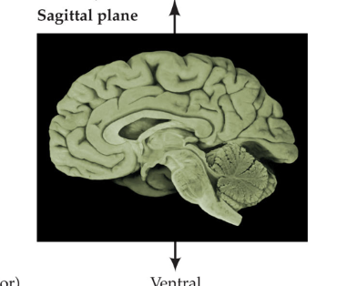
Saggital plane
divides brain into left & right
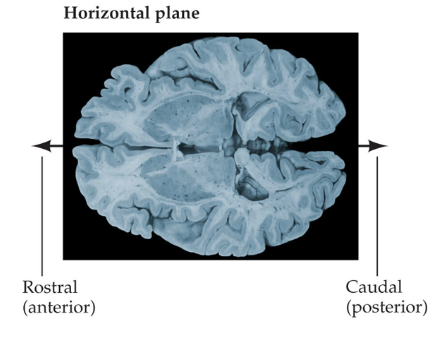
Horizontal plane
divides brain into upper & lower parts
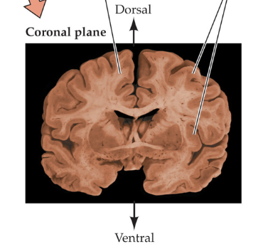
Coronal plane
divides between front and back
Gray matter
outer layers of cerebral cortex, inner layer of spinal cord
contain a preponderance of neuronal cell bodies & dendrites
mostly receives & processes info
White matter
inner layers of cerebral cortex, outer layer of spinal cord
whitish fatty myelin that insulates many axons
mostly transmits info
Gyri
ridges of tissue created by cortex folding
Sulci
crevices separating gyri
Ganglion
cluster of neurons outside of brain
Nucleus
cluster of neurons in brain
Superior

Inferior

Anterior
towards the front

Posterior
towards the back

Dorsal
the top of the neuraxis

Ventral
bottom of neuraxis

Rostral

Caudal

Neuraxis

Proximal
closer to target region
Distal
farther from target region
Lateral
towards the sides
Medial
towards the middle
Thalamus
receives sensory info & relays it to cortex (except olfaction)
Hypothalamus
hormones, motivated behavior
sex
aggression
eating
Basal ganglia
form circuit critical for movement
Limbic ganglia
complex nuclei for emotions
Hippocampus
forming declarative memory/facts
bilateral damage = no declarative memory
Brodmann’s map of the brain
mapped various parts of brain based on function rather than structure
Cerebral cortex
has 6 layers of cells, each layer is has
band of similar neurons
a distinctive pattern of dendrites/axons
Mesencephalon
midbrain
substantia nigra
ventral tegmental area (VTA)
superior colliculus
inferior colliculus
Superior & inferior colliculi
in non-humans, play a big role in vision (especially hunting)
Substantia nigra
uses dopamine for movement
Ventral tegmental area (VTA)
uses dopamine for reward circuit
Rhombencephalon
hindbrain
metencephalon
myencephalon
spinal cord
Myencephalon
medulla oblangata
life-sustaining functions e.g. breathing, heartrate
Metencephalon
pons
cerebellum → coordination
Reticullar formation
large network of neurons that go through hindbrain, midbrain & into forebrain
norepinephrine as neurotransmitter
important for vigillance & attention
Dura mater
outermost, tough layer surrounding brain
visible to naked eye
Arachnoid membrane
looks like spider web
has blood vessels and CSF
below it is subarachnoid space
Pia mater
aka pial layers
microscopic layer just before brain matter
Cerebral spina fluid (CSF)
fills subarachnoid space
keeps brain from collapsing on itself
Hematoma
internal bleeding
Subdural hematoma
internal bleeding in dura mater
blood puts pressure on brain & causes brain damage
may leak out through bottom which is important for life → could die
Carotid arteries
arteries that meet with vertebral artery to go up to brain
found in sides of neck
branch into anterior & middle cerebral arteries
Vertebral artery
artery that meets with carotid artery to go up to brain
towards back of neck
3 main arteries of brain
anterior cerebral artery
middle cerebral artery
posterior cerebral artery
Blood brain barrier
formed by tight junctions between endothelial cells in blood vessels
oxygen, carbon dioxide, some fat-soluble molecules pass through freely
some important substances (e.g. glucose) need active transport by transporter protein
large and charged molecules can’t pass through
Meninges
3 protective layers surrounding brain
dura mater
arachnoid membrane
pia mater
Stroke
clot/narrowing/rupture interrupts blood supply to brain region, causing it to stop functioning/die
Warning signs of stroke
sudden numbness/weakness
altered vision
dizziness
severe headache
confusion/difficulty
Gross dissection
yoink out brain to study with naked eye
only shows structure not function
Studying brain structure
individual neurons
nuclei
gray matter
Study connections between structures
fiber tracts: bundles of axons
white matter