repro test 1
 Call Kai
 Learn
Learn Practice Test
 Spaced Repetition
 Match
Match Flashcards
 Knowt Play
1/63

There's no tags or description
Looks like no tags are added yet.
Name | Mastery | Learn | Test | Matching | Spaced | Call with Kai | Chat |
|---|
No analytics yet
Send a link to your students to track their progress
64 Terms
Ovarian cycle – female HPG axis
- Hypothalamus: Releases (GnRH) in pulses.
- Anterior Pituitary: Secretes (FSH) and (LH) in response to GnRH.
- Ovaries: Produce sex steroids (estrogen/estradiol and progesterone) and peptide hormones (inhibin)
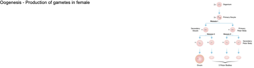
Oogenesis - Production of gametes in female
In utero:
- Gamete form many primary follicles containing each one oogonium
- Each oogonium undergoes mitosis -> each oogonium forms 2 primary oocytes
- Primary Oocytes start meiosis and arrest in prophase 1
After birth, after puberty
- FSH stimulates a few follicles to grow -> every menstrual cycle a few Oocytes complete meiosis I
o Result: 2 cells = Secondary Oocyte and 1st polar body
- Secondary Oocyte develops to metaphase II, rests in this state till after ovulation = haploid oocyte and polar body
- Only if fertilized completion of Meiosis II (casts off second polar body)
Ovarian cycle: follicular phase
- Day 1 of menstruation.
- FSH and LH are released by the pituitary.
- Follicle development: Primordial → Primary → Secondary → Mature (Graafian).
Primordial cell contains an immature primary oocyte surrounded by granulosa cells that multiply as the follicle develops.
The zona pellucida forms around the oocyte.
- FSH → granulosa cells → growth + estradiol + inhibin.
- LH → theca cells → androgens + progesterone.
- Theca-cell androgens are used by the granulosa cells to make estradiol.
- Increasing estradiol promotes development of the dominant follicle, which prepares for ovulation.
ovulation
- Near the middle of the cycle
- estradiol becomes very high.
- the high estradiol level causes positive feedback, resulting in a large LH surge.
- LH surge → follicle wall ruptures → mature follicle is released
Lutel phase
- Antrum contains clotted blood after ovulation -> corpus hemorrhagicum
- Ruptured follicle turns into Corpus luteum after ovulation - Becomes a gland – secrets Progesterone + Estradiol + Inhibin
- Corpus luteum regresses if no pregnancy occurs after 10-12 days -> scar tissue corpus albicans
- If pregnancy occurs Corpus Luteum is supported by hCG = human chorionic gonadotrophic hormone
Menstrual cycle 1. Menstrual Phase
- Lack of progesterone
- Loss of blood due to breakdown of superficial layers of uterine endometrium
- About 30 - 50 ml of mainly arterial blood.
Menstrual cycle : 2. Proliferative phase
- FSH grows follicles and oocytes (in ovaries) -> they produce mainly estrogen
- Estrogen travels through blood to endometrium and thickens and builds it up
- Uterine glands small and round
menstrual cycle 3. Secretory phase
- LH from ovulation and luteal phase
- corpus luteum progesterone and estrogen -> maintains lining and enlarges glands, elongated, & coiled arteries
- Uterine glands secrete uterine milk
Premenstrual phase
- Lack of progesterone
- Glands start breaking down
- Blood vessels leak
- Tissue death
Epithelium of the Cervix
- Mucus secreting simple columnar epithelium on luminal surface
- moist non-keratinized stratified squamous epithelium covering the external surface of the cervix
- Transformation zone - area between columnar and squamous cells
- cervix averages 22 mm in length and 6 mm in diameter at ovulation
Cervical mucus:
- 2 major fractions:
o insoluble gel or mucin
o aqueous phase containing the soluble components (lipids, fatty acids, prostaglandins, proteins, enzyme inhibitors, and immunoglobulins)
- Physical barrier to pathogens
- Immune barrier to pathogens
- Sometimes more or less permeable to sperm
Changes in cervical mucus
1. Early Follicular phase: Mucus thick, sticky
2. Late Follicular phase
- Mucus copious, watery, thin & alkaline → sperm can penetrate mucus
- Dominant influence: Estrogens
3. Ovulatory Period
- Mucus thinnest- wet and clear at time of ovulation
- sperm can easily penetrate through the mucus
4. Luteal phase
- Mucus thick, viscous & cellular
- No ferning pattern when dried
- Dominant influence: Progesterone
Hormonal regulation in the male (HPG-axis)
- Hypothalamus -> GnRH -> anterior pituitary -> LH and FSH
- LH influences Leydig cells to produce testosterone (neg feedback)
- FSH stimulates Sertoli cells to produce ABP and inhibin (neg feedback from inhibin)
The Testes anatomy
- Paired and contained within the scrotum
- Septa divide testis into ~250 lobules
- Each lobule contains 1-4 seminiferous tubules
pathway of sperm
Seminiferous tubules → Straight tubules → Rete testis → Efferent ductules → Epididymis → Vas deferens → (past seminal vesicles) ejaculatory duct → (past prostate and bulbourethral glands) urethra
Seminiferous tubules
- Site of sperm production
- Lined by complex stratified epithelium
Cells in the testes Seminiferous tubules:
in seminiferous tubules
- Spermatogonia
- FSH → Sertoli cells → ABP AND GF → nourishment & protection for developing sperm & contain receptors to bind testosterone
Interstitial space:
- LH → Leydig cells:→ testosterone, -> testosterone diffuses into seminiferous tubules
- Peritubular myoid cells: peristaltic movement
- Capillary cells of blood vessels
Blood-testes barrier
- Separates the testes from the normal circulatory processes
- Tight junctions between adjacent Sertoli cells form the blood testis barrier
o Prevents blood and other body fluids entering the lumen of the seminiferous tubules
o Only allows secretions from Sertoli cells to enter
o Protects developing sperm from the body’s immune system, restricts passage of drugs
Spermatogenesis
- Meiosis: Formation of haploid Spermatids from diploid Spermatogonia
o Stem cells enter into spermatogenesis ~ every 16 days
1. Mitosis: spermatogonia become primary spermatocytes
2. Meiosis I: Reductional division Separation of homologous chromosomes
3. Meiosis II: Equational division Separation of sister chromatids
Spermiogenesis
- remodelling and maturing of spermatids into mature spermatozoa through differentiation
- Round spermatids become elongated spermatids
- Spermatids are nonmotile
- Then spermiation = release of spermatozoa into lumen of seminiferous tubules
- sertoli cells provide nutrients
- 4 phases
1. Golgi phase
- Spermatid almost spherical
Well developed golgi apparatus
- Small vesicles of golgi fuse -> form secretory pro- acrosomic granules
- Vesicle fusion continues till a large acrosomal vesicle is formed
Proximal centriole (PC) is region where tail will attach
cap phase
- Golgi migrates to caudal end
Distal Centriole (DC) forms Axoneme (AX) = flagellum
- Acrosomic vesicle flattens and forms distinct cap:
o Outer acrosomic membrance (OAM)
o Inner acrosomic membrane (IAM)
- Acrosomal contents -> enzyme
Acrosomal Phase
- Nucleus elongates
- Acrosome covers majority of anterior nucleus
- Neck is formed
maturation phase
- Mitochondria assemble in midpiece
- Excess cytoplasm phagocytosed by Sertoli cells
Spermiogenesis after remodeling
- Mature spermatozoa released from Sertoli cells into lumen of seminiferous tubule
- Spermatozoa are now mature but lack motility
- Non-motile spermatozoa transported to epididymis in testicular fluid secreted by the Sertoli cells by peristaltic contraction
Sperm anatomy and functionality
- Head & acrosome for enzymatic penetration of the egg at fertilisation
- Midpiece containing the mitochondrion
- Tail for forward propulsion
- Sperm is stored in epididymis till needed
- During ejaculation sperm musculature in vas deferens rhythmically contracts
- Young men produce ~300.000 sperm a minute = 400 million/day
- Degradation of sperm which is not ejaculated
Erectile tissue in the penis
- Corpus spongiosum: surrounds urethra
- Corpus cavernosa: paired dorsal erectile bodies
- During arousal
o nerves in penis release vasoactive intestinal peptide (VIP) and nitric oxide (NO) → vasodilation
o increased blood flow to arteries in corpus cavernosa
o "helicine arteries“ empty into corpora cavernosa enclosed by the trabeculae engorging it with blood
o swelling = erection & compression of veins and venules to maintain erection
Semen Accessory gland secretions
- Seminal Vesicles: ~65% of seminal fluid, alkaline, prostaglandins, clotting proteins & fructose (ATP)
- Prostate: ~30% of seminal fluid, contains enzymes & secrets milky, slightly acid fluid
- Bulbourethral glands: <5% of seminal fluid, thick, clear mucus to lubricate glans penis, alkaline -> neutralizes traces of acidic urine in urethra
NORMAL SEMEN ANALYSIS
- colour : white, opalescent
- pH : 7.35 - 7.50
- volume : 2 to 6 ml
- sperm count : > 40 million/ml (sterile if < 20mio/ml)
- motility : > 60 % with good forward progression
- morphology : > 60 % normal form
endocrine disruptors
- Organic compounds with hormone-like activity e.g. Pesticides
- Two distinct modes of action:
o direct impact on males after puberty
o When substances in mother’s bloodstream can disrupt embryonic and fetal development
Meiosis I - Reductional division
- Prophase I: chromosomes condense, spindle forms, nuclear membrane dissolves, homologous chromosomes pair and form synapsis, crossing over
- Metaphase I: homologous chromosomes align at equator, spindle fibers attach to centromers
- Anaphase I: homologous chromosomes separate -> drawn to opposite poles by spindle
- Telophase I: spindle disappears, nuclear membrane forms
- Cytokinesis: cytoplasm separates, formation of two separate cells
Meiosis II – Equational Division = Mitotic division of haploid cells produced in Meiosis I
- Prophase II: nuclear membrane dissolves, spindle forms
- Metaphase II: chromosomes align at equator
- Anaphase II: Sister chromatids separate and get drawn to opposite poles by spindle
- Telophase II: spindle disappears, nuclear membrane forms
- Cytokinesis: cytoplasm separates, formation of two separate cells
Introduction of variation - Crossing over
- During Prophase I: Pairing structure of homologous chromosomes: Tetrad = Synapsis = Bivalent
- Crossing over between non-sister chromatids to exchange of genetic material
- Several chiasmata depending chromosome length
Introduction of variation Independent assortment
- meta-phase I: Random alignment at equator and subsequent disjunction/segregation to either pole -> distribution into the daughter cells is random
- Anaphase I: Either of homologous chromosomes drawn to the poles
- In Anaphase II: Either of chromatids drawn to the poles
Aneuploidy
abnormal number of individual chromosomes in a cell
Monosomy X (Turner Syndrome):
Karyotype: 45, X
- Affects 1:2000 females
- Individuals are genetically female
- The only viable monosomy in humans
- No sexual maturation during puberty -> sterile
- Short stature and normal intelligence
- Often congenital abnormalities -> webbed skin of neck
Trisomy X – Triple X Syndrome
Karyotype: 47, XXX
- Affects 1:1000 females
- Individuals are genetically female
- Healthy and fertile
Usually cannot be distinguished from normal female (phenotypically normal)
Klinefelter Syndrome:
Karyotype: 47, XXY
- Affects 1:1000 males
- Genetically male with male sex organs
- Unusually small testes & sterile
- Breast enlargement and other feminine body characteristics
- Usually tall
- Normal intelligence
Double Y: Karyotype: 47, XYY
- Affects 1:1000 males
- Genetically male
- Nondisjunction in the second meiotic division of father
- Normal fertility and sexual development
- Individuals usually phenotypically normal but often taller than average
Patau Syndrome
(trisomy 13): Karyotype: 47, +13
- Affects 1:5000 live births
- Serious eye, brain, circulatory defects as well as cleft palate
- Children rarely live more than a few months
Edward’s Syndrome
(trisomy 18): Karyotype: 47, +18
- Affects 1:10,000 live births
- Almost every organ system affected
- Children rarely live more than a few months
Down Syndrome
(trisomy 21): Karyotype: 47, +21
- Affects 1:800 live births
- Altered phenotype:
- Characteristic facial features: round face, epicanthic folds
- Short stature
- Congenital abnormalities of heart
- IQ often mildly impaired
maternal age and down syndrome
- Risk seems to be associated with delay in completion of prophase I in female gametes
- Down Syndrome is highly correlated with age of mother (risk at 40 years ~1%)
- Newer studies – paternal age important also: up to ¼ of Down Syndrome cases found to originate from paternal non-disjunction
Polyploidy
- More than two complete sets of chromosomes, e.g. tetraploids (4n)
- Often due to duplication of genome without subsequent mitosis
Sperm migration
- Ejaculate of 1.5- to 5.0-ml of semen containing 200 - 500 million sperm -> deposited during coitus in posterior vagina
- Rapid phase: Some sperm reaches fallopian tubes after ~5min through coordinated vaginal, cervical, and uterine contractions
- Sustained Phase: Most sperm only reaches distal fallopian tubes after hours to days after undergoing capacitation
- Spermatozoa undergo capacitation & acrosome reaction before fertilization can occur
- Capacitation in isthmus of fallopian tube
Sperm activation in the female reproductive tract
1. Capacitation
- Acquisition of hypermobile tail
- Sperm head changes to gain capacity to fertilize
2. Acrosome reaction
- Sperm head modified
- Release of enzymes to penetrate zona pellucida
Chemical changes in sperm during capacitation
1. Membrane modification
- Cholesterol leaching through female tract albumin
- Destabilizes the sperm plasma membrane -> permeable to Ca2+
2. Modulation of enzymatic activity through
- Increase of intracellular Ca2+ -> modulating signaling pathways
- Increase of intracellular HCO3- -> increased intracellular pH -> activation of PKA
3. Protein modification
- Hypermobile flagellum
- In sperm head so they can interact with zona pellucida
Changes of Temperature and pH, sperm pathway
- Optimal pH for sperm viability is between 7.0 - 8.5
- Normal vaginal pH is only 3.5 to 4.0, but seminal fluid and cervical mucus in vagina are alkaline and act as buffers
- Vaginal pH rises to 7.0 within just seconds after ejaculation Temperature:
- Low in testis and epididymis allowing maturation (35°C)
- Seminal plasma 37°C
- Increased temperature in fallopian tube (38.5°C)
Sperm survival
- 200 and 500 million sperm deposited in posterior vagina
- Majority of sperm do not reach site of fertilization -> only ~2000-3000 spermatozoa reach oocyte
o destroyed by vaginal acid
o drain out of vagina
o cannot penetrate cervical mucus
o get destroyed by leukocytes in uterus
o some sperm defect
o half go in the fallopian tube not containing an oocyte
Fertilization time period
- Secondary Oocyte has to be fertilized within 12-24h after ovulation
- Takes ovum 72 hours to reach the uterus -> sperm has to fertilize the egg in ampulla of the fallopian tube
- Sperm viable for up to six days
Steps of fertilization 1-3
1. Sperm penetration of cumulus cells
2. sperm receptors attach to zona pellucida (jelly coat) protein 3 (ZP3) triggering acrosome reaction
3. Acrosome reaction induced by calcium and progesterone + other factors
- Membrane surrounding the acrosome fuses with the plasma membrane of the sperm's head -> exposure of acrosome contents:
o Digestive enzymes
o Antigens which can bind oocyte membrane
Steps of fertilization 4-6
Acrosome vesicle fuses with the zona pellucida and releases digestive enzymes which soften the glycoprotein matrix of the zona pellucida -> sperm can penetrate
Sperm entry into perivitelline space and binding to exposed docking proteins on the oocyte membrane
membrane fusion between 1 sperm and ovum -> sperm nucleus enters cytoplasm (cell is now diploid)
Steps of fertilization 7-8
Cortical reaction: enzymes released preventing polyspermy
A calcium-dependent exocytotic process in which the content of secretory granules is released into the perivitelline space immediately after fertilization to prevent polyspermic fertilization
Cortical granules are specialized secretory vesicles positioned near the oocyte plasma membrane
Fusion of sperm and oocyte membranes activates intracellular signaling pathways -> ER of oocyte releases calcium into cytoplasm
Calcium wave triggers exocytosis of cortical granules -> enzyme content secreted into perivitelline space
Released enzymes modify the zona pellucida proteins -> structural change of zona pellucida = “zona hardening”
Formation of a specific coating on oocyte membrane with less receptors for sperm binding
Permanent barrier to sperm entry gradually established = slow block of polyspermy in many animals
hardening of zona pellucida - block to polyspermy
Formation of a zygote
- After the sperm penetrates the secondary oocyte, the oocyte completes meiosis II, forming the ovum and second polar body.
- Sperm and ovum nuclei swell, forming pronuclei
- The DNA in each pronucleus replicates.
- The pronuclei approach each other and a mitotic spindle forms between them.
- Chromosomes of the pronuclei mix and feritilisation is complete, the cell now called a zygote is ready for first cleavage
Contributions to the zygote female
- Female Oocyte:
o 1 nucleus with 23 chromosomes and large amount of cytoplasm
o Granules for cortical reaction to block polyspermy, mRNA and proteins for fertilization, cleavage, cell fate determination, embryo axis orientation
o Mitochondria, nucleolus, centriole pair, ribosomes
o Surrounded by zona pellucida = extracellular matrix – protection
o Corona radiata – nourishment
male contributions ot zygote
o 1 nucleus with 23 chromosomes
o Very few mitochondria and other proteins
Events after zygote formation
- Aim: Quickly grow early embryo
- First Cleavage: Formation of a two-cell embryo by mitosis
o First mitotic division 24h after fertilization -> results in two cell embryo – each cell is now called a blastomere
Cleavage and cell cycle events
- Zygote undergoes further cleavage = rapid cell division without growth
o Cell cycle consists mainly of S phase and M phase (DNA synthesis and mitosis)
o Very little protein synthesis (skips G1 and G2)
o Oocyte brought lots of cytoplasm with proteins and organelles for these cleavages to occur
- Cleavage into 4 cell embryo: 4 blastomers
- Cleavage into 8 cell embryo = 8 blastomers
Understanding early cell fate
- Lineage tracing of embryos developed in vivo after labeling a blastomere by photoconversion at the two-cell stage
- Labeled embryos developed in vivo to the blastocyst stage
- Distribution of cells not equally
Morula: Is a solid ball of cells formed after the zygote undergoes cleavage.
- Typically occurs around 3-4 days after fertilization (~72h)
- Consists of 16-32 blastomeres derived from the zygote, surrounded by zona pellucida
- Blastomeres of the morula undergo compaction, flattening and develop polarity with inner and outer aspects
- Morula then develops into the blastocyst
Blastocyst
- Blastocyst: consists of ~100 cells
o Inner cell mass › Becomes embryonic disc, which will form embryo and three or four extraembryonic membranes
o Trophoblast › Display immunosuppressive factors › Participate in placenta formation
o Blastocoel › Fluid filed cavity
- Blastocyst enters uterine cavity ~ 4-5 days after fertilization
Hatching
- Blastocyst sheds zonal pellucida – enables further growth
- Hatching process typically occurs around day 5 - 6 after fertilization.
Implantation
- Blastocyst implantation begins day 6-7
- Blastocyst nourished by uterine secretions
- Blastocyst Adhesion: Trophoblast cells of Blastocyst adhere to site with proper receptors and chemical signals
- Trophoblast cells proliferate and form two distinct layers
o Cytotrophoblast: inner layer of cells
o Syncytiotrophoblast: › cells in outer layer lose plasma membranes, becoming multinuclear mass › Send out long protrusions that invade and digest endometrium
- Blastocyst fully implanted in endometrium by day12 after fertilization
Hormones during Implantation of the Blastocyst
- Implantation completed ~12 days after ovulation (~day 26-28 of menstrual cycle); about same time menstruation would occur
- human chorionic gonadotropin (hCG) is secreted by trophoblast cells and later chorion
o Corpus luteum is maintained by hCG to prevent menstruation
o Corpus luteum -> becomes endocrine gland -> continues secretion of progesterone and estrogen
o hCG levels rise until end of month 2
o Decline as placenta begins to secrete progesterone and estrogen