Case 5: Novak B. Part 1 - Atherosclerosis + Angina
 Call Kai
 Learn
Learn Practice Test
 Spaced Repetition
 Match
Match Flashcards
 Knowt Play
1/74

There's no tags or description
Looks like no tags are added yet.
Name | Mastery | Learn | Test | Matching | Spaced | Call with Kai | Chat |
|---|
No analytics yet
Send a link to your students to track their progress
75 Terms
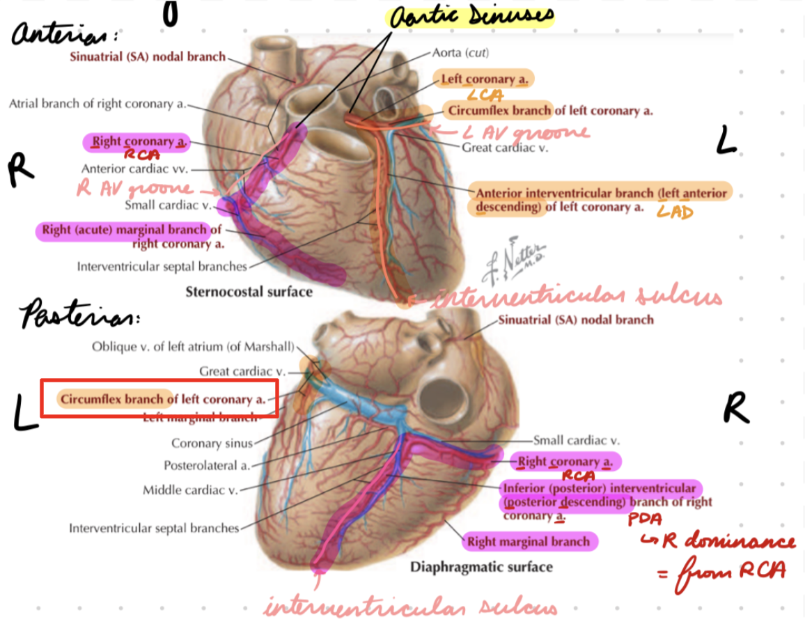
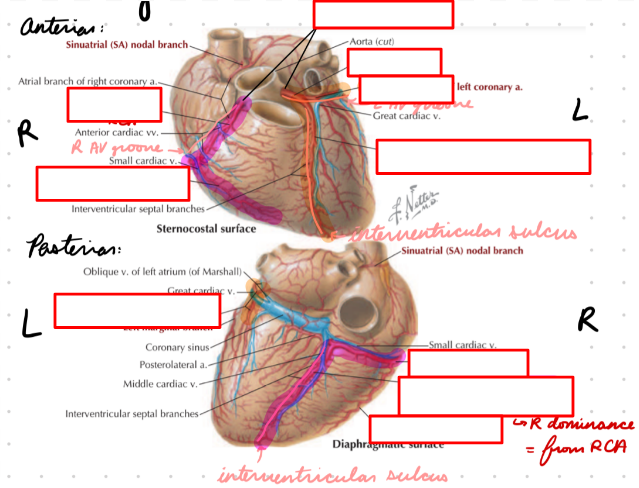
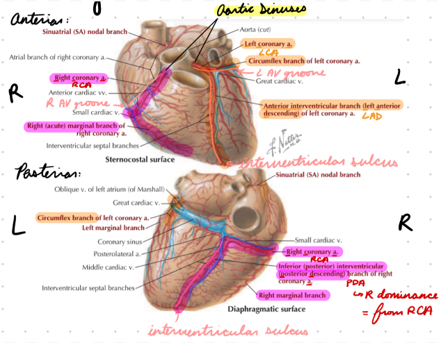
Coronary Arteries
Branch off from aortic sinuses
Above aortic valve on ascending aorta
Blood enter during diastole
Valve open (systole) = Block sinus = No blood flow
Right: 50% diastole
Left: 75% diastole (stronger contraction)
Right Coronary Artery (RCA)
Right aortic sinus → Right AV groove
Divide into posterior descending artery (PDA) and marginal branch
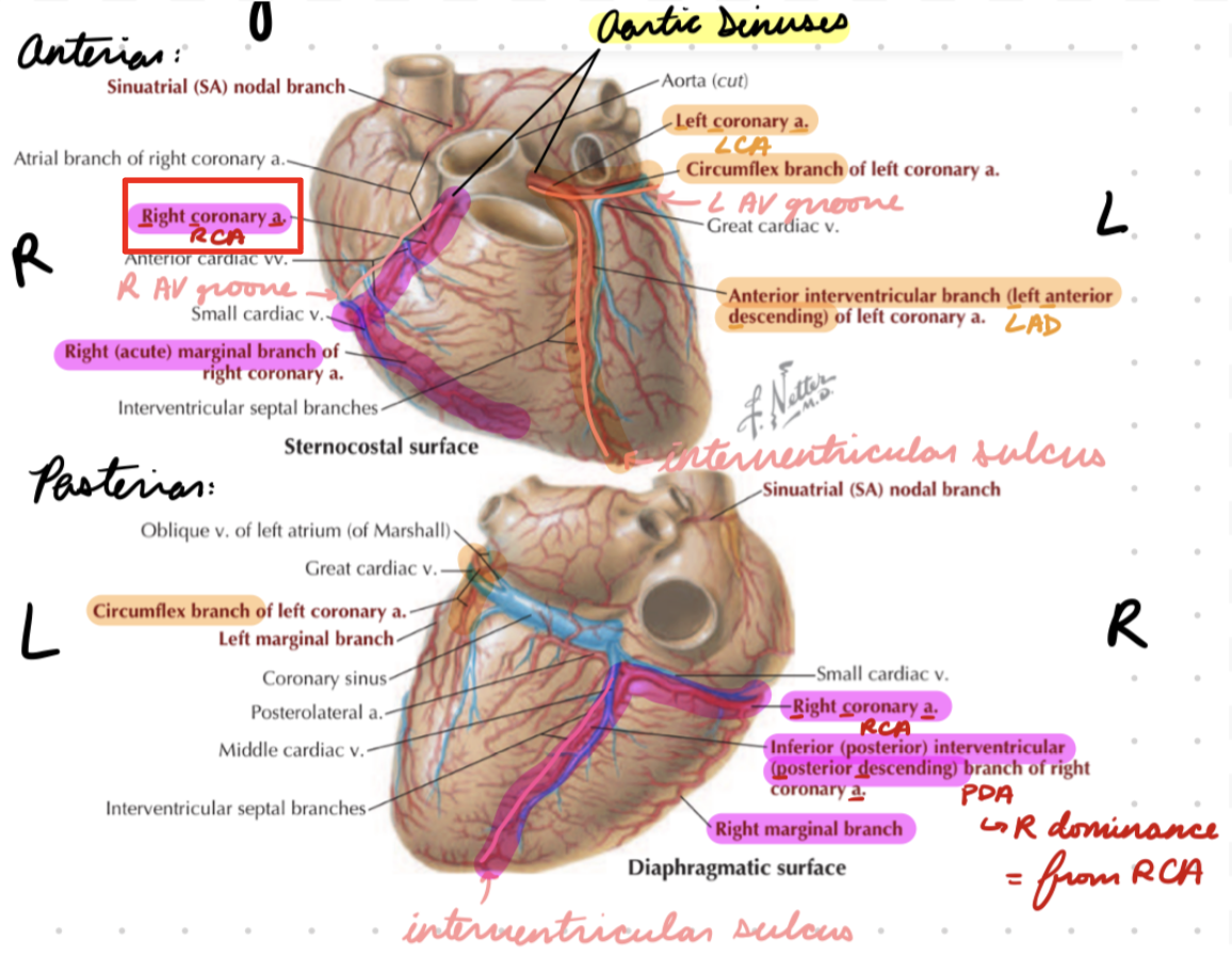
RCA: PDA
In posterior interventricular sulcus (between ventricles)
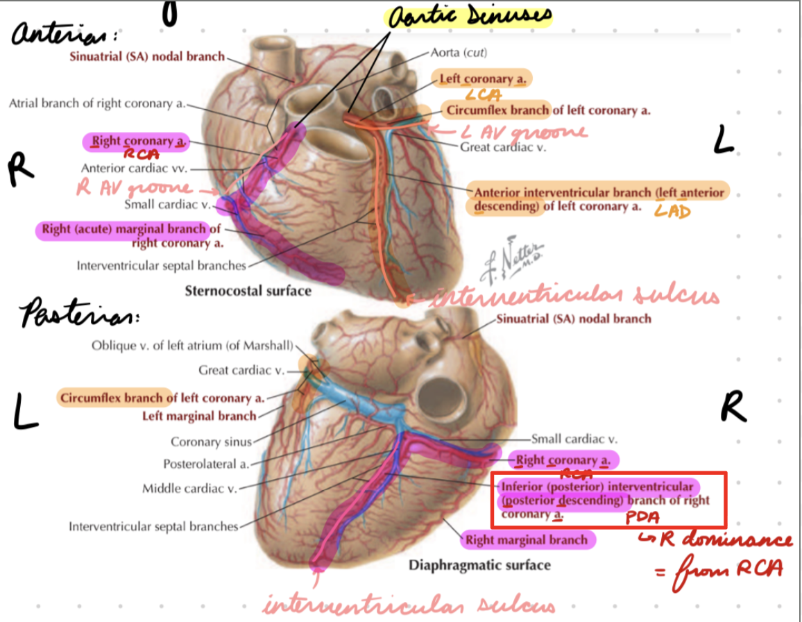
RCA: Marginal
Around acute (right) side of heart

Left Coronary Artery (LCA)
Left aortic sinus → Left AV groove
Divide into left anterior descending (LAD) and circumflex branches
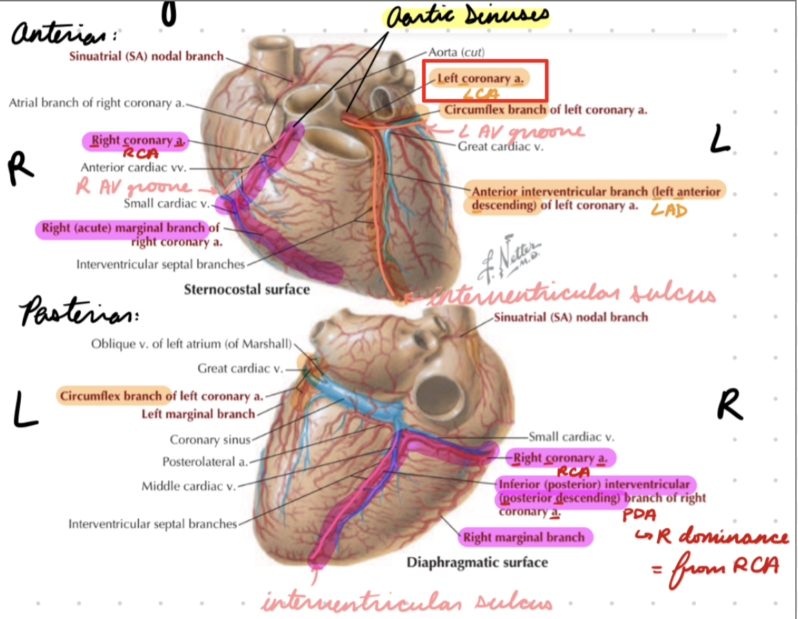
LCA: LAD
In anterior interventricular sulcus (between ventricles)
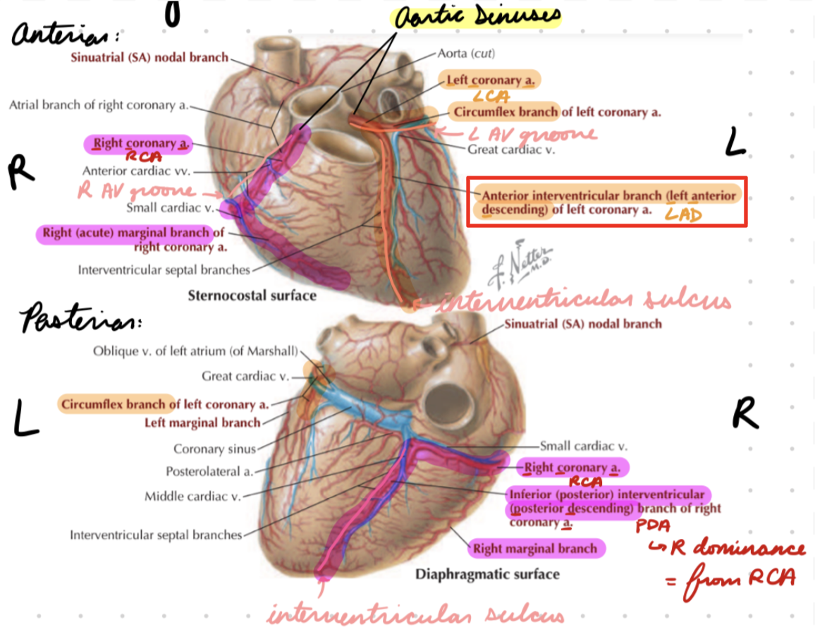
LCA: Circumflex
Around obtuse (left) side of heart
Coronary Veins
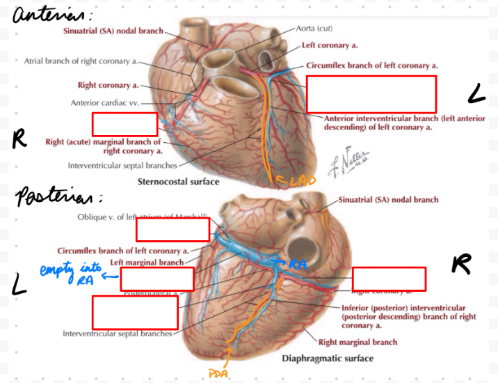
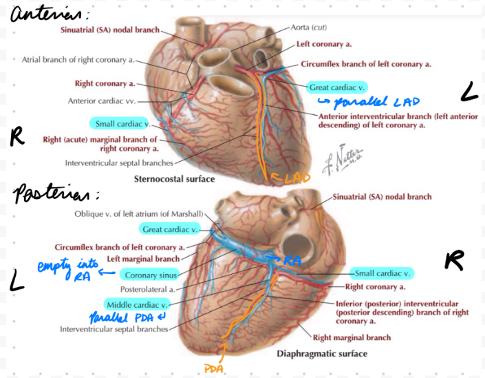
Empty into coronary sinus on posterior AV groove → Into right atrium
Great Cardiac Vein
Parallel LAD
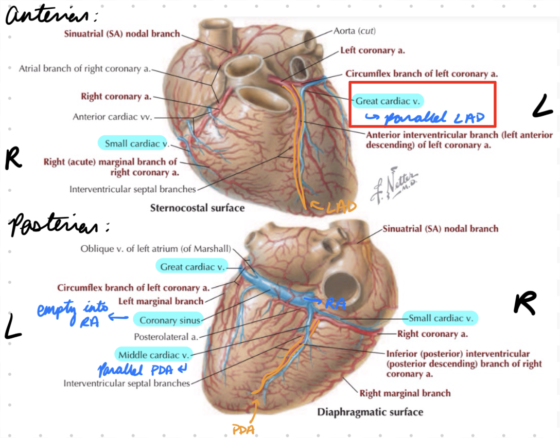
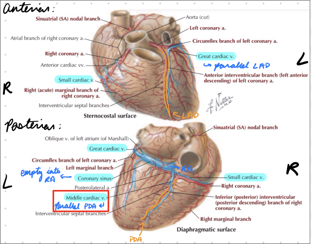
Middle Cardiac Vein
Parallel PDA
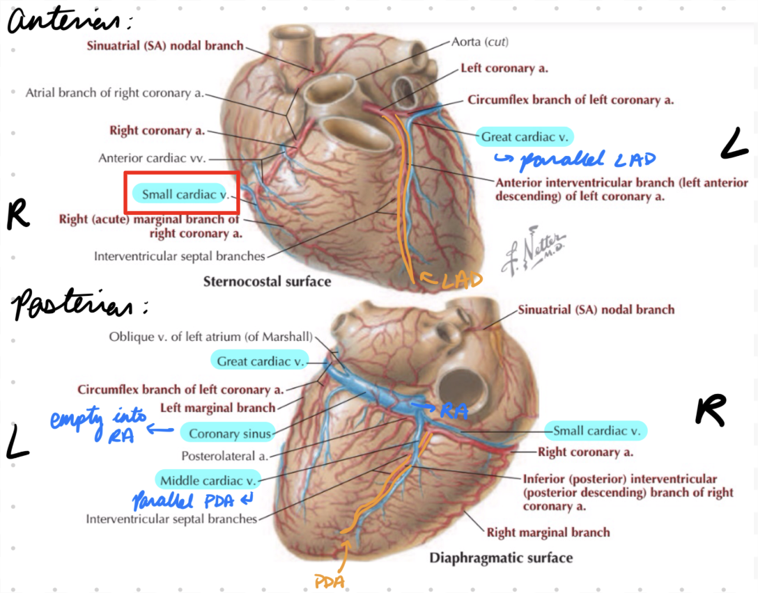
Small Cardiac Vein
Parallel marginal branch
Thebesian Cardiac Veins
Smallest
Empty blood into all 4 heart chambers
Mostly right atrium
Coronary Dominance
Right-Dominant: PDA from RCA
Left-Dominant: PDA from LCA
Higher risk
Occlusion = left heart failure
Determinants of Coronary Circulation
Sympathetic innervation
Metabolites
Mechanical compression
Determinants: Sympathetic Innervation
Minor role
Decrease Blood Flow: Release norepi = Bind alpha-adrenoceptors = Vasoconstriction
Increase Blood Flow: Release epi and norepi = Bind beta-adrenoceptors = Vasodilation
During increased myocardial demand
Determinants: Metabolites
Active Hyperemia: Increased metabolic activity = Increase O2 demand = Release vasodilator substances
Hypoxia: Vasodilation = Increase blood flow
Adenosine: Released from cardiac myocytes = Bind adenosine receptors in coronary vascular smooth muscle = Vasodilation
Determinants: Mechanical Compression
Systole = Occlude/compress blood vessels = Reduce blood flow
Diastole = Reactive hyperemia = Increase blood flow and O2
O2 Use in Muscles
Oxidative phosphorylation convert substrate to energy (ATP)
Low O2 = Anaerobic respiration (glycolysis)
Glycogen/Glucose → Pyruvate → Lactate
Produce less ATP
Ex: Ischemia
Heart Muscle: Energy Substrate
FFA: 70-90%
Other nutrients: 10-30%
Glucose, lactate
Heart Muscle: O2 Consumption
Directly related to cardiac minute work
Cardiac Minute Work = CO x Aortic pressure
Component 1: Volume work (CO)
Component 2: Pressure work (aortic pressure) → More energy demanding
Greater in LV
Same CO
Aortic pressure > Pulmonary pressure
Heart Muscle: Increase Volume Work
Increase CO = Small increase in O2 consumption needs
Ex: Exercise
Heart Muscle: Increase Pressure Work
Increase aortic pressure = Large increase in O2 consumption needs
Ex:
Aortic stenosis → Increased afterload
Stiff aortic valve = Require more pressure to pump into aorta
Systemic/pulmonary hypertension
Increased arterial pressure = Increase aortic/pulmonary trunk pressure
Compensation for Increased Pressure Work
Ventricular hypertrophy
Laplace Law: Increase wall thickness (hypertrophy) = Increase pressure generated (overcome aortic/pulmonary trunk pressure) = Ventricular failure
Heart Muscle: ATP Use
Na+-K+ ATPase: Maintain RMP
Ca2+ pump in sarcoplasmic reticulum: Relaxation
Myosin ATPase: Relaxation (myosin detachment)
Skeletal Muscle: Energy Source
FFA: At rest and light exercise
Carbs: During strenuous exercise
Glucose, glycogen
Skeletal Muscle: O2 Consumption
Increased myoglobin (O2 reserve) to supplement during low O2
Arterial Wall
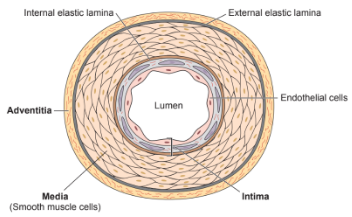
3 layers
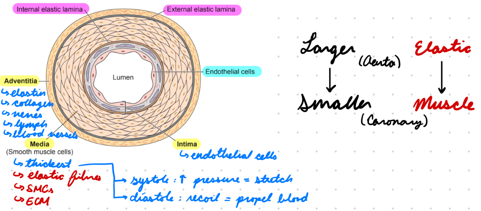
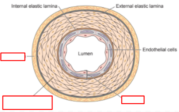
Intima: Innermost
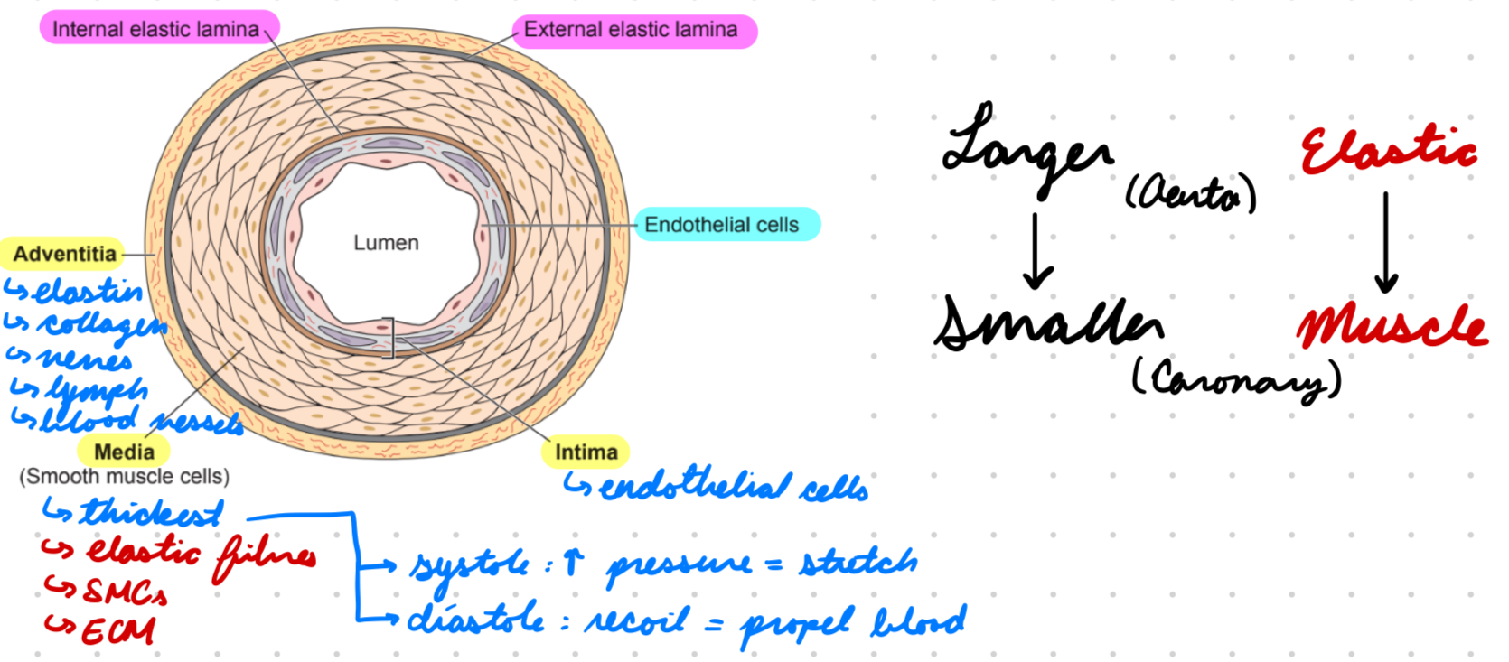
Media: Middle
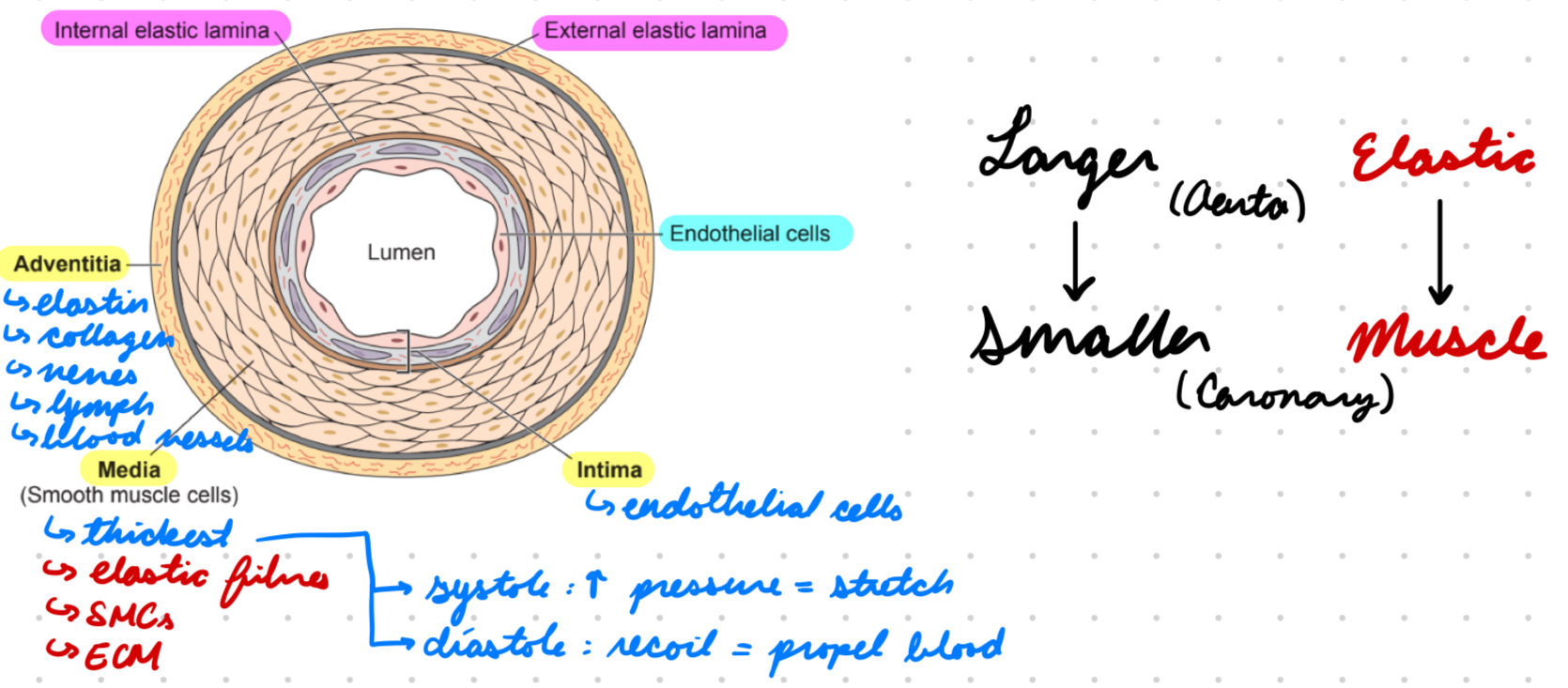
Adventitia: Outer
Arterial Wall: Intima
Endothelial cells
Barrier between blood and vessel wall
Arterial Wall: Media
Thickest layer
Contractile and elastic functions
Systole: Stretch from high pressure
Diastole: Recoil to propel blood forward
Contract = Increase resistance = Decrease flow
Contain:
Elastic fibres
Smooth muscle cells
ECM
Arterial Wall: Adventitia
Elastic tissue + collagen
Nerves
Lymphatics
Blood vessels for arterial wall
Larger vs Smaller Arteries
Larger (aorta): Most elastic fibres
Smaller (coronary): Muscular, less elastic fibres

Athersclerosis: Description
Inflammation and plaque buildup in arterial wall
Atherosclerosis: Epidemiology
More common in males
Risk Factors:
Older age
Comorbidities
Hypercholesteremia
Dyslipidemia (high total cholesterol, low HDL)
Diabetes
Hypertension
Fam history
Smoking
Atherosclerosis: Etiology
Lipid, cholesterol, calcium plaques in arteries
Occlude lumen = Decrease blood flow
Athersclerosis: Pathogenesis
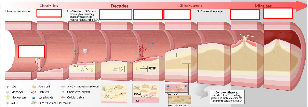
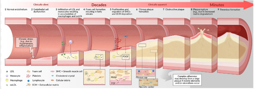
Chronic stress on arterial endothelium from hypertension and turbulence
Endothelial cell dysfunction = Increase permeability
Increase inflammatory cell invasion
Platelet adhesion to damaged wall
Release inflammatory mediators (cytokines) and platelet-derived growth factors (PDGF)
PDGF increase SMC proliferation and fibrosis
Chronic inflammation
Macrophages and SMCs ingest cholesterol (LDL) → Foam cells
Foam cells accumulate → Fatty streaks = Early atherosclerosis
Foam cells and SMCs increase ECM deposition (collagen) in intima
PDGF-triggered from platelets and endothelial cells
Form fibrous plaque (atheroma) under endothelial cells
Tunica intima calcifies
Positive Remodelling: Unstable plaque + Same lumen diameter
Thin fibrous cap + large lipid core, prone to rupture, weak outer layer
Negative Remodelling: Stable plaque + Decrease diameter
Thick fibrous cap + small lipid core, not prone to rupture, stiff outer layer
Plaque ruptures
Thrombogenic material (collagen) released into bloodstream → Thrombus
Coronary Atherosclerosis
Plaque formation in coronary arteries
Restrict blood flow to myocardium → Ischemia
Non-Coronary Atherosclerosis
Plaque formation in arteries outside heart (periphery)
Common in:
Abdominal aorta
Popliteal arteries
Carotid arteries
Circle of Willis (arteries at base of brain)
Atherosclerosis: Investigations
Blood test
Imaging
Atherosclerosis Investigation: Blood Test
Complete lipid profile
High cholesterol
High LDL and VLDL
Low HDL
Atherosclerosis: Imaging
Coronary CT angiography (CCTA)
Visualize arteries and plaques
With or without contrast
Carotid ultrasound
Assess artery thickness and plaques
Atherosclerosis: Clinical Presentation
Coronary: Angina and chest discomfort
Non-Coronary: Decreased O2 reaching brain and retinas
Transient ischemic attack (TIA): Sudden neurological deficits
Unilateral weakness and numbness
Speech disturbances
Monocular blindness
Ischemic stroke: Persistent neurological deficits
Motor and sensory loss
Aphasia (communication disorder)
Vision changes
Bowel ischemia
Atherosclerosis: Treatment/Management
Lifestyle changes
Pharmacological
Athersclerosis: Lifestyle Changes
Life’s essential 8 framework
Nutrition: Dietary approaches to stop hypertension (DASH) diet
Balanced diet
Low sugar, red meat, added fats
Increase physical activity
Avoid nicotine
Sleep
Normal weight
Low non-HDL cholesterol
Normal blood glucose
Decrease BP
Smoking cessation
Atherosclerosis Treatment: Pharmacological
Statins
+ nonstatin lipid-lowering therapy for high-risk
Antiplatelet therapy
Ex: Aspirin
Atherosclerosis Pharmacological: Statins
Ex: Rosuvastatin
MOA: Inhibit hepatic HMG-CoA reductase = Decrease cholesterol synthesis
Atherosclerosis Pharmacological: Non-Statin Lipid-Lowering Therapy
Ezetimibe: Inhibit cholesterol absorption
PCSK9 Inhibitors: Abs against enzyme degrading LDL receptors
Ex: Evolocumab, Alirocumab
Bile Acid Sequestrants: Inhibit bile acid reabsorption
Ex: Cholestyramine, Colesevelam
Atherosclerosis Prevention
ABCDS
A: Aspirin
B: BP control
C: Cholesterol management
D: Diabetes management
E: Exercise
S: Smoking cessation
Angina: Description
Paroxysmal (short, sudden, recurrent) chest discomfort, tightness, and pressure
Angina Types
Stable: Reproducible symptoms
Predictable severity and frequency
Symptoms subside with rest/nitroglycerin
Unstable: Longer symptom duration
No response to rest/nitroglycerin
Variant: Transient
From coronary artery spasms
Angina: Epidemiology
Risk factor: Atherosclerosis
Angina: Etiology
Myocardial ischemia
Angina: Pathogenesis
Decreased O2 delivery to myocardial tissue from coronary arteries due to plaque formation + coronary artery stenosis
Plaque formation from atherosclerosis
Stable plaque = Stenosis
Stiff arteries increase resistance to blood flow = Decrease flow
Low O2 delivery = Ischemia
Reversible: Salvage tissue by restoring O2 supply (angina)
Irreversible: Tissue necrosis (scarring)
Angina: Investigations
Imaging
ECG
Angina: Imaging
Coronary Angiography: Determine plaques and stenosis
Echocardiogram: Determine heart abnormalities (dilation, hypertrophy, inflammation)
Angina: ECG
Stable: Usually normal
Ischemia: ST-segment depression, T wave inversion
Angina: Clinical Presentation
No chest tenderness
Gradual intensity increase
Radiation to adjacent areas
Left arm
Neck
Jaw
No changes with respiration
Questions to Ask:
Nausea
Dyspnea
Dizziness
Sweating (diaphoresis)
Angina: Treatment/Management
Pharmacological
Nitrates
Beta-blockers
Ca2+ channel blockers
Angina Pharmacological: Nitrates
Ex: Nitroglycerin
MOA: Increase NO = Vasodilation = Increase blood flow to heart
Angina Pharmacological: Beta Blockers
Ex: Metoprolol, Propranolol
MOA: Block beta-1 receptors = Vasodilation = Increase blood flow to heart
Angina Pharmacological: Ca2+ Channel Blockers
Ex: Amlodipine
MOA: Block calcium entry into cardiac muscles = Decrease cardiac contraction
Ranolazine: Inhibit late Na+ current = Decrease Na+ and Ca2+ in cell = Increase myocardial relaxation
Acute Coronary Syndrome (ACS): Description
Conditions causing acute myocardial ischemia
STEMI
NSTEMI
Unstable angina
ACS: Clinical Presentation
Acute chest pain (angina)
Dyspnea
Pallor
Nausea and vomiting
Anxiety
Dizziness/lightheadedness and syncope
ACS: Investigations
ECG
Bloodwork
Echo (TTE)
ACS: ECG
STEMI: ST elevation in 2 contiguous leads (same anatomical area)
NSTEMI: ST depression and transient deviations, T wave inversions
ACS: Bloodwork
CBC
Basic metabolic panel
Coagulation panel
Troponin: Increased = Cardiomyocytes under stress (MI)
ACS: Echo
Not routine
For abnormal symptoms
ACS: Treatment/Management
Pharmacological
O2 therapy
ACS Treatment: Pharmacological
Aspirin
Nitroglycerin
Beta-Blocker: Vasodilator
RAAS Inhibitor
NO NSAIDS
ACS Pharmacological: Aspirin
MOA: Anti-platelet = Increase blood flow through stiff/plaque-occluded arteries
ACS Pharmacological: RAAS Inhibitor
ACE Inhibitor (ramipril) or ARB (lorsartan)
MOA: Decrease water retention = Decrease BP
ACS Management: O2 Therapy
Cyanosis
Resp distress/failure
SpO2 < 90%