vertebral column ppt PT700
1/45
There's no tags or description
Looks like no tags are added yet.
Name | Mastery | Learn | Test | Matching | Spaced |
|---|
No study sessions yet.
46 Terms
vertebral column functions
stability
mobility
protection
vertebral column
made up of 33 vertebra
7 cervical
12 thoracic
5 lumbar
5 sacral (sacrum) → fused
4-5 coccygeal (coccyx) → fused

primary curve
vertebral column curve we are born with
thoracic spine
sacrum
“kyphotic” = curves outward

secondary curve
vertebral column curve that develops as we begin to lift head and bear weight during development
cervical spine
lumbar spine
develops after birth
“lordosis” = inward curvature
lordosis
cervical & lumbar spine
sagittal plane
normal & abnormal
abnormal increase in lumbar spine curvature
kyphyosis
thoracic spine and sacrum
sagittal plane
normal & abnormal
abnormal increased curvature of thoracic spine
common in geriatric population
scoliosis
most commonly occurs at thoraco-lumbar spine
frontal and transverse plane of motion
always abnormal lateral curvature
anything over 8 degrees = abnormal
common in pre-adolescent females
vertebral body
anterior cylindrical structure
bodies will become larger going from cervical vertebrae → lumbar due to increased body weight which needs to be supported
intervertebral discs
connect adjacent vertebral bodies
discs create height = creating openings for rami to go through
clinical: stenosis
stenosis
abnormal narrowing of a bodily passage or opening due to disc height in vertebral body
This narrowing can restrict nerves
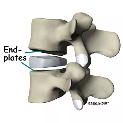
vertebral end-plate
hyaline cartilage on superior and inferior surfaces of body
connects to intervertebral discs
vertebral arch
spinous process
transverse processes
laminate
pedicle
superior articular process
inferior articular process
zygapophpysial joint
pars articularis
lamina
connects spinous and transverse processes on vertebral arch
pedicle
connects transverse processes of vertebral arch to vertebral body
laminectomy
clinical case → surgery to gain access to spinal, usually done in response to excessive pressure/pinching in spinal cord
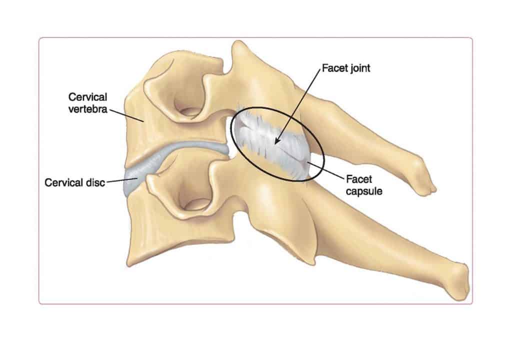
facet (zygapophysial) joint
formed by an inferior articulating process from the vertebra above and a superior articulating process from the vertebra below
connecting will keep vertebral column connected
can become inflamed = nerve entrapment
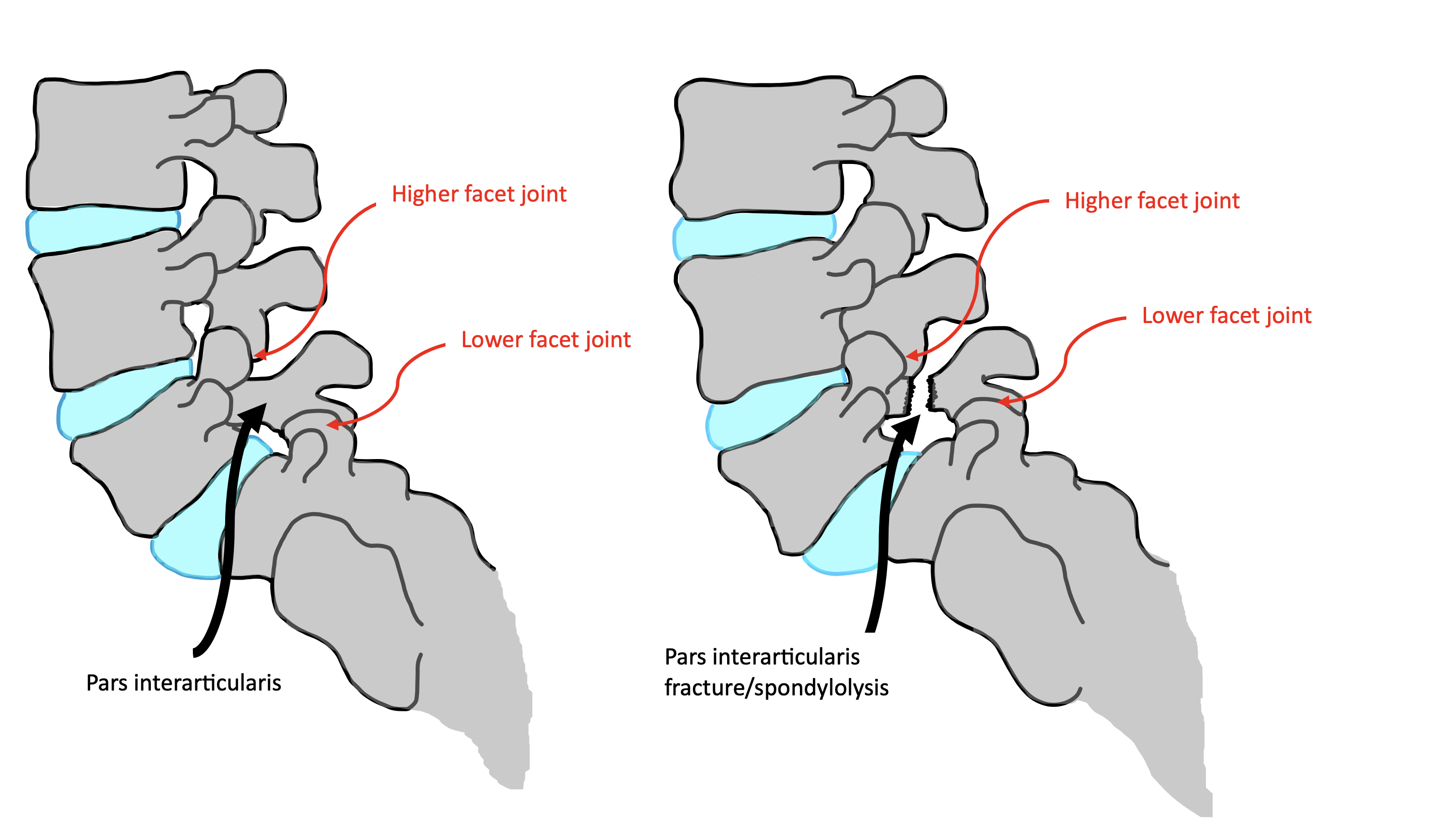
pars articularis
on vertebral arch, b/w superior and inferior articular facets
commonlyl fractured due to stress of gravity/weight
“scotty dog fracture”
vertebral foramen contents
spinal cord
ant and post roots
meninges
blood vessels
cerebrospinal fluid
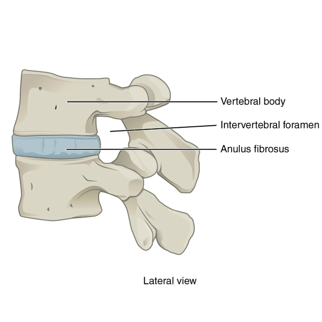
intervertebral foramen
b/w adjacent vertebrae, acts as an opening
place where anterior and posterior roots join to form spinal nerve
bounded by:
vertebral bodies
pedicles
intervertebral discs
facet joint
herniated disc
occurs when the soft, jelly-like center *nucleos pulposes” of a spinal disc pushes through a tear in the outer layer
pinches roots and/or spinal nerve
radiating pain to associated dermatome and/or muscle weakness
dependent upon what is being compromised
disc “pops off”
facet joint arthritis
swelling at facet joint
pinches roots and/or spinal nerve
radiating pain to associated dermatome
spina bifida
congential defect in walls of spinal canal caused by lack of union b/w the laminae
lumbar region most common
may cause spinal dysfunctionc
cervical spine
7 vertebrae → lordotic (secondary) curve
C1 - “atlas”
C2 - “axis”
C3-C7 - typical
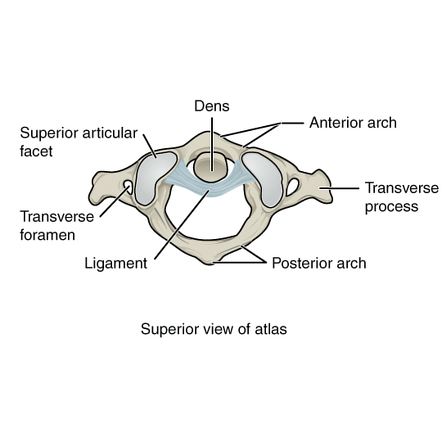
atlas (C1)
ring-shaped, acts like a washer for skull to move on axis
no body, no spinous process
groove for vertebral artery, courses over C1 to get to brain
has largest transverse process of all cervical vertebrae
alantoaxial joint (AAJ)
C1 (facet for dens) resting on C2 (dens process)
superior articular process of C2 articulates with inferior articular facet of atlas (C1)
atlantoocipital joint (AOJ)
articulation spot b/w base of skull and cervical spine
occiput of skull to superior articular facet of C1
axis (C2)
has dens (aka odontoid process)
projects superior from body
articulates with atlas (AAJ)
has a “bifid” spinous process
typical cervical vertebra
C3-C6
bifid spinous process
transverse foramen → acts as passage for vertebral A.
uncinate processes
C7 vertebra
has a transverse foramen like C3-C6, but vertebral A. does not pass through it
has uncinate process
uncinate processes
found on C3-C7
superior surface of body has raised lateral lips of bone which articulate with the body above it
forms uncovertebral joint
uncovertebral joint
found in the cervical spine (C3-C7) and are formed between the uncinate processes of the vertebrae below and the lateral edges of the vertebral bodies above
spine stability and movement
vertebral artery
passes through all cervical transverse foramen except C7
after passing through C1, makes 2 right angle bends to pass through foramen magnum
supplies posterior brain
“beauty parlor stroke syndrome”
beauty parlor stroke syndrome
hypertension of c/s, could cause comrpomised in vertebral artery
could lead to clots → CVA
thoracic spine
primary curve
12 vertebrae
rib facets on transverse processes (unique)

transverse costal facet
small, concave surface located on the transverse process of a thoracic vertebra, specifically designed to articulate with the tubercle of a rib

superior costal facet
a site where a rib forms a joint with the top of a vertebra

inferior costal facet
bony surface on the body of a thoracic vertebra that articulates with the head of a rib.
Specifically, it's the lower of the two facets on the vertebral body that, in combination with the superior costal facet of the vertebra below
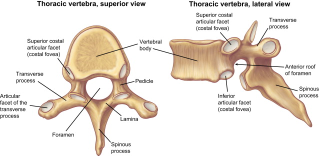
demifacet
a half facet, specifically a smooth, concave surface on a thoracic vertebra, that forms part of the joint where the head of a rib connects
rib facets on transverse processes
articulates with tubercles on ribs
absent on last 2-3 thoracic vertebrae
thoracic spine spinous processes
long, slender and inferiorly directed
lower = longer
“giraffe” shape
lumbar spine
5 vertebrae
lordotic curve
secondary curvature
short, blunt, rounded
“moose head”
lumbar spine 5th body (L5)
wedge-shaped, form fit for sacral articulation
lumbar spine: vertebral canal
spinal cord ends at L2
cauda equina continues inferiorly
spinal tap b/w L4-L5
cauda equina syndrome
medical emergency → “saddle region” paresthesia
loss of bladder and/or bowel control
can cause permanent paralysis
spondylolysis
fracture of pars articularis
“scotty dog fracture”
spondilolisthesis
one body slips off the one below it
anterior: top vertebrae slips forwards
retro/posterior: top slips backwards