Pathology of Superficial Structures
 Call Kai
 Learn
Learn Practice Test
 Spaced Repetition
 Match
Match Flashcards
 Knowt Play
1/45

There's no tags or description
Looks like no tags are added yet.
Name | Mastery | Learn | Test | Matching | Spaced | Call with Kai | Chat |
|---|
No analytics yet
Send a link to your students to track their progress
46 Terms
A superficial epidermal cyst may also be referred to as an
epidermal inclusion cyst, an epidermoid cyst, or possibly a sebaceous cyst
True sebaceous cysts are
uncommon
Superficial epidermal cysts are most likely found in the
scalp, face, neck, trunk, or back
Superficial epidermal cysts are potentially the result of an
obstructed hair follicle
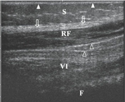
sonographic appearance of skin layers and muscle in the thigh. Longitudinal image of the epidermis and dermis (white arrowheads), the subcutaneous fat (S), the rectus femoral muscle (RF), connective tissue (open arrowheads), the vactus intermedius muscle (VI), and the femur (F).
clinical findings of a superficial epidermal cyst
Visible, palpable mass just under the skin
Pain and redness in the area
sonographic findings of a superficial epidermal cyst
Anechoic, hypoechoic, complex, or hyperechoic cyst or mass
Pseudotestis appearance
A ganglion cyst is a common mass found along the dorsal aspect of the
hand and wrist
Large ganglion cysts in the wrist have been referred to as —, because individuals in the past have used large books—like the Bible—to reduce the cyst by slamming the large book against it.
Bible bumps
clinical findings of a ganglion cyst
Palpable mass most often located along the dorsal aspect of the hand or wrist
Can be painful
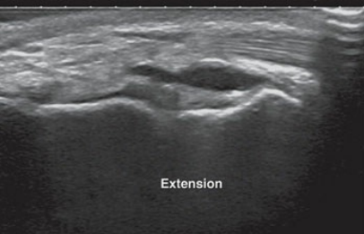
Ganglion cyst. This small cyst was located in a patient’s wrist and was determined to be a ganglion cyst.
sonographic findings of a ganglion cyst
Noncompressible, anechoic mass with acoustic enhancement
May contain debris or septations
Lipomas are usually — and may be observed during a physical examination
compressible
clinical findings of a superficial lipoma
asymptomatic
Obvious mass under the skin
compressable
sonographic findings of a superficial lipoma
Most likely an isoechoic mass as compared to the surrounding tissues
Typically have an oval shape
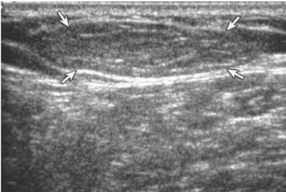
Lipoma. This lipoma (between arrows) appearing as an oval hyperechoic mass is noted just under the skin.
clinical findings of a superficial hemangioma
asymptomatic
Red or reddish-purple, raised mass on the skin
sonographic findings of a superficial hemangioma
Blood flow may be detectable with color Doppler
Hypoechoic
A Baker cyst, or —
popliteal cyst
A Baker cyst, or popliteal cyst, is located in the popliteal fossa,
behind the knee
These cysts are common and a result from the accumulation of 508 synovial fluid from a weakening in the joint capsule of the knee, as seen in conditions such as —
rheumatoid arthritis or osteoarthritis.
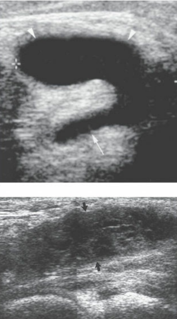
Baker cysts. A. An uncomplicated Baker cyst (between calipers and arrows) appears as an anechoic mass in the popliteal fossa. B. A large Baker cyst (arrows) in a patient with rheumatoid arthritis contains some echogenic debris and pannus
A Baker cyst may be seen in the presence of rheumatoid arthritis or other
knee joint issues.
clinical findings of a baker cyst
asymptomatic
focal tenderness in the popliteal fossa
sonographic findings of a baker cyst
Anechoic mass with posterior enhancement
Complicated Baker cysts may contain echogenic fluid, debris, or septations
A pilonidal cyst is most often found within the
natal cleft
a pilonidal cyst is also referred to as — and may also be seen within the fingers or toes
pilonidal sinus
Pilonidal means
“nest of hair.”
pilonidal cysts are comprised of
loose hairs and skin debris
Patients who are more prone to pilonidal cyst include those who
sit for an extended amount of time and hairdressers
During World War II, the pilonidal cysts were linked with soldiers who had to endure long, bumpy jeep rides, and thus they were termed
“jeep disease.”
clinical findings of a pilonidal cyst
Edema, warmth, and pain in the area of the cyst
Bloody drainage from the cyst may be present
sonographic findings of a pilonidal cyst
Complex, subcutaneous mass
Hyperemia around the mass
Hypoechoic tract may be seen extending from the cyst to the skin surface
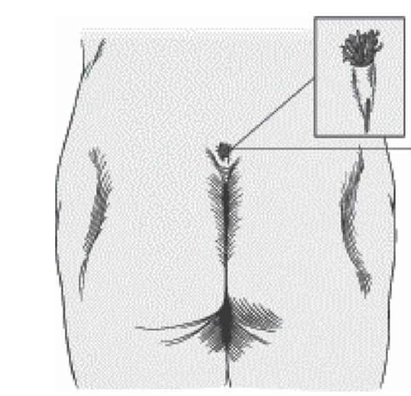
Typical location of a pilonidal cyst.
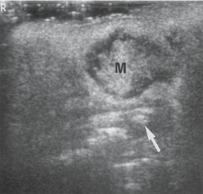
Pilonidal cyst. This pilonidal cyst appeared as a complex mass (M) at the tip of the distal spine (arrow) in this infant.
— is infection and subsequent inflammation of the skin and subcutaneous tissue
Cellulitis
cellulitis is most often caused by
Staphylococcus aureus and Streptococcus pyogenes
A hidden abscess is termed an
occult abscess
abscesses are focal fluid collections that are often complex and have a — that can be detected with color Doppler
peripheral rim of hyperemi
When cellulitis is identified, the sonographer should look carefully for a — that may represent abscess development
complex focal fluid collection
clinical findings of cellulitis
Red, tender, warm area of the skin
Possible elevated white blood cell count (especially with abscess development)
sonographic findings of cellulitis
Hypoechoic, edematous strands within the soft tissue (cobblestone appearance)
sonographic findings of a superficial abscess
Hypoechoic, edematous strands within the soft tissue (cobblestone appearance)
Focal fluid collection that is often complex, denoting the abscess
A peripheral rim of hyperemia may be detectable with color Doppler
Malignant melanoma accounts for up to — of skin cancers.
11%
Melanoma is also the most likely — to metastasize to the subcutaneous fat
primary tumor
sonographic findings of metastatic melanoma
solid, hypoechoic mass