Layperson versus Anatomical Equine and Food Animal Terms for Palpation Instruction
 Call Kai
 Learn
Learn Practice Test
 Spaced Repetition
 Match
Match Flashcards
 Knowt Play
1/85

There's no tags or description
Looks like no tags are added yet.
Name | Mastery | Learn | Test | Matching | Spaced | Call with Kai | Chat |
|---|
No analytics yet
Send a link to your students to track their progress
86 Terms
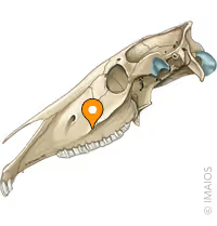
Facial Crest (Description)
Longitudinal ridge extending from the zygomatic arch along the maxilla.
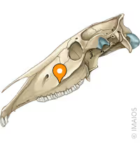
Facial Crest (Function/Use)
Masseter Muscle Insertion Anatomical landmark for venipuncture, nerve blocks, and dental procedures.
Forelock (Description)
A lock of mane hair growing from the poll forward.
Forelock (Function/Use)
Aesthetics and insect control.
Throat latch (Description)
Ventral area where the head and neck meet.
Throat latch (Function/Use)
Site where swelling may be observed in some disease processes/syndromes and in some disciplines, the bridle may be buckled here.
Poll (Description)
Dorsal area where the head and neck meet- Atlanto-occipital joint, soft tissue including nuchal ligament.
Poll (Function/Use)
The poll is important to head movement, balance, coordination, posture and performance.
Mane (Description)
Thick, coarse hair growing along the dorsal aspect of the neck from the poll to the withers.
Mane (Function/Use)
Aesthetics, and may help with temperature regulation, protection from rain, and insect control.
Crest (Description)
The arched ridge of the dorsal aspect of the neck where the mane grows, from the poll to the withers
Crest (Function/Use)
Can be assessed during a physical exam to judge BCS and for certain disease processes. (cresty neck scoring)
Withers (Description)
The ridge created by the dorsal spinous processes of thoracic vertebrae at the base of the neck between the shoulder blades.
Withers (Function/Use)
Insertion of the supraspinatous and nuchal ligament. This is where a horse's height is measured, and we assess this area for pain, and conformation.
Croup (Description)
The top round area from the point of the hip to the tailhead area.
Croup (Function/Use)
We assess this area for pain, and conformation.
Flank (Description)
The soft area along a horse's side just behind the ribcage but cranial to the hind limb.
Flank (Function/Use)
This area is assessed during physical exams for respiration BCS, and borborygmi.
Sheath
Prepuce
Sheath (Description)
Protective double layered tissue covered with skin around a horse's penis.
Sheath (Function/Use)
This tissue protects the penis. This area must be included in assessment and routine care.
Knee (EQUINE ONLY)
Carpus
Knee (EQUINE ONLY) (Description)
This composite forelimb joint is composed of radiocarpal, intercarpal and carpometacarpal joints.
Knee (EQUINE ONLY) (Function/Use)
The primary forelimb joint absorbing heavy impact.
Stifle (Description)
Complex joint made of the articulation of the femur, tibia and patella.
Stifle (Function/Use)
This hindlimb joint is important for strength, coordination, and the stay apparatus.
Chestnut (Description)
Area of keratinized tissue on the medial aspect of the forelimb, just proximal to the carpus, and just distal to the hock on the hindlimb.
Chestnut (Function/Use)
Thought to be evolutionary remnants. Require appropriate care.
Ergot (Description)
Hardened growth of skin along the back of the fetlock joint on all four legs.
Ergot (Function/Use)
Thought to be evolutionary remnants. Require appropriate care.
Hock
Tarsus
Hock (Description)
Tarsocrural, talocalcaneal, tarsometatarsal, proximal intertarsal and distal intertarsal joints
Hock (Function/Use)
Complex joint composed of both high and low motion joints. Common site of osteoarthritis in the horse.
Cannon (Description)
In the forelimb: third metacarpal bone In the hindlimb: third metatarsal bone
Cannon (Function/Use)
This bone is a weight bearing, shock absorbing bone.
Splint Bones (Description)
Two small vestigial bones which run along the medial and lateral aspects of the cannon bone.
Splint Bones (Function/Use)
Thought to be evolutionary remnants. Although vestigial, they can become injured and are part of our assessment in the horse.
Fetlock joint
Metacarpo(tarso) phalangeal joint
Fetlock joint (Description)
This joint helps bear weight in the horse and absorb impact during movement.
Fetlock joint (Function/Use)
Effusion of this joint is possibly an indicator of pathology.
Long pastern bone
P1 (proximal phalanx)
Long pastern bone (Description)
May be palpated and viewed radiographically.
Long pastern bone (Function/Use)
This bone provides shock absorption and joint stability.
Pastern joint
Proximal interphalangeal joint
Pastern joint (Description)
Low motion join in the distal limb.
Pastern joint (Function/Use)
This joint provides shock absorption and joint stability.
Short pastern bone
P2 (middle phalanx)
Short pastern bone (Description)
May be palpated and viewed radiographically.
Short pastern bone (Function/Use)
This bone provides shock absorption and joint stability.
Coffin joint
Distal interphalangeal joint
Coffin joint (Description)
Within the hoof capsule. May be viewed radiographically.
Coffin joint (Function/Use)
This joint absorbs concussive impact and helps roll the foot forward.
Coffin bone
P3 (distal phalanx)
Coffin bone (Description)
The distal-most bone, fully enclosed in the hoof.
Coffin bone (Function/Use)
The bottom most bone, fully enclosed in the hoof, plays a role in bearing weight, shock absorption, and conformation.
Coronary Band (Description)
Highly vascularized tissue area where the haired skin transitions into the hoof wall.
Coronary Band (Function/Use)
This is the main growth center for the hoof. Assessed in every patient, this area can be involved in many disease processes.
Hoof (Description)
Living keratin structure made up of the wall, frog, and sole.
Hoof (Function/Use)
The function of the hoof is to bear the weight of the horse, absorb shock and aid in blood circulation.
Paralumbar fossa (Description)
The soft, sunken, triangular hollow between the last rib, hip bone, and transverse vertebral processes.
Paralumbar fossa (Function/Use)
Equine patient: This area is assessed for gut sounds (borborygmi). Bovine patient: This area is used to assess the rumen for motility on the left and other GI sounds on the right during a physical exam.
Hooks
Tuber coxae
Hooks (Description)
These are the ileal wings or the wide prominent bony parts of the cranial pelvis.
Hooks (Function/Use)
This area is assessed during a physical exam to determine body condition scoring.
Pins
Tuber ischii
Pins (Description)
These are the bony projection of the ischium on the caudal aspect of the pelvis.
Pins (Function/Use)
This area is assessed during a physical exam to determine body condition scoring.
Thurls (BOVINE)
Greater trochanter
Thurls (BOVINE) (Description)
Area of soft tissue overlying the hip joint between the hooks and pins.
Thurls (BOVINE) (Function/Use)
This area is assessed during a physical exam to determine body condition scoring and to aid in localization of the hip.
Transverse vertebral processes (Description)
The long, broad, flattened, lateral bony projections from the lumbar vertebral bodies.
Transverse vertebral processes (Function/Use)
These processes are not only the dorsal border of the paralumbar fossa, but also are the area for the paravertebral nerve block.
Brisket (BOVINE)
Pectoral Muscle
Brisket (BOVINE) (Description)
Lower chest area used in body condition assessment.
Brisket (BOVINE) (Function/Use)
This area helps determine body condition.
Tail vein
Coccygeal vein
Tail vein (Description)
This is the vein running along the ventral surface of the tail in the bovine patient.
Tail vein (Function/Use)
This is a common vein for access for obtaining a blood sample.
Supramammary LN (Description)
These are the lymph nodes that drain the mammary glands in the cow.
Supramammary LN (Function/Use)
Palpated during physical exam, inflammation could indicate disease of the mammary glands.
Claws (lateral and medial)
Medial and lateral digit
Claws (lateral and medial) (Description)
The cloven hoof species have two claws or digits each with a hoof capsule.
Claws (lateral and medial) (Function/Use)
These areas are the main weight bearing areas and can be affected by trauma, or disease processes leading to lameness.
Dewclaws
Accessory digit
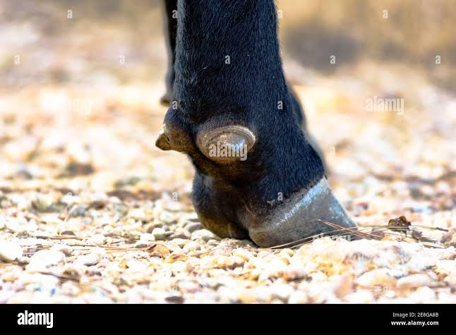
Dewclaws (Description)
Vestigial structures on the lower back part of the leg just above the claws in cattle.
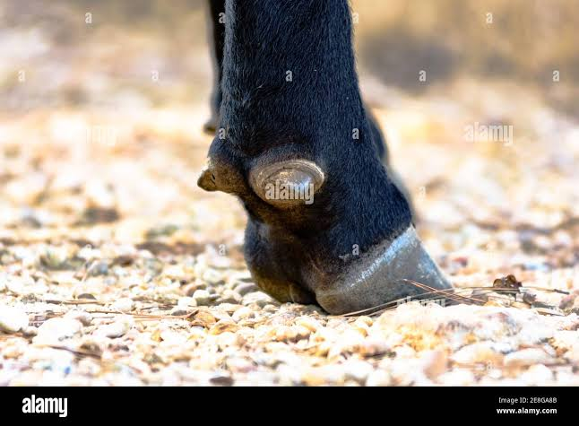
Dewclaws (Function/Use)
Although they are mostly non-weight bearing they can be the source of pain due to trauma, or disease processes leading to lameness.