ZOO3744 Exam 3
1/202
There's no tags or description
Looks like no tags are added yet.
Name | Mastery | Learn | Test | Matching | Spaced | Call with Kai |
|---|
No analytics yet
Send a link to your students to track their progress
203 Terms
What are some of the main properties of light?
It is a kind electromagnetic radiation where energy is proportional to frequency. Common properties are wavelength, frequency, and amplitude.
What is visual light?
Electromagnetic radiation from 380-750nm
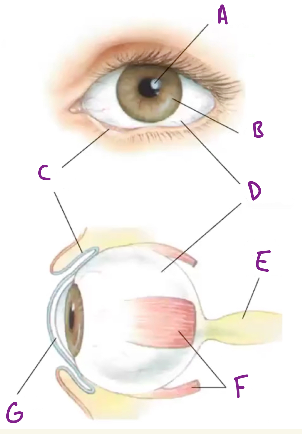
Name the following parts of the eye
A) Pupil B) Iris C) Conjunctiva D) Sclera E) Optic Nerve F) Extraocular muscles G) Cornea
What is the pupil of the eye?
An opening where light enters the eye
What is the sclera of the eye?
The white of the eye
What is the iris of the eye?
The part that gives color to the eye and controls pupil size.
What is the cornea of the eye?
The glassy transparent external surface of the eye that refracts light and helps focus it on the retina
What is the retina of the eye?
The part of eye that contains neurons sensitive to light and transmits visual signals to target (coverts light to electrical signals).
What is the ciliary body of the eye?
The part that adjusts the refractive power of the lens and produces fluid that fills the front of the eye.
What is the optic nerve of the eye?
A bundle of axons that come from the retina.
What is myopia and what causes it?
This means someone is nearsighted, too much refraction causes light to focuses in front of the retina
What is hyperopia and what causes it?
This means someone is farsighted, too little refraction causes light to focus behind the retina
Why are ciliary muscles important?
They aid in altering the shape of the cornea called accommodations to make vision more clear.
How many classes of neurons are there in the retina?
5
What are photoreceptors?
Specialized neurons of the eye (rods and cones) that contain membranous disks with light sensitive photopigment.
What is the path light hits when it enters the eye?
Ganglion layer → Inner nuclear layer (bipolar neurons) → Outer nuclear layer (photoreceptor cells)
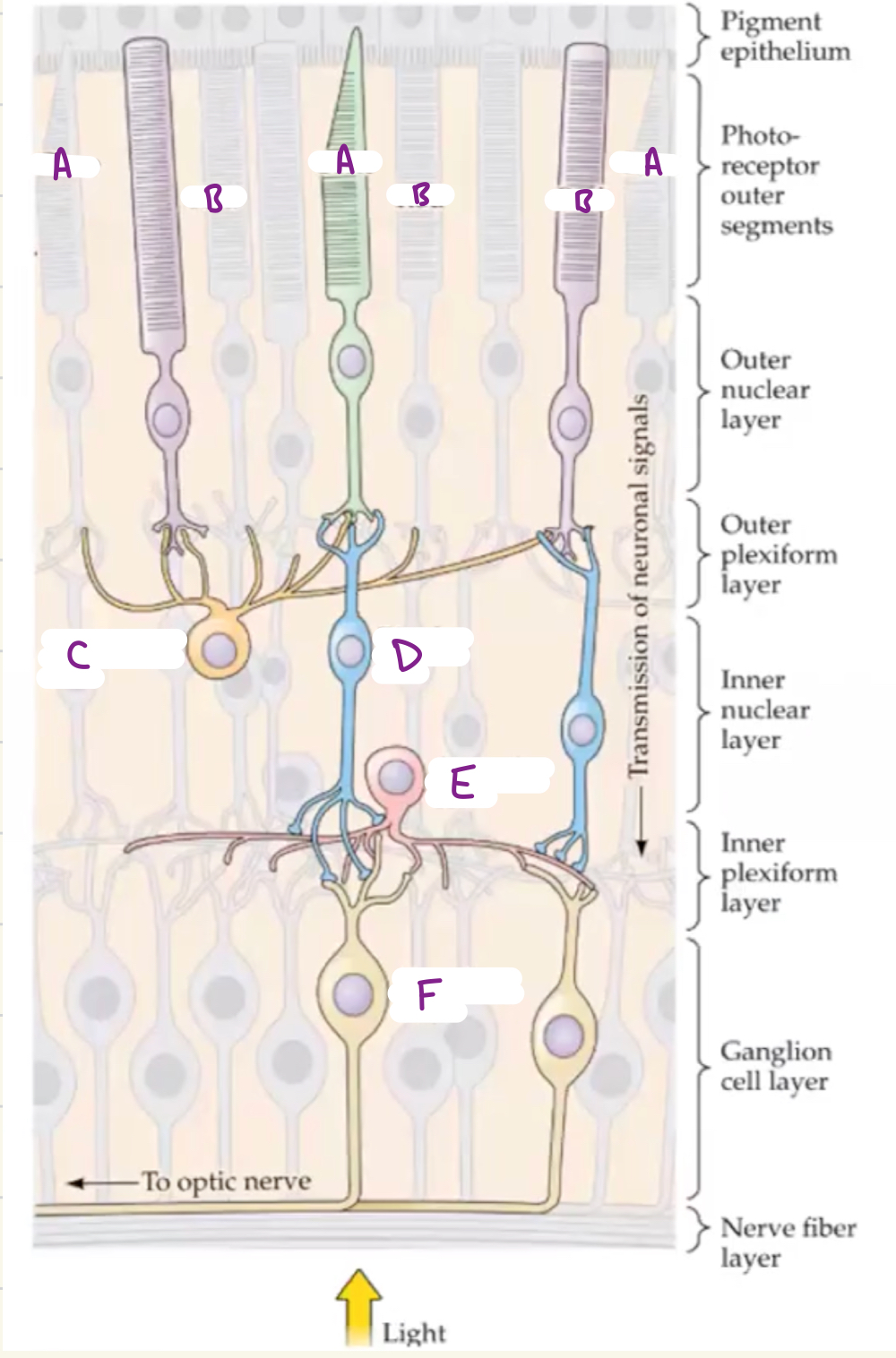
What are the following labeled sections of the retina?
A) Cone, B) Rod, C) Horizontal cell, D) Bipolar cell, E) Amacrine cell, F) Ganglion cell
What cells to photoreceptors make connections with?
Bipolar cells and Ganglion cells
What do bipolar cells do?
Retinal neurons that link photoreceptors to ganglion (excitatory)
What do ganglion cells do?
Retinal neurons that transmit visual information to the brain
What do horizontal cells do?
Receive input from photoreceptors and project to other photoreceptors/bipolar cells.
What do amacrine cells do?
Receive input from bipolar cells and project to ganglion cells, bipolar cells, and other amacrine cells
What is the direct path of information flow from the eye to the brain?
Photoreceptor → Bipolar cell → Ganglion cell
What is phototransduction?
The process of turning light into an electrical signal.
What happens when light hits a photoreceptor?
It triggers hyper-polarization of the membrane potential along with a graded release of neurotransmitter into the postsynaptic space.
What are some characteristics of rods?
Long, cylindrical outer segment with many disks which cause them to do better with low light
What are some characteristics of cones?
Short, tapered outer segment with fewer disks which cause them to do better in high light.
Rods are over ____ times more sensitive to light than cones
1000
What specifically to the disks in photoreceptors contain that help process light?
They contain photopigment 11-cis-retinal coupled to 7-pass transmembrane proteins called opsins.
What specific opsins are found in rods and cones?
Rods = rhodopsin and cones = cone opsins (3 kinds for color)
What happens when a photon of light is absorbed by a photoreceptor?
Cis-retinal shifts to trans-retinal triggering (decreasing cyclic GMP) the downstream signaling cascade closing Na+ and Ca2+ channels (hyperpolarization as K+ still leaves).
What happens if photoreceptors are in the dark?
Cyclic GMP levels are high so Na+ and Ca2+ channels are kept open and K+ channels are kept open causing depolarization
What happens if photoreceptors are in the light?
Cyclic GMP levels are low so Na+ and Ca2+ channels are closed and K+ channels are kept open causing hyperpolarization
How are rods and cones distributed in the retina?
There are more rods than cones meaning they are usually more dense, but cones are more common in the fovea.
What is the fovea?
Pit in the retina where the outer layers are pushed aside, leaving an area of highest visual acuity.
Do rods and cones exhibit the same convergence patter?
No, rods are highly convergent (one rod bipolar cell receives 15-30 rod inputs) and cone bipolar cells receive input from one cone.
How do rods highly convergent pattern affect its function?
It improves the rods ability to detect light (as well as function in low light) but reduces spatial resolution.
How do cones low convergent pattern affect its function?
It creates maximum resolving power in cones, meaning high visual acuity in the fovea.
What part of the retina handles the phototransmission of color?
Cones through the use of different opsins: Red (long wavelength), green (medium wavelength), and blue (short wavelength)
What causes colorblindness?
When there is loss of genes that encode cone pigments. These genes (specifically for red and green) lie close together on the X chromosome.
What adaptations have been made to the eye to handle both dark and light environments?
Dilation of pupils, regeneration of rhodopsin, and adjustment of functional circuitry
What is the visual receptive field?
Region of visual space in retinal neurons and brains visual centers where light alters activity or firing pattern.
In the retinal processing pathway, what do ON ganglion cells do?
They increase discharge rate to increase in light or depolarize in response to light.
In the retinal processing pathway, what do OFF ganglion cells do?
They increase discharge rate to decrease in light, or hyperpolarize in response to light.
How do the ON and OFF bipolar receptive cells differ from each other?
ON produces a depolarization and OFF produces a hyperpolarization in response to light, as well as differ in glutamate downstream signaling.
What happens after a horizontal cell receives a synaptic input?
Release of glutamate from photoreceptor depolarizes horizontal cells, but GABA release from horizontal cells hyperpolarizes photoreceptor terminals.
How do signals from the retinal ganglion transmit to the brain?
Optic nerve → Optic chiasm → Lateral geniculate nucleus → Optic radiation → Primary visual cortex
What is the superior colliculus?
Part of the midbrain that coordinates head an eye movements
What is the suprachiasmatic nucleus?
Found above the optic chiasm it regulates circadian rhythms.
What are the 3 kinds of ganglion cells?
P cells, M cells, and K cells
What do P ganglion cells do?
They project to parvocellular layers, which deal with detail and color
What do M ganglion cells do?
They project to magnocellular layers, which deal with motion and visual detection of motion.
What do K ganglion cells do?
They project to koniocellular layers, which are not well understood
How many layers is the primary visual cortex?
6 layers, pyramidal neurons in layers 2,3,5 and 6 (excitatory) and spiny stellate neurons in layer 4
What are ocular dominance columns?
Segregated termination patterns in the primary visual cortex (in layer 4)
How do the 2 signals produced in each eye integrate?
Once signal reaches the primary visual cortex in ocular dominance columns, individual inputs will be integrated into one.
What are the Extrastriate Visual Areas?
Area outside the primary visual cortex that participate in vision. They do not receive input from the LGN
What is the ventral stream known for in the sight pathway?
High resolution vision and high level object recognition (what pathway) consisting of V1, V2, V3, V4, and IT
What is the dorsal stream known for in the sight pathway?
Spatial aspects of vision, analysis of motion and speed (where/how pathway) consisting of V1, V2, V3, MT, and MST
What is area MT known for in the sight pathway?
Most cells here are direction-selective and respond more to the motion of objects than their shape
What is area V4 known for in the sight pathway?
Shape and color perception
What is area IT known for in the sight pathway?
Receptive fields respond to a wide variety of colors and abstract shapes (important for both visual perception and visual memory)
What is arhromatopsia?
Clinical syndrome caused by damage to area V4 which causes partial or complete loss of color vision
What is the oldest and most common sensory system?
Chemical sensation
What are the main components of chemical sense?
Gustation (taste), Olfaction (smell), and chemoreceptors
What is the taste pathway?
Taste buds → Taste cells → Cranial nerve → Gustatory nucleus → Ventral posterior medial nucleus of thalamus → Insular taste cortex
Where can you find taste cells in the body?
The tongue, palate, pharynx, and upper esophagus
What is triggered on top of taste reception when taste cells are activated?
Salivation and swallowing
What helps prevent taste overstimulation?
The threshold for tastant to simulate response is high
What actually detects tastants?
Taste papillae which have taste buds lining them that contain taste cells.
What is the breakdown of taste receptor cells?
Apical ends → Microvilli → Taste pore
Are taste buds neurons?
No, they are epithelial cells that release neurotransmitters onto afferent neurons.
What are some properties of taste receptor cells?
They are selectively sensitive to specific tastes, most receptor cells respond primarily to just one basic taste
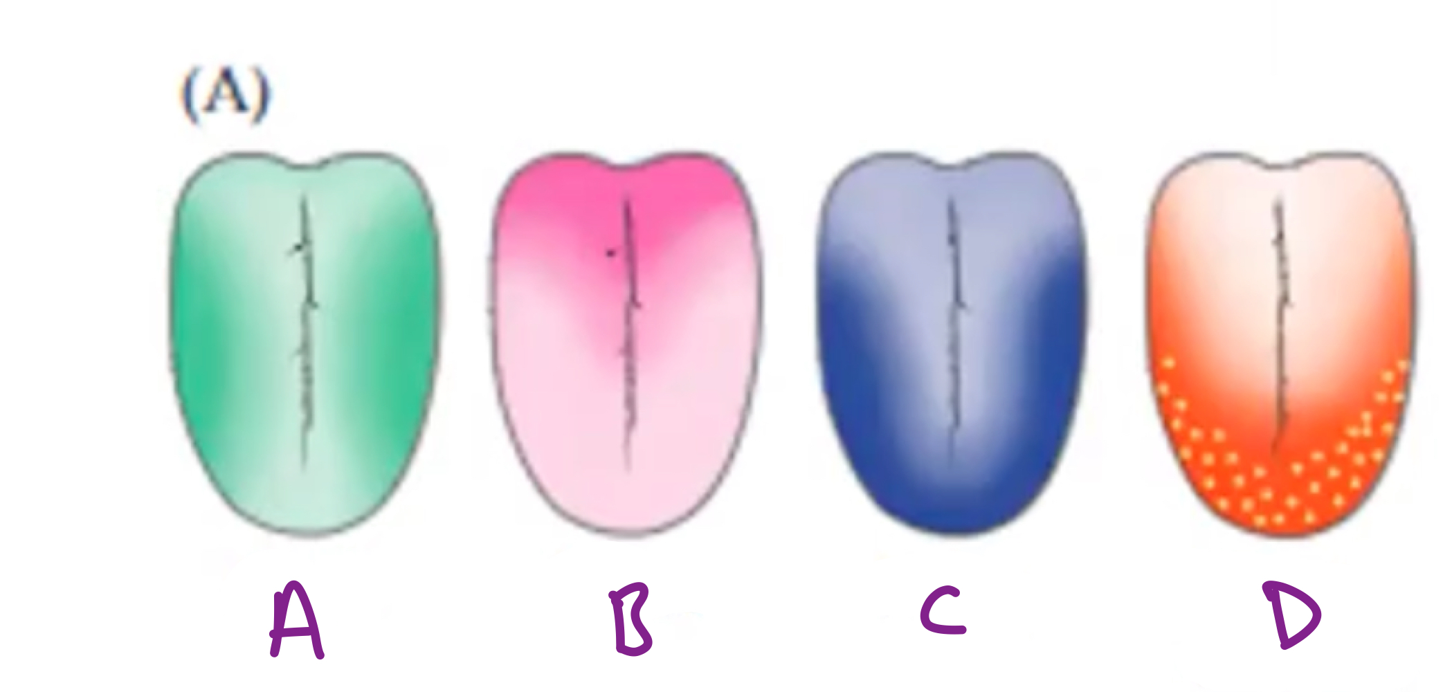
Identify which taste receptor cells are primarily found in each region.
A) Sour, B) Bitter, C) Salty, D) Sweet/Umami
What are the ways tastants can begin taste transduction?
They can pass directly through ion channels, bind/block ion channels, or bind to G-protein receptors to activate secondary ion channels.
What are some characteristics of taste transduction?
It’s initiated at the apical end and electrical signals conduct intracellularly to release neurotransmitters (thought to involve serotonin, ATP, and GABA).
What nerves receive the synapses initiated by taste transduction?
Cranial nerves
What do salty taste receptor molecules respond too?
Ions (sodium and other small cations)
What do sour taste receptor molecules respond too?
Protons (acidity)
What do sweet receptor molecules respond too?
Sugar molecules
What do bitter/umami receptor molecules respond too?
Amino acids
Where are taste receptors concentrated?
On the apical microvilli of taste cells
Which one of the 5 classes of taste receptor molecules are the most sensitive to avoid toxins?
Bitter
What is neuronal coding?
How the identity, concentration and pleasurable vs aversive value of a tastant is represented by firing action potentials
What is the labeled line hypothesis for neuronal coding?
Individual taste receptor cells for each stimuli
What is the population coding hypothesis for neuronal coding?
Roughly labeled lines contribute to a large number of broadly tuned neurons that contribute to the temperature and textural features of food
What are odorants?
Molecule that activates the transduction process in neurons for smell (Olfactory axons constitute olfactory nerve)
What is the cribiform plate?
A thin sheet of bone through which small clusters of axons penetrate, coursing to the olfactory bulb
Why do humans have weaker smell ability compared to many animals?
Due to the small surface area of the olfactory epithelium
What is the olfactory pathway?
Olfactory receptor cells → Olfactory bulb → Olfactory tract (Pyriform cortex, etc.) in temporal lobe
What is special about the olfactory pathway compared to the other sensory pathways?
It does not relay on a thalamic relay like the other pathways, instead it is relayed to pyriform cortex which can interface with the thalamus
What is the olfactory epithelium?
Neurons and supporting cells that line ½ the surface of the nasal cavity. These are interspersed with respiratory epithelial cells and lined with mucus
What are olfactory receptor neurons (ORN)?
Olfactory cilia extended into respiratory mucosa that are found in the olfactory epithelium.
How does smell transduction begin?
Odorant binds to the odorant receptor in the cilia or ORN, this triggers action potential from cilia to cell body to olfactory nerve
What type of receptor is an ORN?
G-Protein coupled receptor that is conserved across species, but number of functionally transcribed/translated genes differ.
What specific G-protein does ORNs produce?
Golf
How are specific smells differentiated from each other?
Each receptor cell expresses a single olfactory receptor protein, and each receptor protein binds a specific odorant causing cells to respond with different stimuli
What is the audition system?
The sense of hearing (detect sound and perceive/interpret nuances)
What is the vestibular system?
The sense of balance (head and body location/movement)
How does the external ear impact the auditory pathway?
Gathers and focuses sound