Radiology of the Thorax
1/104
There's no tags or description
Looks like no tags are added yet.
Name | Mastery | Learn | Test | Matching | Spaced | Call with Kai |
|---|
No study sessions yet.
105 Terms
true
True or false: there is already a lot of contrast between tissue types in the thorax so choosing a specific technique is not neccesary
high
The the thorax, a _______ kVp is used
low; high
A high kVp generates a _______ contrast while a low kVp generates a _________ contrast
> 15 cm (such as in large animals)
At what patient thickness would kVp be altered and a grid used?
inspiration
Thorax radiographs should be taken during inspiration or expiration?
-increases radiolucency
-high contrast of vasculature, bronchi, and heart
-minimal/no contact of diaphragm and cardiac silhouette and sternum and cardiac silhouette
-flattened diaphragm
-able to visualize accessory lobe of right lung
Reasons why it is importnant to take images on inspiration:
thoracic inlet; diaphragm
The cranial to caudal boundary for the thorax spans from the _______ _________ to ________
spinous processes; sternum
The dorsal to ventral boundary for the thorax spans from the _______ _________ to ________
2 cm cranial to first rib
Cranial boundary for thoracic imaging
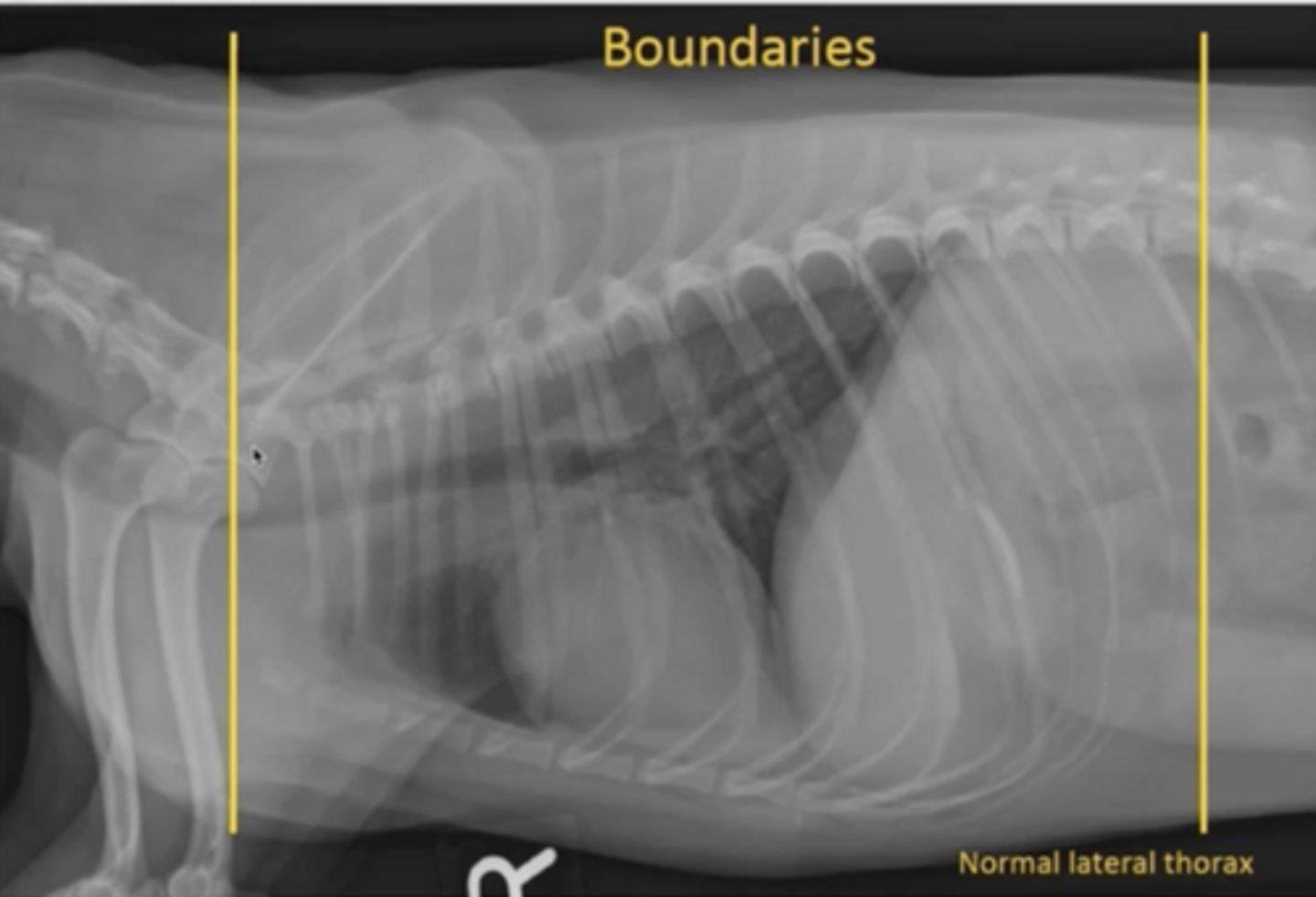
just caudal of L1 vertebrae (caudal to last rib)
Caudal boundary for thoracic imaging

heart
The thoracic radiograph image should be centered around the _________
5th
or 2 fingers caudal to scapula
To center around the heart, center of image should be at the ______ intercostal space or...
more
When taking a radiographic image, the further things are away from the plate, the _______ magnified they become
accesroy lung lobe
In a V/D image, what anatomy is more magnified?
caudal lobe pulmonary vessels
In a D/V image, what anatomy is more magnified?
one large dome
What does the diaphragm look like on a D/V image?
left and right crus are visible as well as the dome
What does the diaphragm look like on a V/D image?
D/V
Is this a D/V or V/D image?
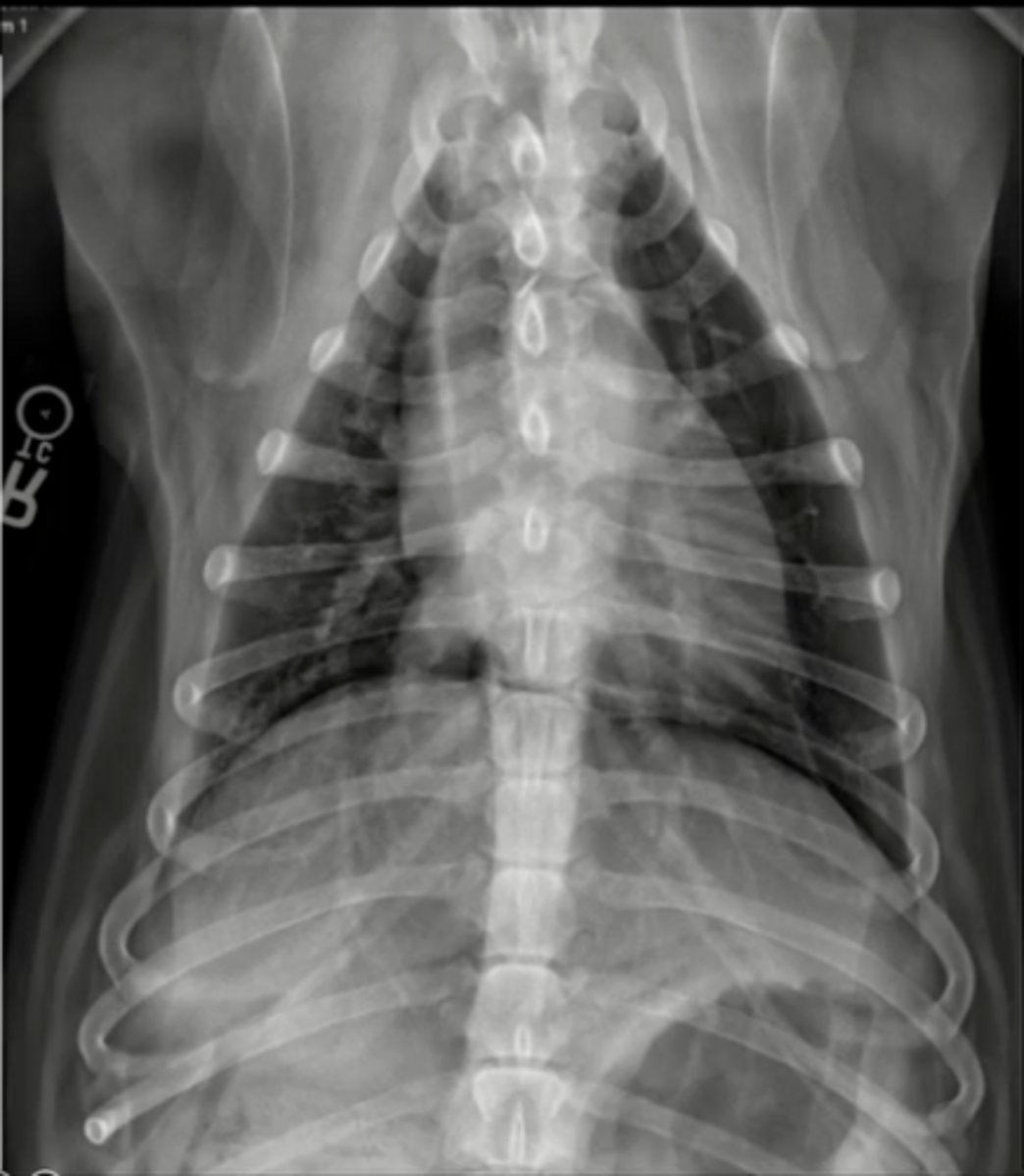
V/D
Is this a D/V or V/D image?
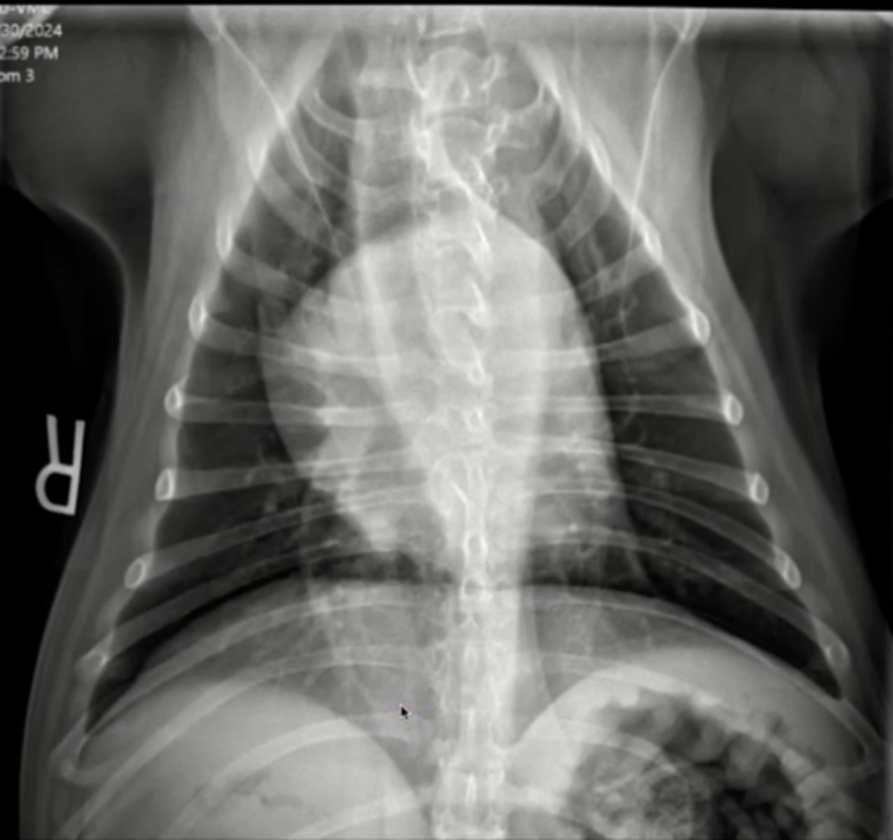
left and right crus and the dome
What does the diaphragm look like on a D/V image?
true *and since they are so large, multiple images may have to be taken to encompass the entire area
True or false: large animal thoracic radiographs (such as adult horse) are only taken standing
The legs cannot be pulled away and can cover some of the thoracic anatomy
What is a disadvantage to taking images while standing?
right lateral, left lateral, and V/D
A _________ view thoracic sereis is considered a full study
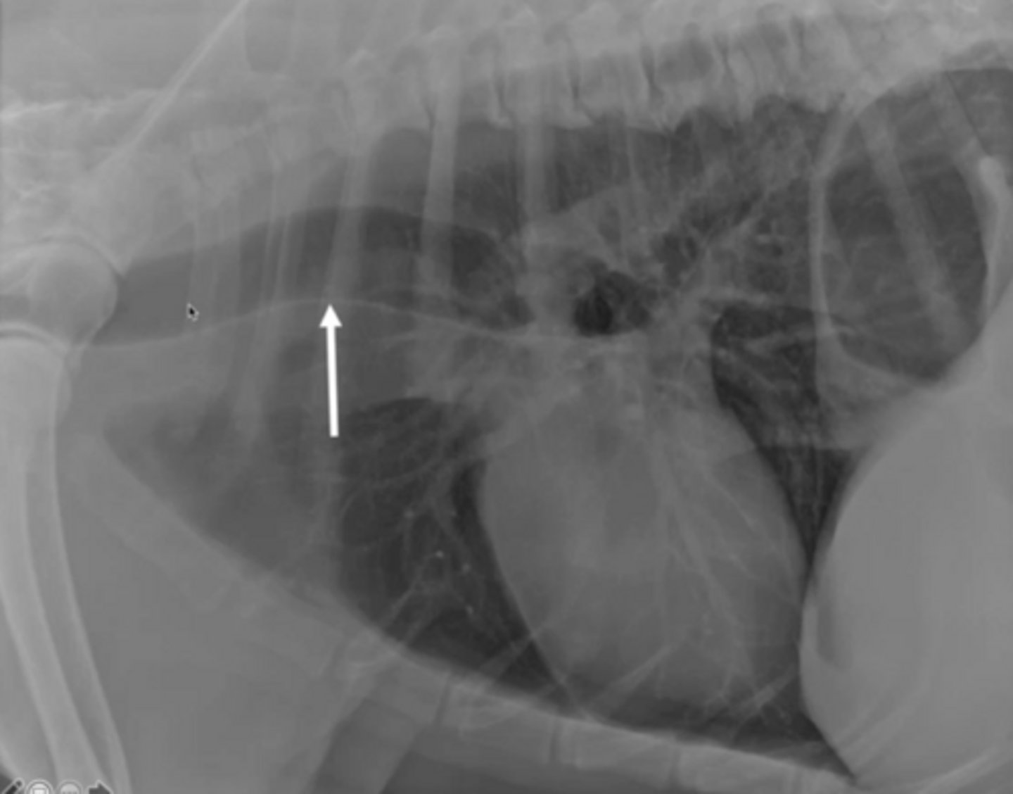
dorsal elevation of the trachea due to incorrect positioning of the head (ventroflexion)
The arrow is pointing to what appears to be a pathology, but what is it really?
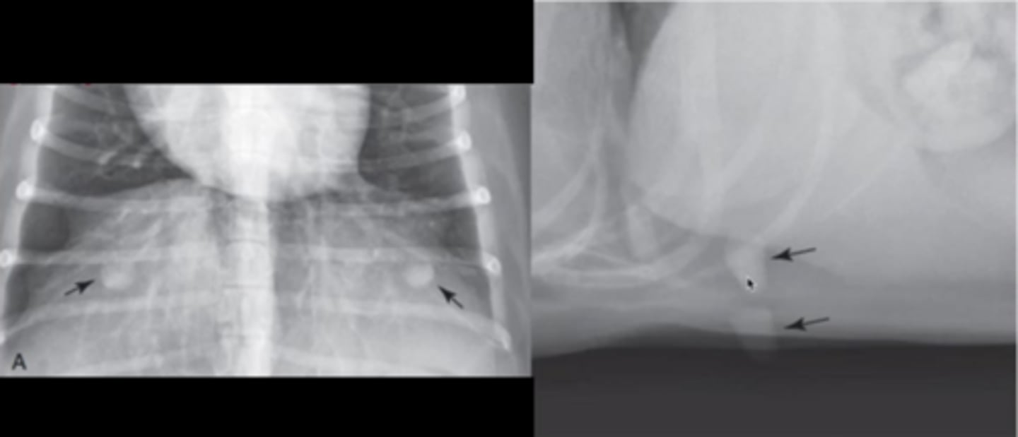
cutaneous lesions/nipples that could be mistaken for nodules in the lungs
The arrows are pointing to what appears to be a pathology, but what is it really?
left lung
For a right lateral view (recumbency), which lung is better visualized?
right lung
For a left lateral view (recumbency), which lung is better visualized?
right lung
For a right lateral view (standing), which lung is better visualized?
left lung
For a left lateral view (standing), which lung is better visualized?
spine of scapula
1

trachea
2
thoracic vertebrae
3

carina of trachea
structure I forgot to label

aorta
4

left crus of diaphragm
5

right crus of diaphragm
6

caudal venal cava
7

pylorus of stomach
8

blood vessels
9

sternum
10

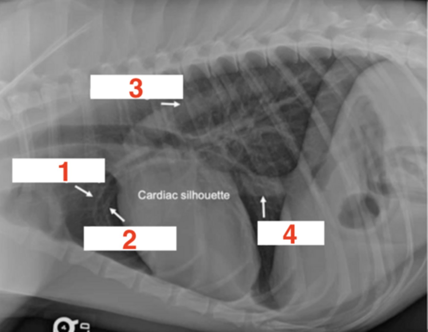
pulmonary artery
1

pulmonary vein
2

descending aorta
3

cadual vena cava
4
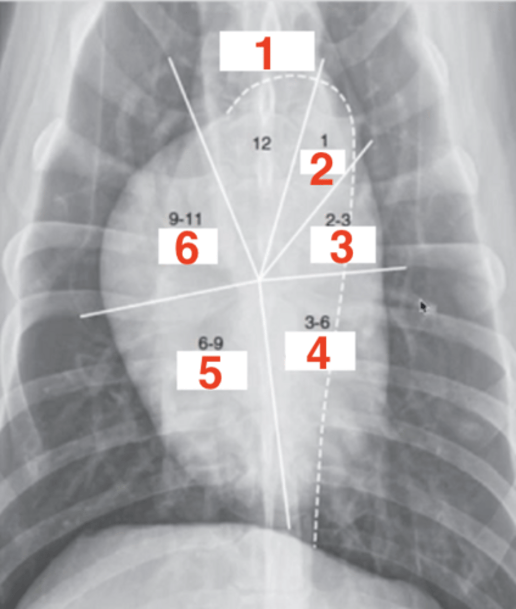
1.) left atrium
2.) left ventricle
3.) right ventricle
4.) right auricle, aorta, pulmonary atery
Six structures that correlate with the cardiac silhouette clockface anaolgy in a lateral view:
12-2
On a lateral recumbant image, the left atrium is what on the clock face?
2-5
On a lateral recumbant image, the left ventrice is what on the clock face?
5-9
On a lateral recumbant image, the right ventricle is what on the clock face?
9-12
On a lateral recumbant image, the right auricle, aorta, and pulmonary atery are what on the clock face?
1.) aortic arch
2.) pulmonary trunk
3.) left auricle
4.) left ventricle
5.) right ventricle
6.) right atrium
Six structures that correlate with the cardiac silhouette clockface anaolgy in the V/D view:
11-1
On a V/D image, the aortic arch is what on the clock face?
1-2
On a V/D image, the pulmonary trunk is what on the clock face?
2-3
On a V/D image, the left auricle is what on the clock face?
3-5
On a V/D image, the left ventricle is what on the clock face?
5-9
On a V/D image, the right ventricle is what on the clock face?
9-11
On a V/D image, the right atrium is what on the clock face?
left atrium
What chamber cannot be seen on the V/D view, but can be seen on the lateral view?
cats; left atrium is seen at the 1-2 clock face region along with the pulmonary trunk
What is the exception to the left atrium not being seen on a V/D view?
aortic arch
1

pulmonary trunk
2

left auricle
3

left ventricle
4

right ventricle
5

right atrium
6
correlates the size of heart to size of dog
Vertebral heart scale
tracheal bifurcation to apex of heart
How is the heart length measured for a vertebral heart scale
widest part perpendicular to length line
How is the heart width measured for a vertebral heart scale
8.7-10.7
Normal range for VHS in dogs
6.9-8.1
Normal range for VHS in cats
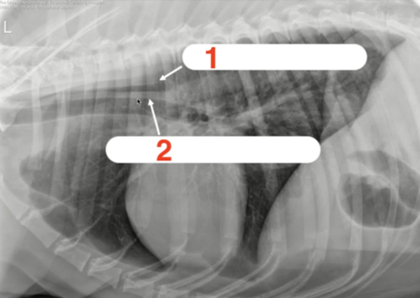
left pulmonary artery
1

bronchus
2
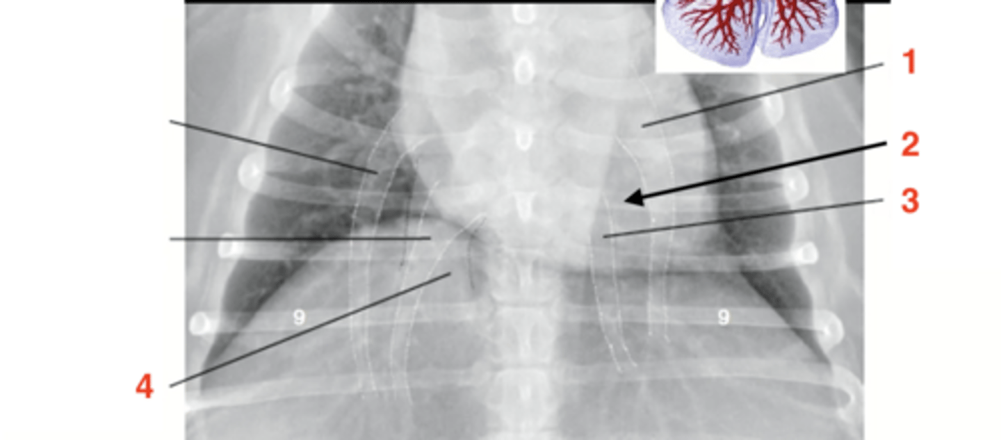
left pulmonary vein
3

caudal vena cava
4 (black dots)

9th; square
*rectangle could indicate vessel is too large
As the pulmonary artery and vein cross the _______ rib, they should create a ________ shape
left lateral (can visualize the right lung better)
Is this a left or right lateral image?

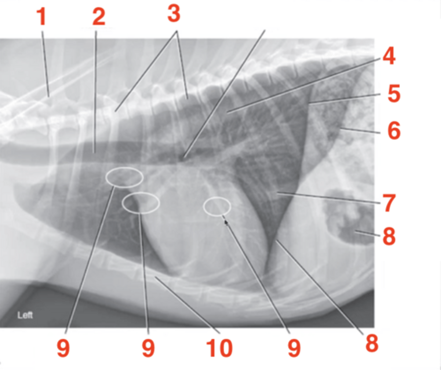
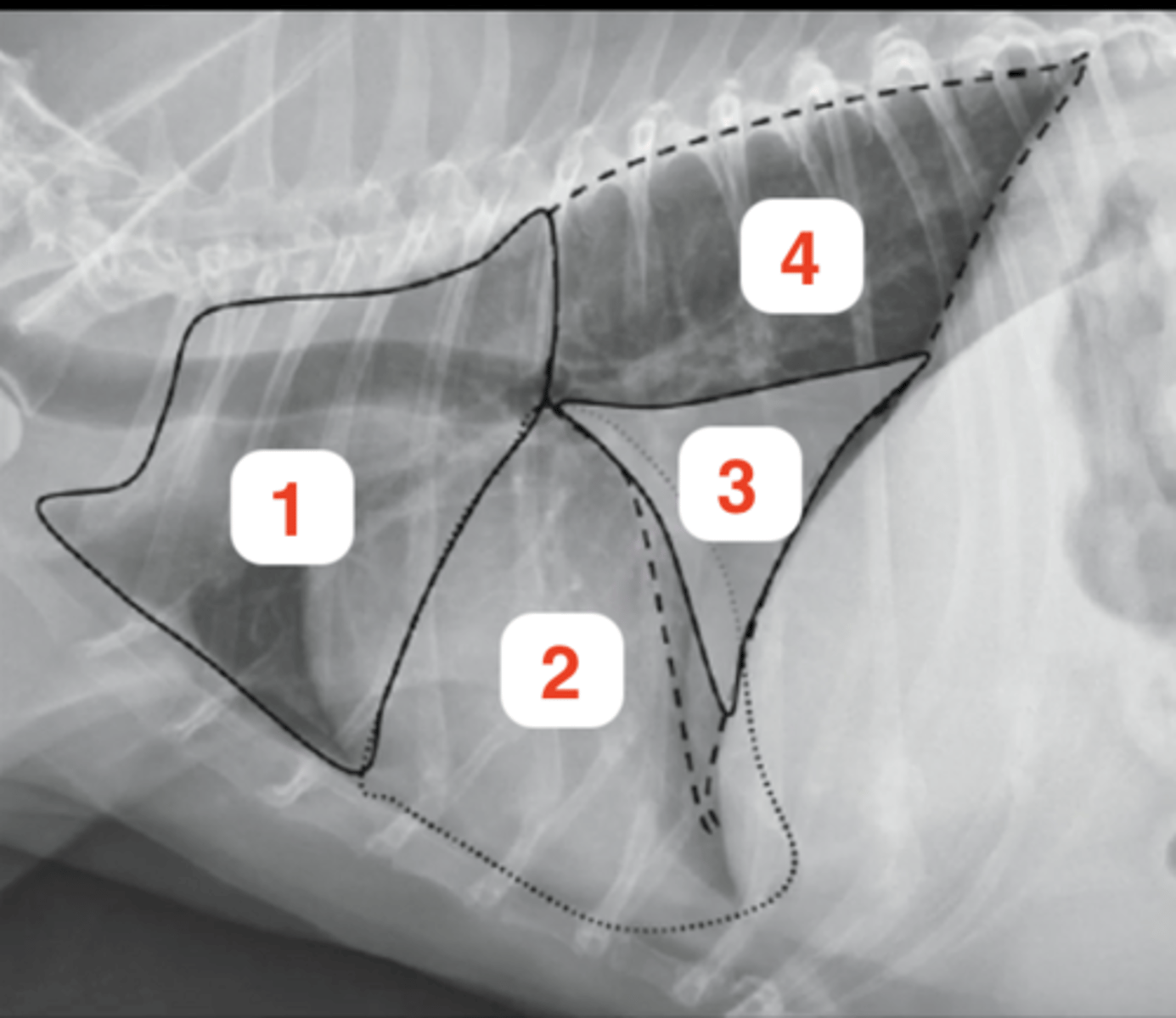
cranial lobe of right lung
1

middle lobe of right lung
2

accessory lobe of right lung
3

caudal lobe of right lung
4
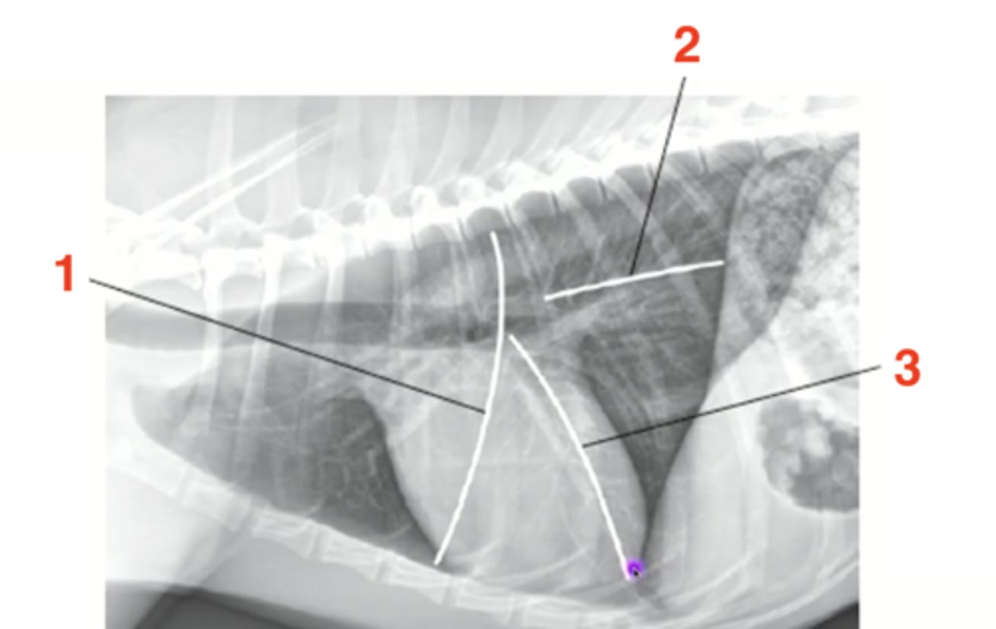
caudal margin of right cranial lobe
1

dorsal margin of accessory lobe
2

caudal margin of right middle lobe
3
border betwen cranial and caudal parts of left cranial lobe
1

caudal margin of left cranial lobe
2

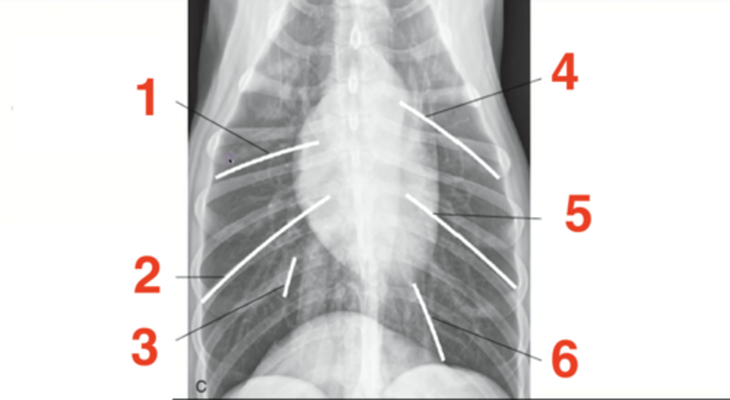
border between right cranial and right middle lobes
1

border between right middle and right caudal lobes
2

right lateral most aspect of accessory lung lobe
3

border between cranial and caudal parts of left cranial lobe
4

border between left cranial and left caudal lobes
5

left margin of accessory lung lobe
6
true
True or false: "field" is not appropriate terminology when discussing the lungs
true
True or false: normally the esopghaus is NOT seen
when there is gas within the esophagus
When can the esophagus be seen?
looks like the trachea walls are thickened, but it is actually the trachea and esophagus superimposd on each other
tracheoesophageal stripe sign
gas within esophagus
1
tracheoesophageal stripe sign
2

false! should not normally be seen
True or false: it is normal to see the pleural space
cupula is prominent with central bulge
What does the diaphragm look like on a D/V image
triple bump; cupula + 2 crura
What does the diaphragm look like on a V/D image
parallelling crura
What does the diaphragm look like on a right lateral image