BMS1052 - W1: Foundations of Neurobiology
1/22
There's no tags or description
Looks like no tags are added yet.
Name | Mastery | Learn | Test | Matching | Spaced |
|---|
No study sessions yet.
23 Terms
What are the two major classes of cells in the nervous system?
Neurons and Glia
What are secretory vesicles?
Why are these important for neurons?
A special package that allows neurons to release something out into the extracellular space.
Important for neurons because they package up neurotransmitters and they can use that to influence other neurons. (basis of communication)
What are neuronal processers/ neurites?
parts of neuron which protrude away from cell body - these allow neuron to communicate with other neurons
basically, axons and dendrites (which are unique to neurons)

Do neurites contain mitochondria?
Explain.
The rough endoplasmic reticulum (which contains mitochondria) does not extend into the neurites.
This means that there is no protein being made in these neurites.
What makes neurons different from other cells?
They have a specialised structure (neural processes that extend away from the cell body.)
They have specialised function (mechanisms for electrical and chemical communication)
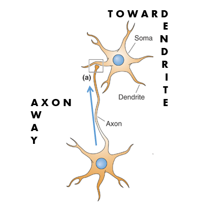
Axons and dendrites
axons and dendrites contain a range of specialised ion channels that aid communication
Electrical signals (information) are communicated
towards the cell body via dendrites
Away from the cell body via axons
Explain what these ‘electrical signals’ or ‘information’ is.
In the brain, this refers to chemical and electrical signals that represent some property about the internal or external environment.
What are the two ways that neurons can transmit information?
(chemical) Information flow is electrical along dendrites and axons.
(electrical) Information is transmitted through action potentials
Why is the structure of neurons (specifically axons and dendrites) dynamic?
This is because these axons and dendrites are how neurons connect to other neurons. So this means that the cytoskeleton needs to be dynamic as it needs to help control movement of the axons and dendrites and maintain the shape of the neurons once connections have been made with other neurons.
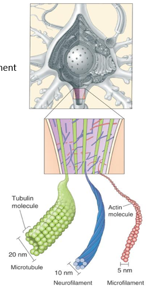
What are the three main components that make up the cytoskeleton?
Microtubules
the thickest filaments that run longitudinally down neurites
critical for mediating intracellular transport (movement of proteins and waste products within the neurons)
have specialised proteins - microtubule-associated proteins (MAPs) which help regulate their assembly and functions
Neurofilaments
smaller than microtubules
provides structural support and regulate the diameter of axons
Microfilaments
crosslinked between microtubules and the membranes
formed from a molecule called actin (which is the same molecules that helps muscles contract)
What is transported in axons?
In what two ways is material transported within the axon?
Physically transported:
proteins (so they can be incorporated into the cell membrane or serve other specilised processes)
neurotransmitters
metabolites
(Information/ electrical signals are not transported physically)
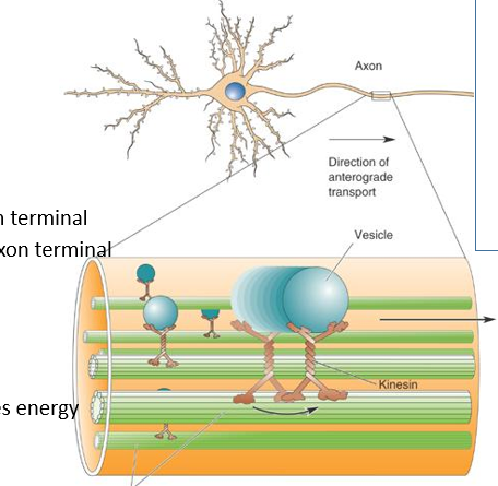
Transport direction:
-anterograde (forwards/ towards axon terminal) via kinesin
-retrograde (backwards/ away from axon terminal) via dynein
1) Fast axoplasmic transport
Kinesin molecules (motor proteins) basically walk along the microtubules (carrying a vesicle). Highly energy consuming process
proteins synthesized in the soma are actively transported towards the axon terminal (anterograde?)
metabolic waste products from axon terminal are transported to cell body (retrograde?)
1 m/ day
2) Slow axoplasmic transport
-mechanisms unclear (it’s not simply passive diffusion - does require energy, but the speed are quite different to that of fast transport)
0.1-10mm/ day
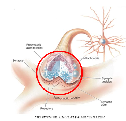
Axon terminal:
no microtubules within axon terminal
lots of internal vesicles (the little blue bubble looking things circled in the image) which contain chemical messengers (neurotransmitters) which can be released to impact of a downstream neuron.
membrane of axon terminals is very protein dense (as the process that controls this sort of mechanisms require a lot of specialized proteins)
lots of mitochondria
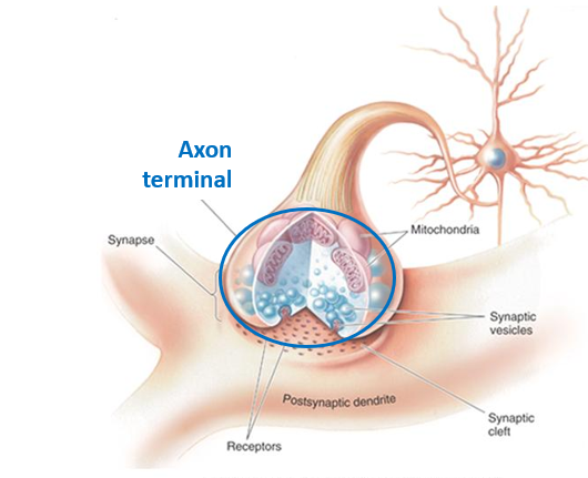
What is the synapse?
The axon terminal is just part of the synapse - which is the overall name for the special connection between two neurons
synapse contain a:
pre-synaptic membrane (which releases vesicles containing neurotransmitters)
synaptic cleft (small gap between membranes of two neurons)
post-synaptic membrane (neurotransmitter binds to specialized protein and is converted into an electrical signal)
What are dendritic spines?
In many neurons, dendrites receive axonal inputs on dendritic spines - small extensions of cell membrane where axon of another neuron comes into proximity
Length and density of the spines is very important
We have thousand of dendritic spines
If all these dendritic spines require proteins, does protein synthesis occur in the dendrites?
Yes it does.
Proteins are transported along axons because there is no protein synthesis in the axons, so dendrites and axons are quite different.
This suggest that whatever is happening inside synapses can control protein synthesis in dendrites.
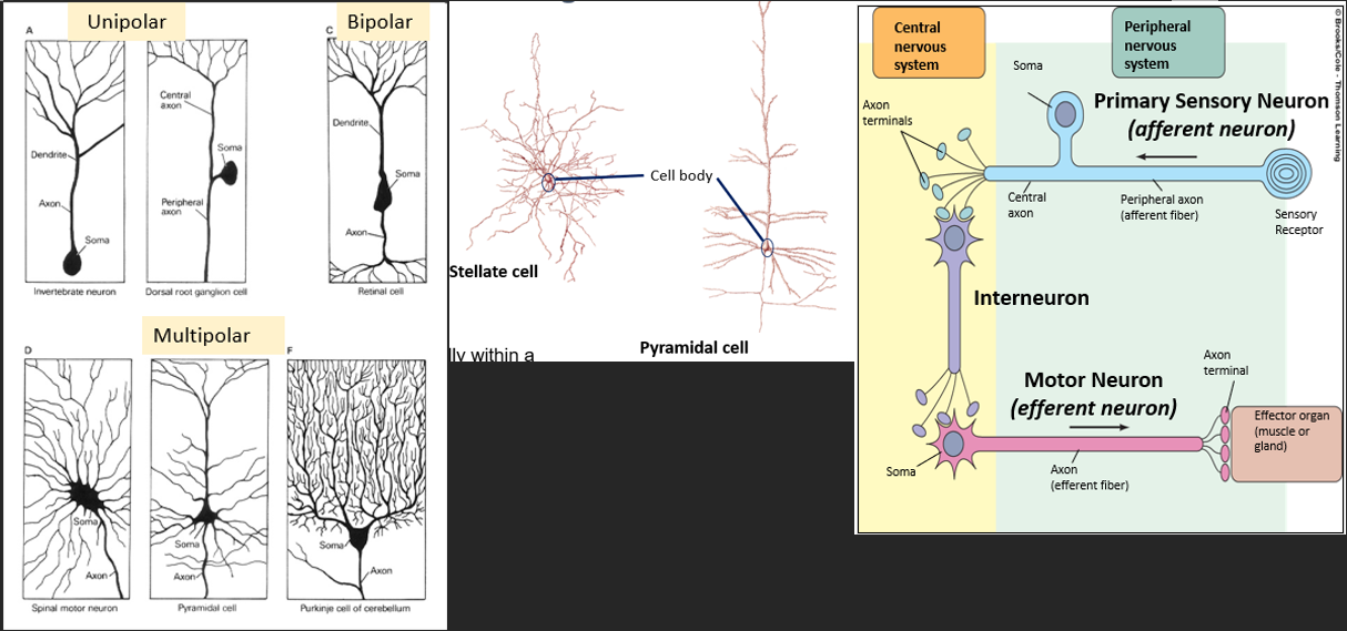
What are 5 ways that neuronal structure and function varies?
Number of neurites
(unipolar, bipolar, multipolar)
number of connection coming into the soma
Shape and dendrites
stellate cell - collate information from other neurons in a local region (as they are more compact)
pyramidal cell - long, can extend many metres so they are involves in sending information around body.
axon length
connections
Sensory neuron - input is taking information in from external environment, output is another neuron
Interneuron - receives input from another neuron and outputs to another motor neuron
Motor neuron - receives from another neuron, but outputs to an effect (muscle or gland)
neurotransmitter
nature of the type of neurotransmitter they release can also be different
What do glia cells do?
Glia support neuronal function and may be involved in information transmission
What are the 4 main types of glia?
Myelinating glia (increase reliability and velocity of axonal signals)
Found in the central nervous system and peripheral nervous system
Oligodendroglia (CNS)
Schwann cells (PNS)
Astrocytes (a range of supporting roles)
Microglia (The brain’s immune system)
Ependymal cells (produce cerebrospinal fluid)
Myelinating glia:
We need reliable and rapid electrical signal conduction along long and thin axons. Axons are leaky to charge/ ions).
The solution is to electrically insulate between the inside of the axon and the cytosol and extracellular space.
-Oligodendroglia (CNS) and Schwann cells (PNS) generate myelin sheaths around axons.
But Schwann cells tend to have a 1:1 relationship - so in the PNS they wrap myelin around just one singular neuron.
However, we can’t completely insulate between the axon and cytosol. What is the solution?
Regular gaps in the myelin (nodes of Ranvier) that exposes regions of membrane with high density of channels (in particular voltage gated channels) so they can communicate directly with the cytosol.
Astrocytes:
The most common of all the glia (they fill in much of the brain space that is not occupied by neurons and blood vessels) - in close proximity to neurons
can influence neurite growth (the way axons and dendrites grow and move in the nervous system)
can regulate the chemical content of extracellular space (e.g. buffer K+ around the extracellular space - so if the K+ concentration is too high in one region, they can take up K+ and buffer/ move it through their internal processes and release it somewhere else).
Contain glycogen and capable of gluconeogenesis
Can affect blood flow
May contribute to or impair nervous system repair (can contribute to glial scarring, which can prevent regeneration or regrowth of neurons)
The astrocyte (astrocyte process) is found in close proximity to the synapse and can influence what occurs in the synapse. (Because of this close proximity, they can regulate neurotransmitter that’s in the synapse).
-So when the neurotransmitters are released from the vesicles, normally they would target the receptors of the postsynaptic terminal, they can actually be modulated by the astrocyte.
-can unsheathe small arterioles and capillaries (meaning they can modulate blood flow by changing the diameter of those blood vessels
Microglia:
-form about 10-15% of the glial population in the nervous system
-they are essentially the specialized immune system within the brain
include macrophages (which remove debris associated with dead/ degenerating cells)
fight inflammation within the brain
Ependymal Cells:
Ependymal cells line the fluid filled ventricles and they produce CSF
found within ventricles (inside brain)
produce cerebrospinal fluid (CSF) which fills the ventricles and also extends down the spinal cord. And it also leaks out and surrounds the brain.