SKELETAL SYSTEM
 Call Kai
 Learn
Learn Practice Test
 Spaced Repetition
 Match
Match Flashcards
 Knowt Play
1/154

There's no tags or description
Looks like no tags are added yet.
Name | Mastery | Learn | Test | Matching | Spaced | Call with Kai | Chat |
|---|
No analytics yet
Send a link to your students to track their progress
155 Terms
Skeletal System
Accounts for approximately 20% of the body weight
Axial Skeleton, Appendicular Skeleton
Main divisions of skeletal system
Axial Skeleton
central core of the human body that consists of 80 bones along the body’s main axis to protect vital organs
Axial Skeleton
It forms the framework that supports your head, neck, and torso
Appendicular Skeleton
consists of 126 bones that form the upper and lower limbs, shoulder girdles, and pelvic girdle
Appendicular Skeleton
comprises the body’s appendages and the connection points linking them to the central trunk
Bones
Provide support, protection, movement, mineral storage, and blood cell production
Cartilages
Connective tissue that provides support and flexibility
Cartilages
Important during skeletal development
Tendons
Connect muscle to bone, Made up of dense regular connective tissue
Ligaments
Connect bone to another bone, Made up of dense regular connective ti
206 bones
how many bones in total
Support
Bones are suited for bearing weight and are the major supporting tissues of the body [Functions of the Skeletal System]
Support
Cartilage provides firm and flexible support within certain structures [Functions of the Skeletal System]
Support
Ligaments hold the bones together [Functions of the Skeletal System]
Protection
Protects internal organs [Functions of the Skeletal System]
Movement
Bones provide attachment points for muscles and act as levers [Functions of the Skeletal System]
Movement
Joints allow movement between bones [Functions of the Skeletal System]
Movement
Ligaments allow some movement between bones but prevent excessiveness [Functions of the Skeletal System]
Mineral Homeostasis
Bones store and release minerals, especially calcium and phosphorus [Functions of the Skeletal System]
Mineral Homeostasis
Calcium is important for muscle contraction and normal heart function [Functions of the Skeletal System]
Storage
Stores minerals such as calcium and phosphorus [Functions of the Skeletal System]
Storage
Yellow bone marrow stores fat [Functions of the Skeletal System]
Blood Cell Production
Red bone marrow contains hematopoietic tissue responsible for blood cell formation (RBCs, WBCs, platelets)[Functions of the Skeletal System]
Yellow Bone Marrow
mainly stores fat

Red Bone Marrow
Contains hematopoietic tissue responsible for blood cell formation (RBCs, WBCs, platelets)

Compact and spongy
types of bone tissue
Cortical Bone
other word for Compact Bone
Compact Bone
forms the solid outer casing of bone
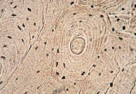
Compact Bone
Dense and strong
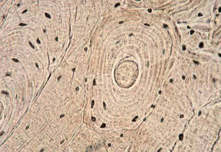
Compact Bone
Contains a large amount of bone matrix

Compact Bone
Organized into structural units called osteons
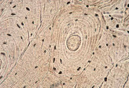
Osteons
main functional and structural unit of compact bone

Volkmann’s Canals
allow blood vessels and nerves to communicate between different areas of bone

Interstitial Lamella
remnants of older osteons (located between osteons)

Cancellous Bone
other word for Spongy Bone
Spongy Bone
Contains numerous spaces

Spongy Bone
More porous than compact bone
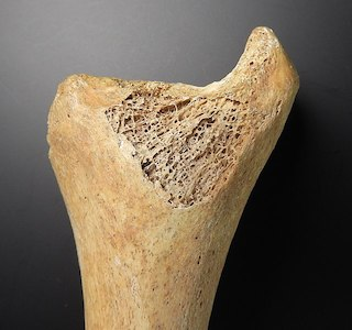
Spongy Bone
Contains less bone matrix

Spongy Bone
consists of trabeculae
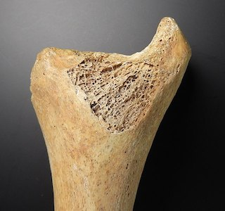
Trabeculae
Thin plates or fragments of bone
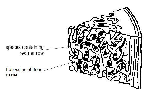
Trabeculae
Form the framework of spongy bone

Trabeculae
Spaces between _____ contain bone marrow
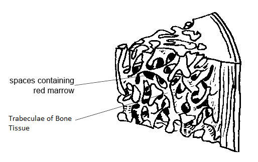
Long Bone
Longer than they are wide
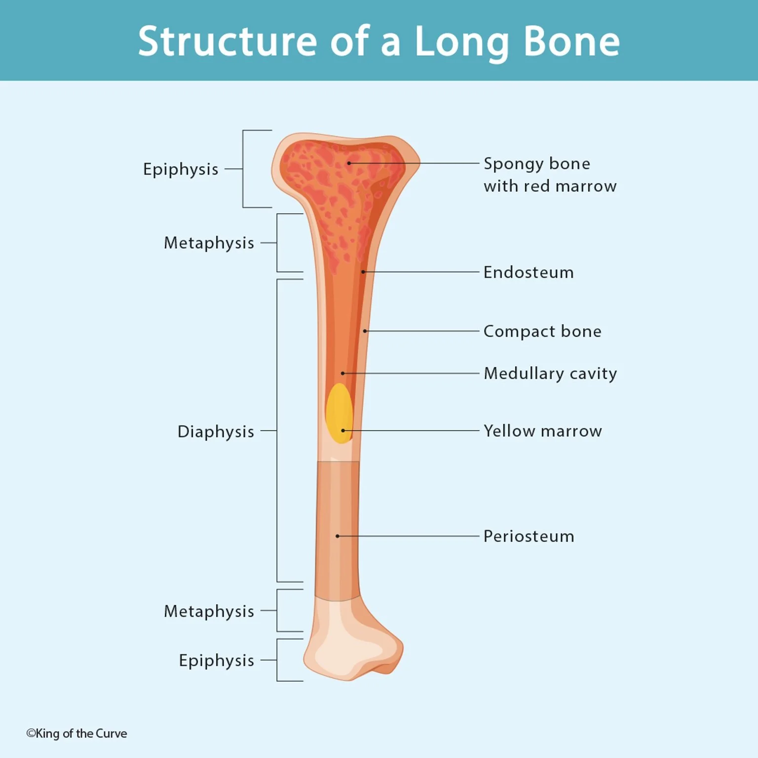
Long Bone
Characterized by a shaft

Short Bone
have approximately equal length and width

Short Bone
No distinct shaft

Short Bone
May appear relatively square or cube-like

Flat Bone
relatively thin and flattened

Flat Bone
Often provide protection and broad surfaces for muscle attachment

Irregular Bone
Do not fit into bone shape categories

Irregular Bone
Have irregular and complex shapes
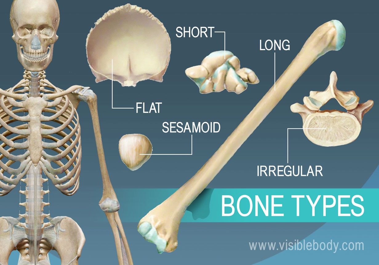
Sesamoid Bones
Bones develop inside tendons

Sesamoid Bones
Commonly found in the tendons of the lower extremities

Diaphysis
The shaft of the long bone, long and elongated
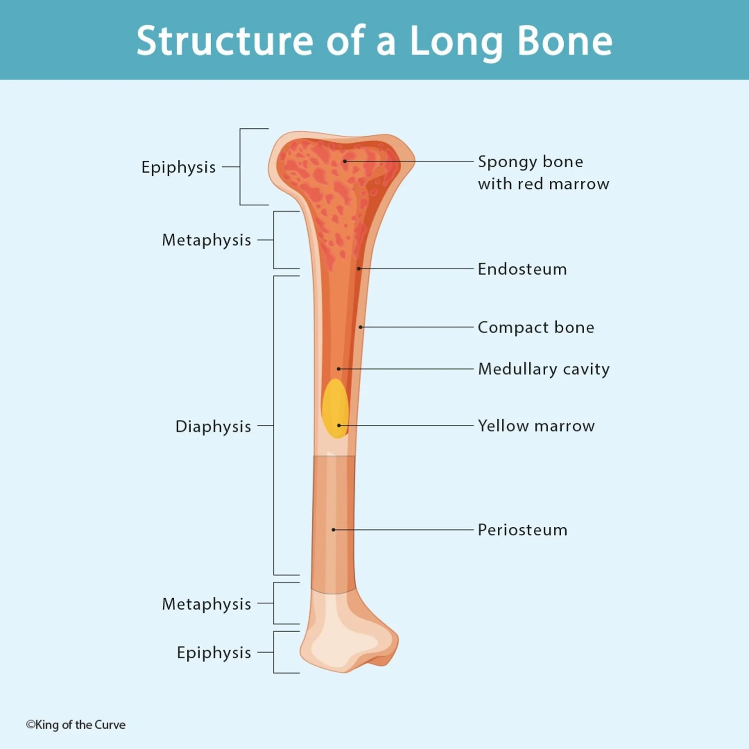
Epiphysis
The two ends of a long bone

Epiphysis
Includes proximal epiphysis and distal epiphysis

Epiphysis
Involved in joint formation

Metaphysis
Region between the diaphysis and epiphysis

Metaphysis
Contains the epiphyseal plate during bone growth

Epiphysis Plate
made of hyaline cartilage

Epiphysis Plate
Allows the bone to increase in length
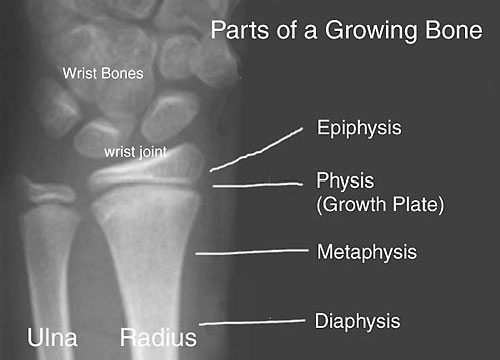
Epiphysis Plate
It lasts until 18-20 years old
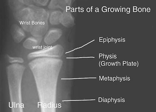
Epiphysis Plate
Once it closes, it become the epiphyseal line

Epiphysis Plate
After closure, the bone can no longer increase in length

Periosteum
External covering/lining of the bone, Contains dense irregular connective tissue

Medullary Cavity
Internal cavity of the bone

Medullary Cavity
Contains bone marrow

Medullary Cavity
Helps lighten the weight of the bone

Endosteum
Internal lining of the bone

Articular Cartilage
Made of hyaline cartilage

Articular Cartilage
Covers the surfaces of the bones involved in joints

Articular Cartilage
Helps reduce friction during movement

Extracellular Matrix
Bone is made of; 25% water, 25% collagen fibers, 50% crystallized mineral salts
Extracellular Matrix
consists of mineral salts and collagen fibers
Mineral Salts
provide rigidity/hardness
Collagen fibers
provide flexibility and help bones withstand stress
Collagen
A tough, rope-like protein
Collagen
Provides flexibility and tensile strength
Proteoglycans
Large molecules consisting of many polysaccharides attached to and surrounding core proteins
Proteoglycans
Important components of connective tissue, especially cartilage
Osteogenesis imperfecta
brittle bone disease, a rare condition caused by an abnormality in collagen fibers
Osteoprogenitor Cells
stem cells of bone
Osteoblasts
Responsible for bone formation
Osteocytes
Mature bone cells
Osteocytes
Surround by bone matrix
Osteocytes
Located in the lacunae
Osteoclasts
Responsible for bone breakdown/resorption
Osteoclasts
Digest and remove bone tissue
Osteoclasts
contain Howship’s lacunae
Osteogenesis
Process of bone formation
Osteogenesis
Initial bone formation begins around the 6th week of embryonic development
Intramembranous Ossification
Bone develops directly from mesenchymal connective tissue membranes
Intramembranous Ossification
Formation of flat bones and some irregular bones
Ossificiation Center Development, Calcification, Trabeculae Formation, Periosteum Development
Steps for Intramembranous Ossification
Ossificaiton Center Development
Mesenchymal cells differentiate into osteoproenitor cells
Ossification Center Development
Osteoprogenitor cells develop into osteoblasts
Calcification
Osteoblasts produce bone matrix
Calcification
Deposition of minerals
Calcification
Osteoblasts became osteocytes as they become surround by bone matrix