Lecture 5: Vision & Eye Movements
 Call Kai
 Learn
Learn Practice Test
 Spaced Repetition
 Match
Match Flashcards
 Knowt Play
1/91

There's no tags or description
Looks like no tags are added yet.
Name | Mastery | Learn | Test | Matching | Spaced | Call with Kai | Chat |
|---|
No analytics yet
Send a link to your students to track their progress
92 Terms
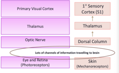
How are the Sensory System & Visual Pathway Similar?
-Both systems carry information from the "periphery" towards the brain to thalamus to primary (sensory or visual) cortex
Retina
-Receives light from the lens and converts it to neural signals that the brain can understand
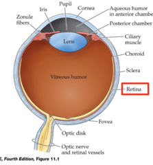
What structures make up the retina?
-Photoreceptors
-Horizontal cells
-Bipolar cells
-Ganglion cells
-Fovea
-Macula
-Plexiform layers
-Pigment epithelium
Photoreceptors
1. Cones
2. Rods
-Cells in the retina that respond to light
Rods (Photoreceptors)
1. Primarily found in peripheral retina
2. Achromatic (without colour)
3. High light sensitivity
4. Nighttime vision, when light levels are low
5. More rods than cones
Cones (Photoreceptors)
1. High acuity and colour vision during day-time when light levels are higher
2. Primarily found in the central retina (fovea)
Optic Disc
-Region where the axons leaving the retina gather to form the optic nerve
-There are no photoreceptors here so it is considered a blind spot
*only ganglion cell axons
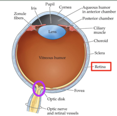
Fovea
-Central fixation point for each eye
-Region of the retina with the HIGHEST visual acuity = full of cones & almost no rods
-Small in size
-Provides input to half of the optic nerves and cells in visual cortex
Does the Fovea function well in dim light?
-No, since there are no rods present in the fovea
-Provides sharp, colour vision
Macula
-Oval region surrounding the fovea
-High visual acuity
-Occupies the central 5 degrees of visual space
How much space does the Fovea occupy?
-Central 1-2 degrees of visual space
Phototransduction
-Receptor activated (photoreceptors)
-Receptive field (cells respond to light)
-Path to brain (via optic nerve)
Are somatosensory and vision transduction the same?
-Receptor detects stimulus
-Receptive field = specific cells respond to the stimulus
-Path to brain
Receptive Field in Visual Pathway
-Defined as the portion of the visual field where light causes excitation or inhibition of the cell
-Each rod & cone has their own
-Each photoreceptor monitors its own spot in the visual field where they respond to light
Receptive Field (Touch)
-Property which allows your brain to determine the location of the stimulus
-Each mechanoreceptor has their own of this and they can be large or small
Ganglion Cells
-Many types which respond to different characteristics such as motion, colour, acuity
*respond to more details than just light
1. Parasol
2. Midget
Parasol Ganglion Cells
-Large cell bodies, large receptive fields, large axons
-Respond to gross stimulus features and movements
Midget Ganglion Cells
-Respond to fine visual detail and colour
-small cell bodies, small receptive fields, smaller axons
-More numerous
Photoreceptors & Ganglion Cells Connection
-Lots of synaptic interactions between the two
-Allows the signal sent to the brain to already be highly processed when it leaves the retina (brain does not just see light or dark)
-Emphasizes the most important aspects of the visual scene which allows the brain to focus on informative visual features
Connection b/w Ganglion Cells & Mechanoreceptors
-Both create specialized information that gets send to the brain
-They provide information about more specific details of the stimulus
-Difference is that one happens at the sensory level (i.e., skin) while the other one happens post sensory level
Path to Brain for Somatosensory vs Phototransduction
-Optic nerve
-PCML
Common Visual Disorders of Eye & Retina
-Age Related Macular Degeneration
-Diabetic Retinopathy
-Glaucoma
-Cataracts
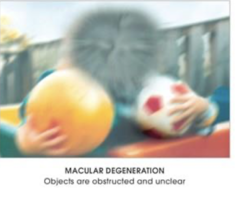
Age-Related Macular Degeneration (AMD)
-Damage to the macula
-Macula contains lots of cones which means it is receiving a lot of details
-Central vision affected
Diabetic Retinopathy
-Excess blood sugar causes damages to retinal blood vessels (which are lined with rods and cones)
-Results in patchy vision loss throughout visual field
-Rods and cones not receiving blood flow
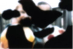
Cataracts
-Loss of lens transparency
-Lens = clear coating on top of the eye
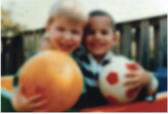
Glaucoma
-Increase intraocular pressure = damage to optic nerve
-Causes peripheral deficits
-Can still see central part but peripheral part is obscured
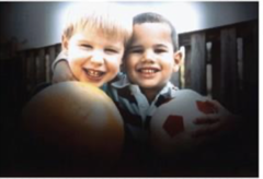
Why does Glaucoma cause peripheral deficits first?
-The increased eye pressure damage the outermost fibers first (fibers from the peripheral retina)
Disorders related to Primary & Secondary Visual Cortex
-Homonymous field deficits (e.g., following stroke)
-Agnosias (e.g., propsopagnosia)
General Pathway of Visual System
-Eye & Retina
-Primary Visual Cortex
-Visual Association Areas
Nasal Retinal Fibers
-Captures the peripheral visual field (peripheral part of the eye)
-The fibers from this part of the optic nerve cross over at the optic chiasm and gets processed contralaterally in the brain
Temporal Retinal Fibers
-Captures the central & contralateral part of the visual world
-Does not cross over at the optic chiasm
Ex: the left temporal retina captures the central/right part of the visual field (shared with the right eye)
Optic Chiasm
-Optic nerves from both eyes meet and partially cross to contralateral side of brain for processing
-Specifically the nasal retinal fibers cross over here while the temporal retinal fibers do not
-About 60% of the fibers (all nasal) cross over
Optic Tracts
-Carries information from both eyes (fibers from both eyes)
-Ex (left optic tract): contains right nasal fibers and left temporal nasal fibers
-And so it carries all the information from the right visual field (which is received from both eyes)
-After the optic chiasm
Optic Nerve
-Carries all visual information from one eye (both nasal & temporal fibers of that eye)
-Bundle of retinal ganglion cells that carries visual information from one eye to the brain
-Originates at the optic disc
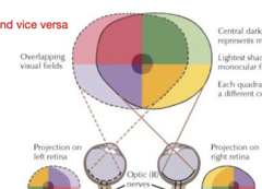
How are images perceived by the retina?
-Already inverted and upside down due to the way the lens focuses light
-This is the whole image, before any crossover
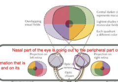
Where does information go from optic nerve?
-through the optic chiasm --> optic tract --> to the LGN (lateral geniculate nucleus) in the thalamus
Lateral Geniculate Nucleus (LGN)
-Located in the thalamus
-Visual relay nucleus
-Receives input from optic tract (from the retinal ganglion cells)
-Goes to the primary visual cortex in the occipital lobe
*Has 6 layers
-3rd order neuron
Ventral Posterolateral Nucleus (VPL)
-Located in thalamus
-Somatosensory relay nucleus for the body
-Receives input from the medial leminscus and spinothalamic tract
-Sends information to the primary somatosensory cortex (postcentral gyrus)
-3rd order neuron
Why are Photoreceptors not considered 1st order neurons?
-They do not send their axons out of the retina, instead they synapse on bipolar cells
-Bipolar cells are the 1st true neurons in the visual pathways (since they connect to the ganglion cells, whose axons leave the retina)
-Photoreceptors do not transmit information through long axons (they do not fire APs)
Optic Radiations
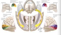
-Axons that carry visual information from the LGN in the thalamus to the primary visual cortex (V1) in the occipital lobe
How is information processed in the primary visual cortex (V1)?
-Input from left visual field is processed in right hemisphere & vice versa
-Superior (upper) visual field is processed in the inferior portion of the visual brain
-Processed upside down
Magnocellular Layers (of LGN)
-Ventral 2 layers
-Information received from Parasol ganglion cells (they synapse here)
-Motion and spatial analysis
Parvocellular Layers (of LGN)
-Information received from Midget Ganglion Cells (synapse on this layer)
-Detailed form and colour
Visual Association Cortex
-From the primary visual cortex (V1), information can go either:
*Parietal/Dorsal stream
OR
*Temporal/Ventral Stream
Parietal/Dorsal Stream
"Where" (things are, how are they moving)
-Analysis of motion and spatial relations
Pathway: Parasol cells --> magnocellular layers --> V1 & V2 --> dorsolateral parieto-occipital cortex (destination)
-Starts in V1
Temporal/Ventral Stream
- "what"(recognition)
-Analysis of form and colour (information from cones)
-Pathway: Midget cells --> parvocellular layers --> V1 & V2 --> Inferior Occipito-temporal cortex (destination)
-Starts in V1
How do neurons encode "WHAT" information from temporal/ventral stream?
-Recognition neurons
-Preferentially respond to different visual stimuli (faces)
-Response is maximal when preferred stimuli (faces) are present
Ex: monkey looked at various pictures, when looking at distorted stimuli, there were fewer neurons firing as opposed to when shown a monkey face
Recognition Neurons
-group (population) of neurons with similar but slightly different tuning preferences works together to perceive complex stimuli
-They are tuned to preferentially respond to specific visual stimuli
-Left IT = objects, words, letters
-Right IT = faces
Lesion in Right Inferior Temporal lobe
-Produces Prosopagnosia
-Inability to recognize faces
-This is the area of the brain which contains recognition neurons which are fine tuned to recognize various parts of a face
Prosopagnosia
-Lesion to the right inferior temporal cortex
-Will recognize eyes, nose, mouth etc but cannot recognize the face as a whole
-Disruption to face-tuned recognition neurons
Lesion in Right Optic Nerve
-Total right eye blindness
Lesion in the Optic Chiasm
-Complete loss of peripheral visual fields from both eyes
Lesion in the Right Optic Tract
-Complete loss of left visual field from both eyes
Lesion in Right Optic Radiations after LGN
-Complete loss of left superior quadrant in both eyes
-Lesion is in temporal lobe of Meyer's Loop = information from superior visual field
Lesion in Right Primary Visual Cortex
-Complete loss of left visual field from both eyes
-The right visual cortex processes all the left visual field information
Four Retinal Projections
-Information from retina projects to 4 important subcortical regions
-Retino-geniculate pathway: (conscious vision)
*LGN
-Extra-geniculate pathways (3): (subconscious)
*Pretectum
*Superior Colliculus
*Hypothalamus
Retino-Geniculate Pathway
-Primary visual pathway for conscious vision
-Carries information from the retina to the primary visual cortex
Extra-Geniculate Pathways
-Retinal projections outside the primary visual cortex, bypass the thalamus
-Subconscious vision
Pretectum
-Controls pupillary light reflex
-Of the extra-geniculate pathway
Superior Colliculus
-Controls orienting eye movements
-Of the extra-geniculate pathway
-Visually guided head and eye movements
Hypothalamus
-Regulates the circadian rhythms
-Of the extra-geniculate pathway
Which muscles are responsible for Volitional Eye Movement?
-Extraocular muscles
Extraocular Eye Muscles
1. Lateral Rectus (lateral eye movement)
2. Medial Rectus (medial eye movement)
3. Superior Rectus (upwards eye movement)
4. Inferior Rectus (downwards eye movement)
5. Superior Oblique (rolling)
6. Inferior Oblique (rolling)
What are eye muscles controlled by?
Cranial nerves (3, 4, 6)
What programs Volitional Movements?
-Primary Motor Cortex (M1)
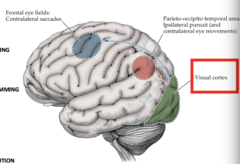
What programs Voluntary Eye Movements?
-Frontal eye fields
-Parieto-occipital-temporal areas
What plans volitional movements?
-Premotor cortex
What dictates where we move our eyes?
-Sensory input drives motor output
-What we see in the outside world
Brainstem Gaze Centres
-These are responsible for the automatic control of our extra-ocular muscles (subconsicously)
-subset of Supra-nuclear centers (above the oculomotor nuclei)
-Direct the movements of the eyes (coordination, adjust for target speed/position, respond to head position)
"Yoke eye movements together"
Where are Supra-Nuclear Centres located?
-Brainstem
-Cerebellum
-Basal ganglia
-cerebral cortex
How are Brainstem Gaze Centers connected Eye Movements?
Via Inter-nuclear pathways to output nuclei which are attached to CNs (3,4,6) --> move extraocular muscles
3 Circuits for Eye Movements with Brainstem Gaze Centers
1. Horizontal Eye movements
2. Vertical Eye movements
3. Vergence eye movements
**all work the same way
What plans eye movement?
-V1 & association areas
-Detects visual targets and analyzes their location and features
-Decides what might be worth looking at
Paramedian Pontine Reticular Formation (PPRF)
Horizontal Brainstem Gaze Center
-Via MLF (inter-nuclear pathway)
-Works on CN 3 & 6 (Lateral & medial rectus)
Ex (look left): CN 6 will pull the LEFT lateral rectus and CN 3 will pull the RIGHT medial rectus = conjugate gaze
Paramedian Pontine Reticular Formation (PPRF) Role
-Commands CN6 to move the ipsilateral eye (lateral rectus)
-via the MLF, it signals the contralateral CN3 to move the other eye (medial rectus)
**without MLF, one eye will move, but other will not
MLF
-Connects ipsilateral CN6 to the contralateral CN3 to coordinate conjugate horizontal eye movements
-communication highway
*allows coordination of both eyes (ensures the other eye also moves)
-Also involved in the vestibular-ocular reflex (head and eye movement)
CN 6 Palsy
-Lesion to CN6 axon
-Depending on which CN6 axon is impaired, there will be trouble shifting that eye to the rightward or leftward gaze
Ex (lesion to right CN6 axon): right eye cannot be pulled to the look towards the right, but left medial rectus will still move towards the right
CN6 Lesion: Right Lateral Gaze Palsy
-Will not be able to conduct rightward gaze
-Right CN6 lesioned (so connected to right lateral rectus and left medial rectus are lost)
-Leftward gaze is normal
Right PPRF Lesion: Right Lateral Gaze Palsy
-Will not be able to conduct rightward gaze
-Right PPRF lesioned (so connections to right lateral rectus and left medial rectus are lost)
-Leftward gaze is normal
Saccadic Eye Movements
-Really rapid eye movement (subconsciously)
-Brings targets of interest into the field of view (on the fovea)
-Once the movement starts, it has to go the whole way, won't stop halfway
-Controlled by either: PPRF (horizontal) OR iMLF (vertical)
2 Types of Saccadic Eye Movements
1. Reflexive (programmed by superior colliculus)
2. Voluntary (programmed by frontal eye fields)
Reflexive Saccadic Eye Movement
-In response to something really loud/startling
Voluntary Saccadic Eye Movement
-I see someone moving in periphery and voluntarily look over there
What do the Superior Colliculus and Frontal Eye Fields Communicate with?
-Brainstem Gaze Centres (PPRF, iMLF) --> horizontal and vertical
What Motor Commands do Superior Colliculus and Frontal Eye Fields send?
-To move eyes in magnitude/direction specified
-Results in eye moving to new position for foveation
Where does the PPRF receive input from in Saccadic Eye Movements?
-Frontal eye fields (voluntary) --> brainstem gaze centre
-Superior Colliculus (reflexive), skips the FEF and go straight here --> brainstem gaze centre
Smooth Pursuit Eye Movements
-Requires moving stimulus, cannot be carried out voluntarily without a stimulus
-Requires processing in visual cortex to analyze what an object is (whether it is of interest)
-Lot of neural processing so cannot follow a fast moving stimulus
-Uses Ipsilateral parieto-occipito-temporal cortex --> Brainstem gaze centre
-Happen via the "WHERE" pathway
Optokinetic Nystagmus (OKN)
-Reflexive eye movement that stabilizes the visual scene when the entire visual field moves (watching passing scenery from a train)
-Eyes slowly follow then quickly snap back
1. Slow phase
2. Fast Phase
Slow Phase of OKN
Smooth pursuit
-Eyes track the moving object/scene to keep it on the fovea
-In the same direction as the moving visual field
Fast Phase of OKN
-Saccade like
-Eyes quickly reset to a new position so the next object can be tracked
-In the opposite direction of the moving visual field
Vestibular Ocular Reflex (VOR)
-Eye movements maintain fixation on target when the head moves so the outside world looks stable
-FOR ANY MOVEMENT OF THE HEAD (up/down, left/right)
-Involves information from the vestibular system and eye muscles
-can follow fast moving stimulus since there does not require conscious input or V1
VOR Head Turn to the Left...What do Eyes do?
-Turn to the right
-CN6 and CN3 activated