Ch 29 + FA Dental Papers
1/3
There's no tags or description
Looks like no tags are added yet.
Name | Mastery | Learn | Test | Matching | Spaced |
|---|
No study sessions yet.
4 Terms
Zetterstrom et al, 2021
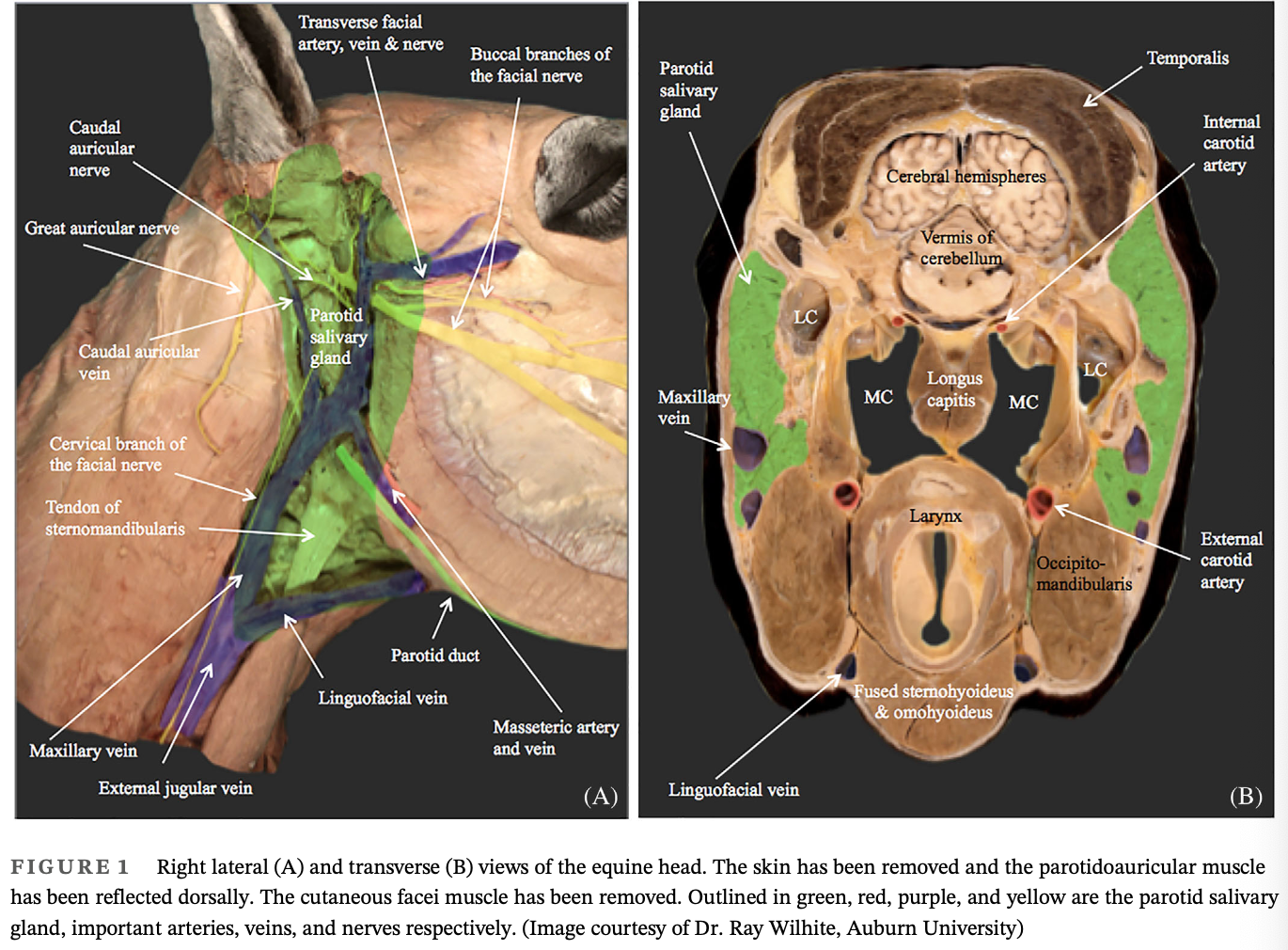
Partial Parotid Sialoadenectomyy
Objectives: To describe the surgical findings, histopathological features, and long-term outcome for a horse with parotid salivary carcinoma
Results:
Swelling below the base of the right ear
Ultrasound examination revealed a mass involving the right parotid salivary gland
Incisional biopsy was consistent with parotid carcinoma
The tumor was marginally excised along with the lateral wall of the guttural pouch which was reconstructed with a porcine small intestinal submucosal (SIS) sheet
Cisplatin beads were implanted in the wound be prior to closure
Postoperative complications included right-sided facial nerve paralysis, difficulty with deglutition of fibrous feeds, and surgical site dehiscence
Wound healing was achieved by second intention
Partial improvement in nerve function was observed within the first 6 months
At 12 months post-parotidectomy no sign of tumor reoccurance or metastatic disease was present and the gelding returned to work
Constant et al, 2019
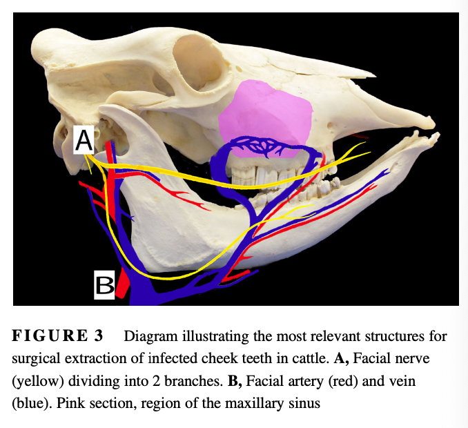
Cheek Teeth Apical Infections in Cattle
Objective: To report the clinical presentation, treatment, and outcome of cattle undergoing surgical extraction of apically infected cheek teeth (CT)
Results:
Main presenting complaints were mandibular swelling and decreased appetite and milk production
7 mandibular and 3 maxillary CT were extracted, 7 molars and 3 premolars that were distributed more frequently on the left dental arcades
Two cattle had no visible external lesions
Radiograph images revealed that lucency surrounded all affected tooth roots
Mandibular teeth were removed by lateral buccotomy with removal of alveolar bone plate or retrograde repulsion and maxillary teeth were removed by repulsion through a maxillary sinus flap
Most common bacterial isolates consisted of anaerobic bacteria (6/11 isolates) and Truperell pyogenes (3/11 isolates)
Most common complicatoins included inability to remove the tooth intact (4 cattle) and surgical site infection (5)
All cattle remained in their herd after treatment
Conclusion: Surgical extraction of CT was achieved in all 9 cattle. The postoperative morbidity was high but without long-term consequences on animal productivity
Locher et al, 2021
Repair of Open Mandibular Pars Incisiva Fractures in Calves
Case Description: 3 neonatal female calves were examined because of mandibular trauma
Clinical Findings: Physical examination indicated that each calf had an open fracture of the mandibular pars incisiva (rostral mandibular fracture) with ventral displacement of the incisors at the affected region. Oral radiographs were obtained for 1 calf and revealed that 5 incisors were fractured at the level of the apical dental buds
Treatment and Outcome: Each calf was anesthetized. The fracture site and surrounding tissues were surgically debrided and flushed with sterile 0.05% chlorhexidine soluation. The laceration in the oral mucosa was closed with absorbable suture in an interrupted horizontal mattress pattern. A Penrose drain was placed during primary closure and removed 4 days later in 1 calf. The fractured incisors were removed during primary debridement in another calf. All calves received perioperative antimicrobials and analgesics. One calf developed mild osteomyelitis of the rostral mandible which resolved with additional surgical debridement and antimicrobial treatment. Two calves developed mild brachygnathia. At time of last follow-up, all 3 calves were eating and growing as expected.
Clinical Relevance: 3 calves with open rostral mandibular fractures were successfully managed by surgical debridement and primary closure of the oral laceration. The procedure was easy to perform, did not require specialized equipment and was less expensive than other repair methods.
Simpson et al, 2019
Frontal Sinusitis in Adult Beef Bulls
Objective: To characterize frontal sinusitis unrelated to standard dehorning procedures in adult beef bulls
Results:
Most common owner complaints were nonspecific signs (separation from the herd, hypo- or anorexia, weight loss, n=10) and suspected horn or sinus infection (7)
Only 8 bulls had nasal discharge and only 7/17 bulls for which the rectal temperature was recorded were febrile
Results of radiography indicated frontal sinusitis in 12/13 bulls with increased opacity of the affected sinus (n=11) noted most commonly
17 bulls were discharged from the hospital alive
One was euthanized for meningitis and suppurative meningoencephalitis extending from the affected frontal sinus was diagnosed on necropsy
Long term follow-up was obtained for 14 bulls all of which recovered fully and 9/13 bucking bulls performed well after treatment
Conclusion: Frontal sinusitis should be considered as a differential diagnosis in beef cattle examined for nonspecific clinical signs and that with appropriate treatment, the prognosis is good for long-term, survival in affected beef cattle