NRS 3016 Chapter 14 Intrapartum Fetal Surveillance
 Call Kai
 Learn
Learn Practice Test
 Spaced Repetition
 Match
Match Flashcards
 Knowt Play
1/49

There's no tags or description
Looks like no tags are added yet.
Name | Mastery | Learn | Test | Matching | Spaced | Call with Kai | Chat |
|---|
No analytics yet
Send a link to your students to track their progress
50 Terms
fetal oxygenation facotrs
sufficient maternal BF and volume to the placenta
normal maternal oxygen saturation
adequate exchange of O2 and CO2
open circulatory path from placenta to fetus through umbilical cord vessels
normal fetal circulatory and OCC
fetal oxygenation alterations: maternal cardiopulmonary
maternal hemorrhage leads to maternal hypotension
supine hypotension (compression of vena cava and aorta)
maternal HTN d/t gestational HTN
lowered oxygen level in mother’s blood (ex: anemia)
fetal oxygenation alterations: uterine activity
placental disruptions (hypertonic contractions, abuptio placentae)
fetal oxygenation alterations: fetal alterations that interrupts BF to umbilical cord
Nuchal cord → around fetal neck or entangled in fetal body parts
knot in umbilical cord
oligohydraminos → lower than normal amounts of amniotic fluid volume
inadequate Wharton’s jelly → cushions umbilical cord (decrease wharton jelly = cut off supply compression of cord)
EFM instrumentation
intermittent auscultation and transducers
uterine activity via tocodynamometer (toco)
external fetal HR obtained via doppler or US
bedside fetal monitoring and transducers: benefits
non-invasive
used during antepartum and intrapartum period (not really for labor)
used with telemetry
no known risks to woman or fetus
continuous recording of FHR and UA (uterine activity)
bedside fetal monitoring and transducers: limitation
limits maternal mobility
frequent repositioning for readable/accurate tracing
may double-count a slow FHR <60bpm or half-count a FHR >180bpm
may record maternal HR if placed over maternal arterial vessel (aorta)
unable to assess intensity of UA
difficult to monitor on obese women and preterm or multifetal gestations
external monitors for FHR/contractions
ultrasound transducer → FHR
tocotransducer (toco) → contraction pattern
internal monitors for FHR/contractions
internal scalp lead (ISL) → FHR by screwing into baby’s head
internal uterine pressure catheter (IUPC): contraction pattern
sterile, flexible catheter that sits between wall of uterus and baby’s head, pressure measured during contraction when everything is squeezes (mmHg)
fetal scalp electrodes (FSE)
internal fetal monitoring: requires cervical dilation and ruptured membranes to directly apply electrodes to fetal presenting part (head or buttt)
internal fetal monitoring benefits
FSR: accurate fetal cardia arrhythmias & FHRs between 30-240bpm, maternal position changes does not affect reading
IUPC: only true accurate measure of UA, allows to use of amnioinfusion (give fluid back thru IUPC if cord is compressed too much)
internal fetal monitoring limitations: FSE & IUCP
FSE: vaginal lacerations, presenting part must be accessible, may record maternal HR in presence of fetal demise, fetal hair can cause inadequate ECG conduction
IUCP: uterine perforation, placental abruption, maternal position change can cause hydrostatic pressure, variations in IUPC types
Both: requires ruptured membranes, cervix must be dilated sufficiently, increased infection risk
fetal monitoring tracing
each tracing has an x & y-axis with an upper and lower graph depicting FHR and contraction pattern
uterine activity: frequency
onset of one contraction to onset of the next contraction (minutes)
uterine activity: duration
onset of one contraction to the end of another contraction (seconds)
uterine activity: intensity-strength
palpation-mild, moderate, or strong, quantitative numbers
in the form of mmHg used only with IUPC
strength is ONLY measured internally or subjectively
uterine activity: resting tone
soft or relaxed when not contracting
contraction variability means
well oxygenated
FHR interpretations
baseline FHR
normal = 110-160bpm
bradycardia <110bpm
tachycardia >160bpm
ideally: baseline FHR is over a 10min period of time
FHR = balance between fetus parasympathetic and sympathetic NS
FHR variability
fluctuations in the baseline FHR that irregular in amplitude and frequency
it evaluates fetal ANS, indicates fetal oxygenation, & most reliable for fetal well-being
variability classifications: absent
amplitude visually undetectable
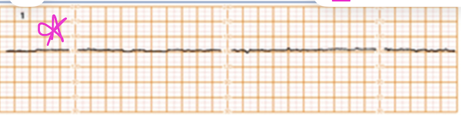
variability classifications: minimal
amplitude range visually detectable but 5bpm or less (sleeping, relaxing drug)
if occurs for longer than 30mins, try to wake baby
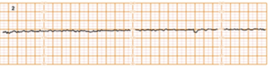
variability classifications: moderate
amplitude range 6-25bpm (normal)
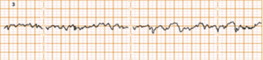
variability classifications: marked
amplitude range >25bpm (big M)

variability classifications: sinusoidal pattern
smooth wave-like (S) appearance, rare but indicates severe fetal compromise

potential variability changes
fetal: sleep cycle (20-30mins), anemia, tachycardia, sepsis, metabolic acidosis from ongoing hypoxic injury
maternal: drug ingestion (narcotics, CNS depressants, Mg sulfate, barbiturates, tranquilizers), general anesthesia, infection
FHR interpretation: periodic
associated with uterine contractions
FHR interpretation: episodic
not associated with uterine contractions
accelerations are what FHR pattern
periodic or episodic (w/ or w/o contraction)
early head compression is what deceleration FHR pattern
periodic (w/ contraction)
late uteroplacental insufficiency is what deceleration FHR pattern
periodic (w/ contraction)
variable cord compression is what deceleration FHR pattern
periodic or episodic (w/ or w/o contraction)
prolonged deceleration is what deceleration FHR pattern
periodic or episodic (w/ or w/o contraction)
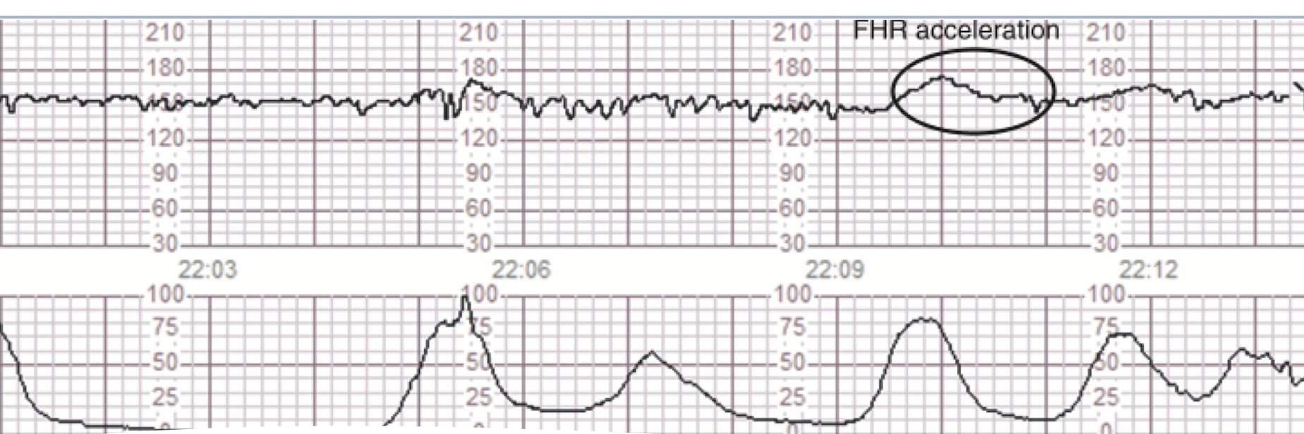
FHR accelerations
normally occur in response to fetal movement, vaginal exams, or contractions
definition: increase baseline of HR (15 beats for 15s)
indicates fetal well-being (sympathetic fetal response) & reassuring sign
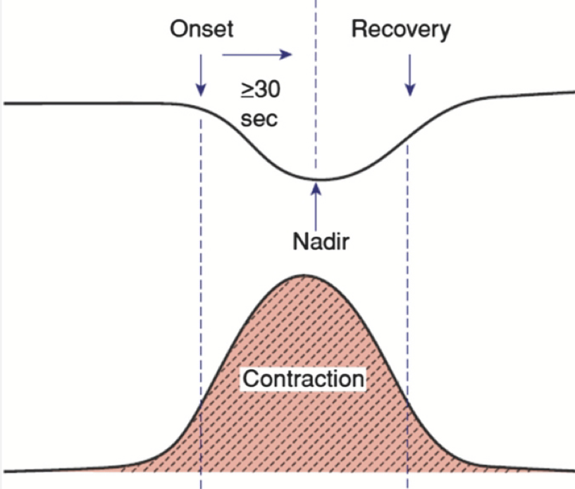
early decelerations
fetal head compression: symmetric in shape, gradual decrease, return to FHR baseline that mirrors the uterine contractions, vagal response during head compression, benign & not associated with interruption of fetal oxygenation
RN: monitor progress
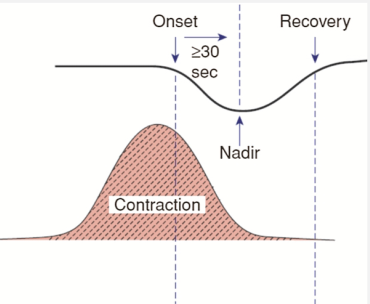
late decelerations
BAD, placental issues
visually apparent, usually symmetric in shape
gradual decrease and return of FHR baseline
drop in FHR begin after the peak of contraction
FHR returns to baseline after the contraction ends
similar to early deceleration but are shifted to the right
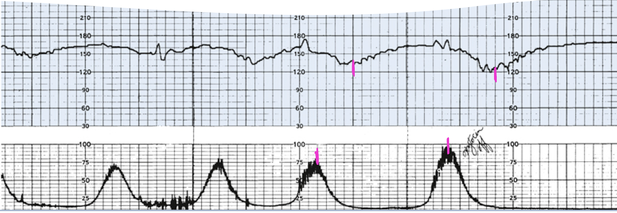
late deceleration
appears before loss of variability therefore indicates transient fetal hypoxia state (maternal hypotension)
one ‘late deceleration’ is a priority intervention by changing pt position
concerning: recurrent (fecurrent) when late deceleration occurs with 50% or more uterine contractions over 20 minutes
associated with tachycardia and loss of variability therefore severe fetal hypoxia
IMMEDIATE delivery
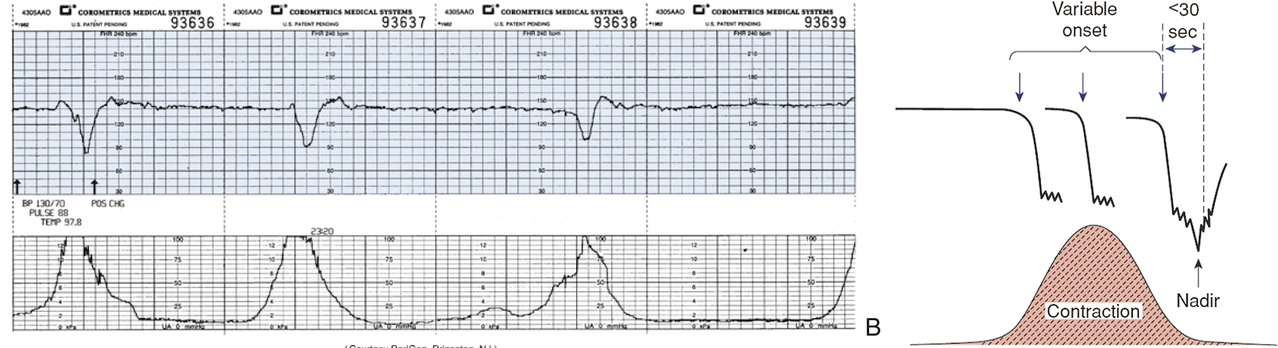
variable deceleration
caused by interruption of oxygenation at umbilical cord d/t compression
visually apparent, abrupt decreases from onset of a deceleration
most frequent type of deceleration during first-second stage of labor
RN: change maternal position
prolonged deceleration
decrease in FHR that lasts a minimum of 2-10min
reflects interruption of oxygen transfer from environment to fetus at one or more points
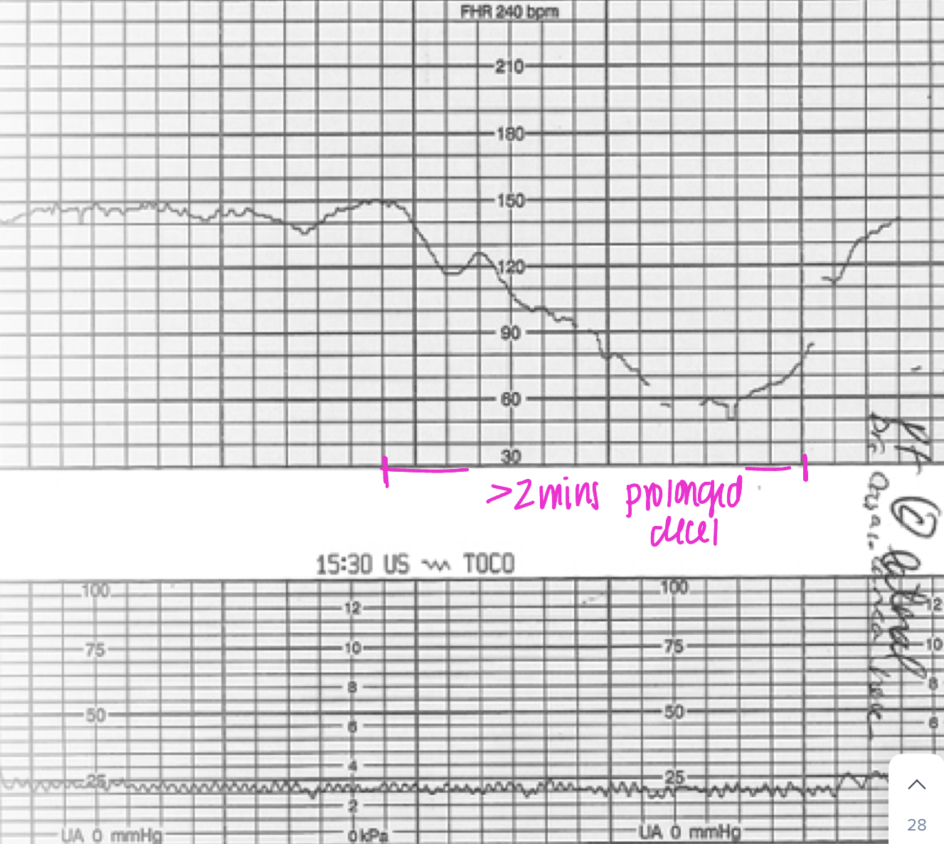
RN: give fluids, more than 10mins = get baby out
most common 3 deceleration patterns mneumonic
VEAL CHOP
variable → cord → reposition
early → head → okay
acceleration → ox → okay
late → placenta → not good
3 tier FHR interpretation
category I: normal fetal acid-base status
category II: indeterminate fetal acid-base uncertain (some interventions)
category III: abnormal fetal acid status (bad)
category I: normal FHR tracing
baseline FHR: 110-160bpm
variability moderate
accelerations/early decelerations absent or present
late & variable decelerations absent
category II: indeterminate FHR tracing
bradycardia w/o absent variability
tachycardia
minimal variability
absence of induced acceleration after fetal stimulation
prolonged decelerations
recurrent late decelerations with moderate variability
category III abnormal FHR tracings
absent variability
recurrent late or variable decelerations
bradycardia
sinusoidal pattern
cannot continue to labor → get to OR
how to improve placental BF and fetal oxygen supply mneumonic
P→ change position
O → administer O2 with non-rebreather
I → increase rate of IV fluids
S → perform sterile vaginal exam-prolapsed cord
O → stop oxytocin (pitocin)
N → notify MD/CNM
*always document FHR category and response to intervention)
approach to FHR management mneumonic
A → assess O2 pathway and identify FHR etiology, changes of maternal/fetal
B → begin conservative measures
C → clear obstacles to delivery
D → determine delivery plan
common FHR corrective measures
maternal repositioning
IV boluses
administering oxygen
reducing UA (stop pitocin)
correct maternal hypotension
modify second stage pushing efforts (see decel with mom pushing)
assessment frequency & documentation
ongoing maternal-fetal assessment of FHR & UA
health information technology (HIT)
improve obstetric patient centered care and safety
ongoing fetal monitoring education and competency
the nurse notes a pattern of late decelerations on the fetal monitor. the most appropriate action is to:
continue observation of this reassuring pattern
notify the physician or midwife
give the mom oxygen by face mask and place pt in side lying position (left)
place the woman in a trendelenburg position
give the mom oxygen by face mask and place pt in side lying position (left)