alimentary overview
 Call Kai
 Learn
Learn Practice Test
 Spaced Repetition
 Match
Match Flashcards
 Knowt Play
1/562

There's no tags or description
Looks like no tags are added yet.
Name | Mastery | Learn | Test | Matching | Spaced | Call with Kai | Chat |
|---|
No analytics yet
Send a link to your students to track their progress
563 Terms
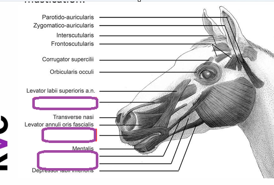
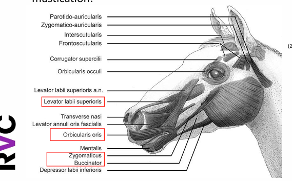
levator labii superioris
to elevate the upper lip and draw it to one side
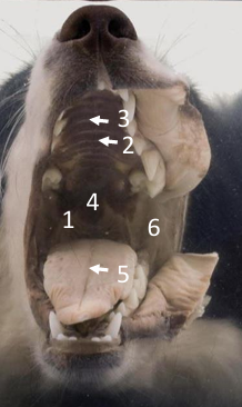
buccal cavity
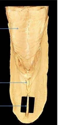
palatine raphe
transverse ridges
soft palate
median sulcus
vestibule
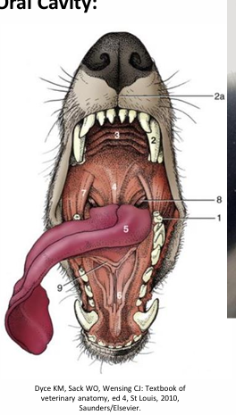
vestibule
canine tooth
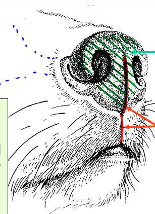
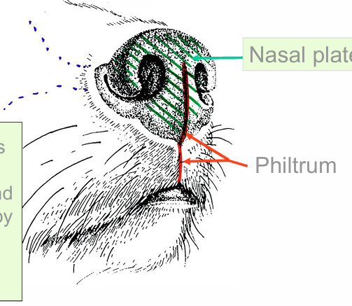
2a. philtrum
hard palate
soft palate
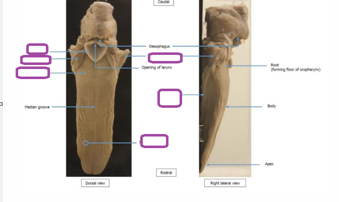
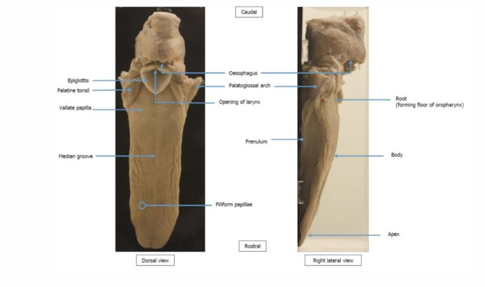
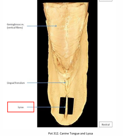
tongue
sublingual caruncle
palatoglossal arch
palatine tonsil
frenulum
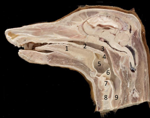
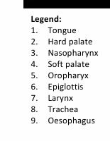
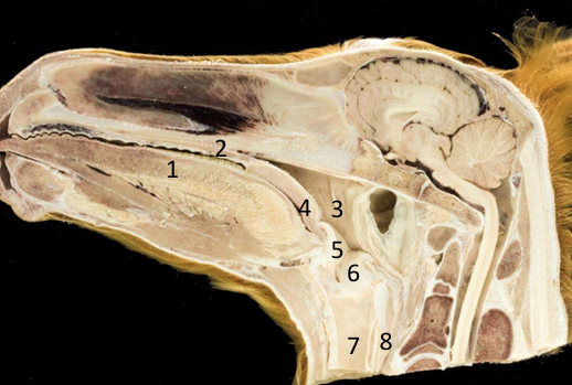
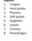
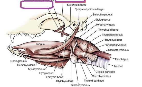
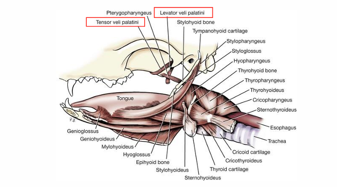
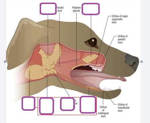
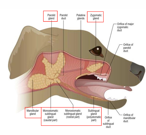
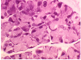
serous gland
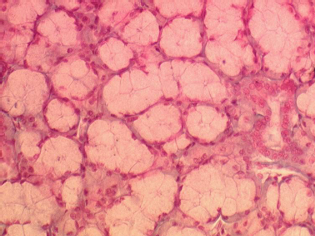
mucous gland
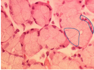
mixed gland
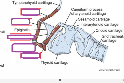
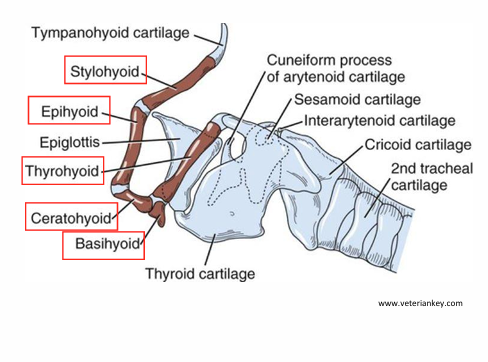
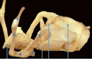
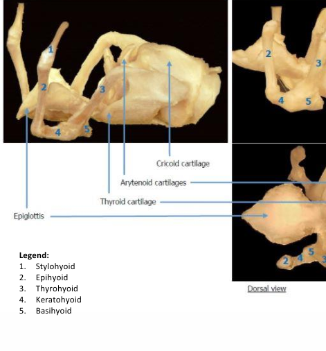
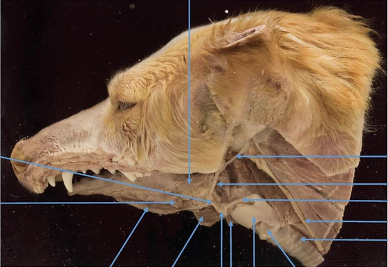
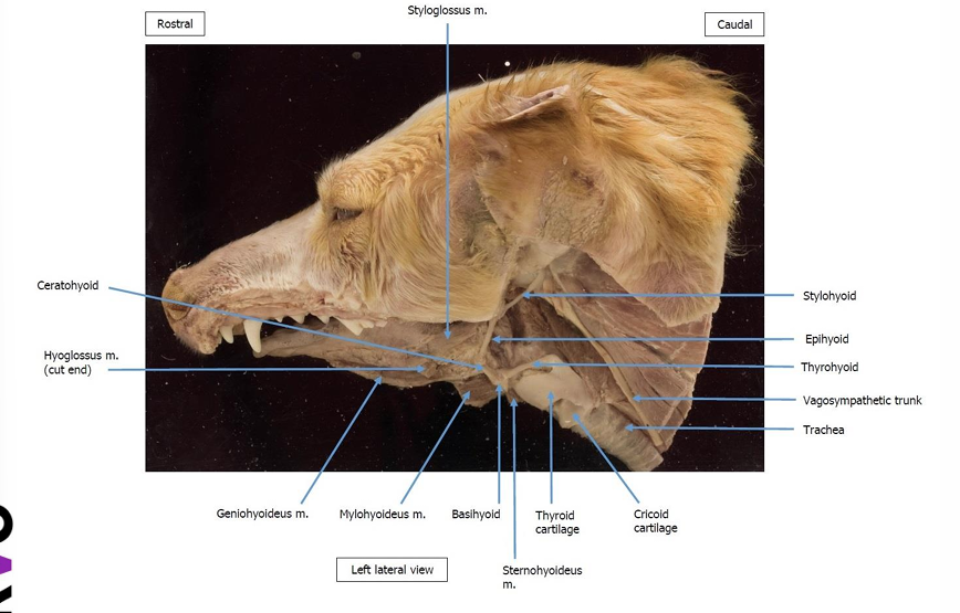
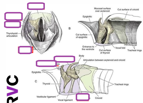
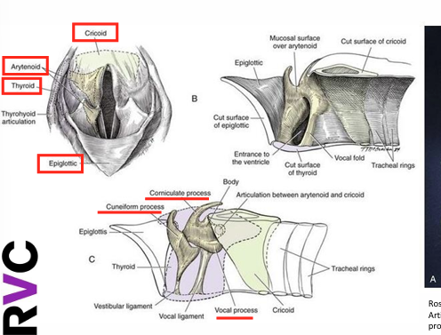
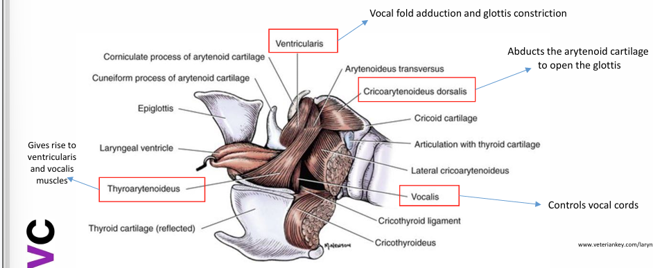
thyroarytenoideus- what does it give rise to
gives rise to ventricularis and vocalis muscles
vocalis
controls vocal cords
cricoarytenoideus dorsalis
abducts the arytendoidal cartilage to open the glottis
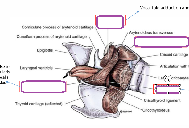
ventricularis
vocal fold adduction and glottis constriction
innervation to the larynx
cranial and caudal laryngeal nerves which originate from the vagus nerve
in equine gastric ulcer syndrome why are particular regions of the stomach affected
non glandular area lacks the protective mucus layer
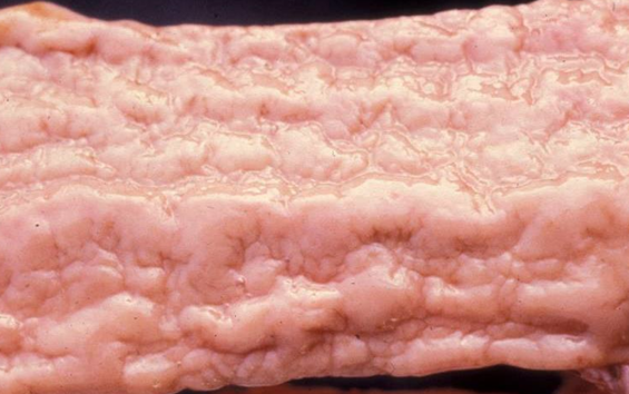
what does this show
thick corrugated mucosa- cobblestone gut
johnes disease
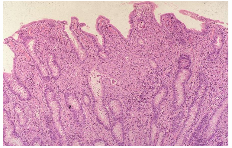
what does this show
shortening and widening of villi which are infiltrated by large numbers ofmacrophages
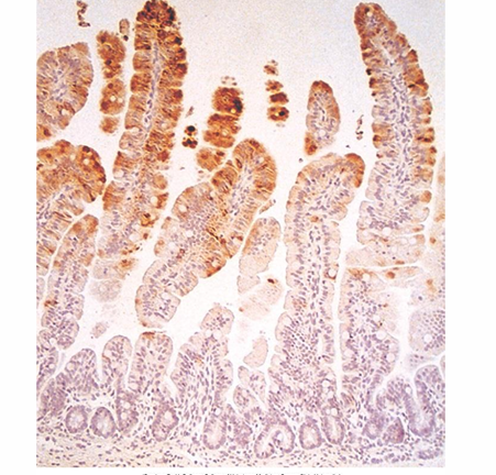
viral antigen
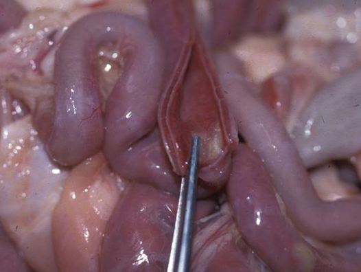
what pathology can be seen here
red brown discolouration of mucosa
parvo
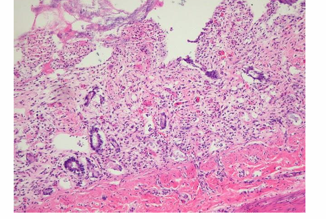
destruction of intestinal crypts and collapse of mucus
parvo
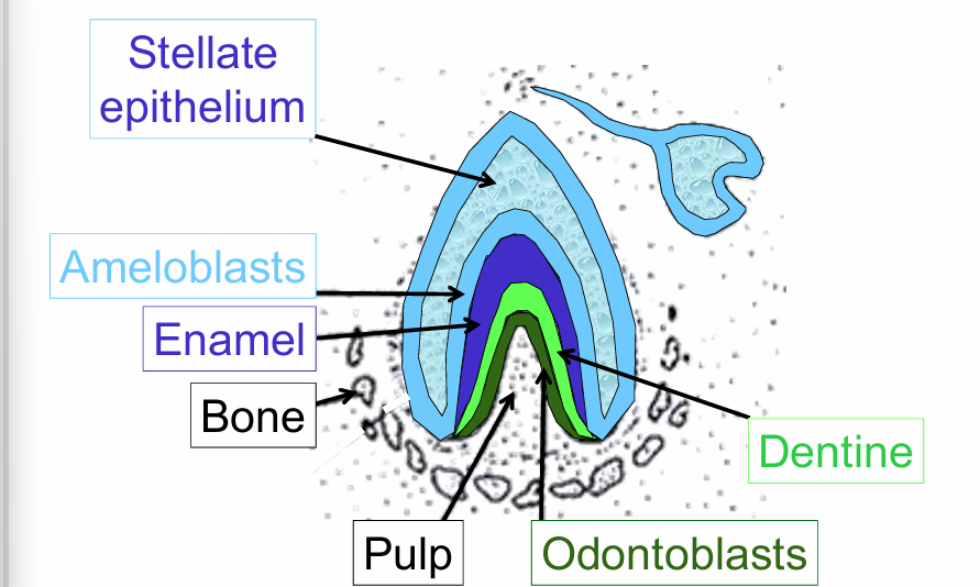
what is the stellate epithelium
star shaped epithelial cells in the enamel organ during development
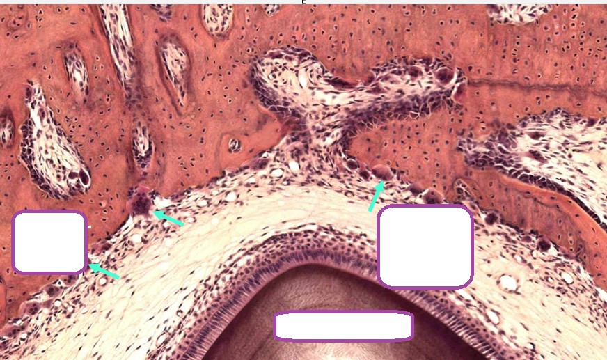
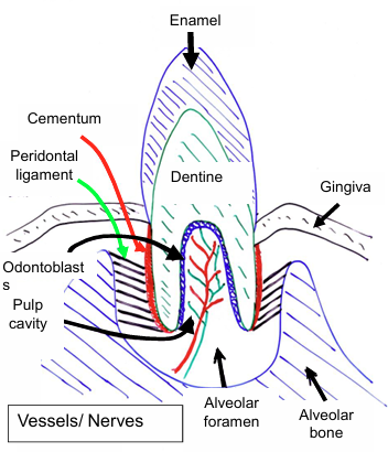
nerve and blood supply of the tooth
Nerve & blood supply: superior and inferior alveolar (arteries, nerves & veins) In dental (pulp) cavity. At least one branch in each major elevation of the crow
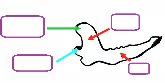
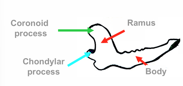
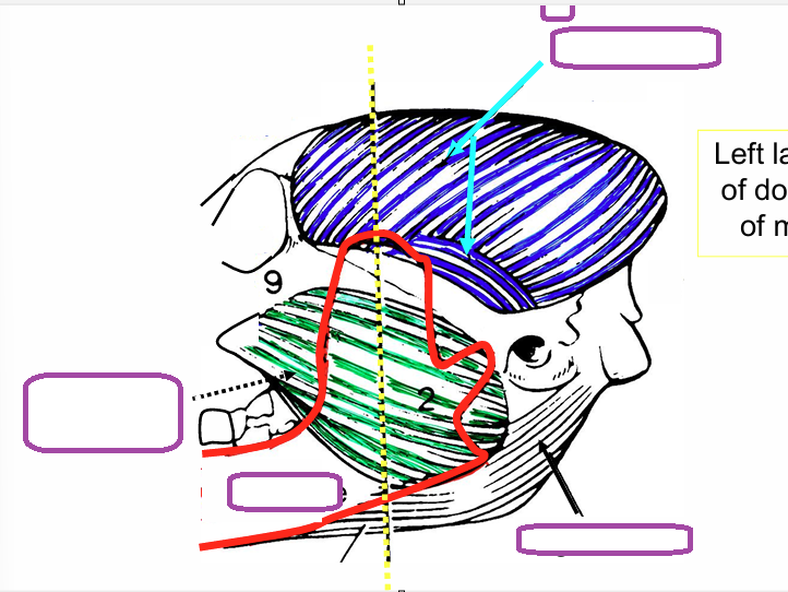
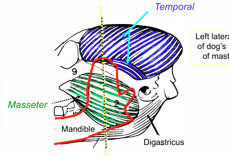
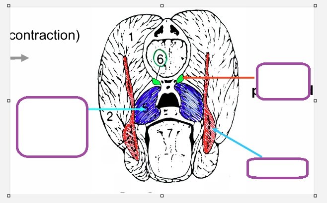
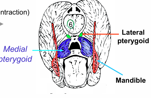
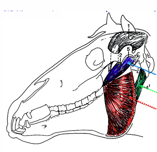
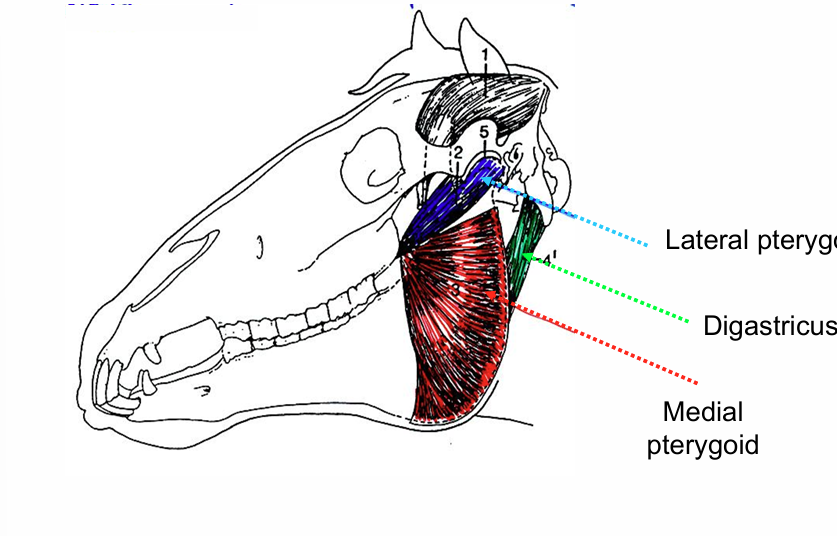
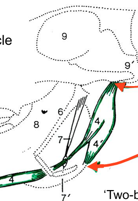
digastrcus
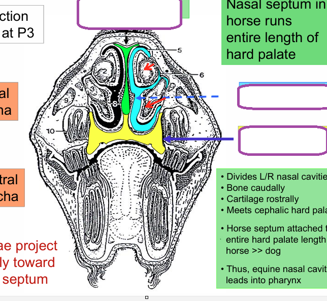
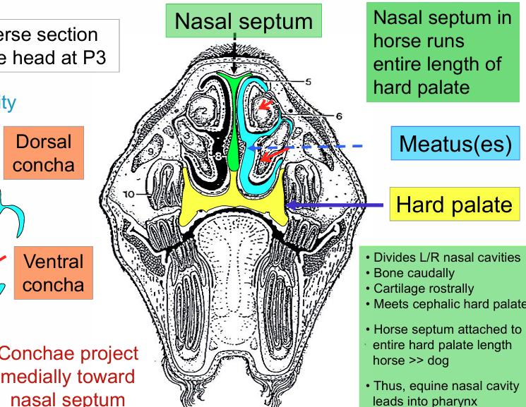
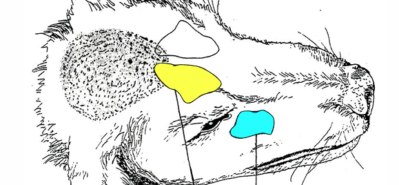
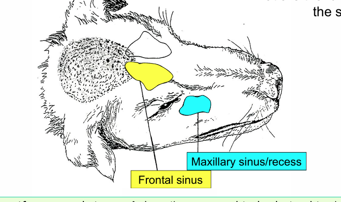
wat type of epithelium covers the hard palate
keratinised stratified squamous
what type of epithelium coves the oral surface of the soft palate
stratified squamous
what glands are present in the soft palate
palatine
what type of epithelium covers the aboral surface of the soft palate
ciliated pseudostratified columnar
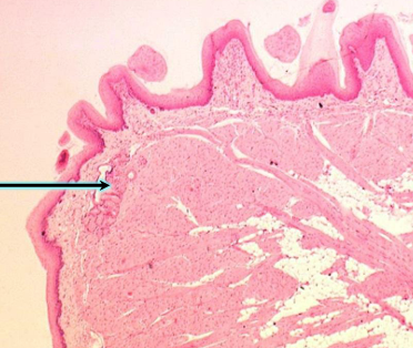
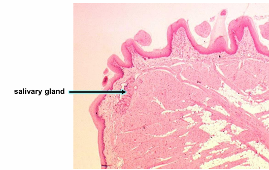
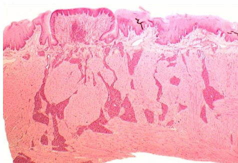
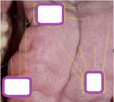
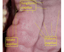
what kind of papillae
circumvallate
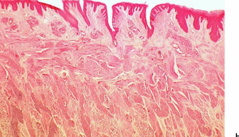
what kind of papillae
foliate
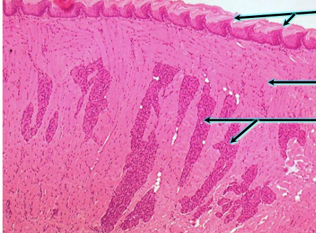
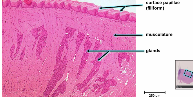
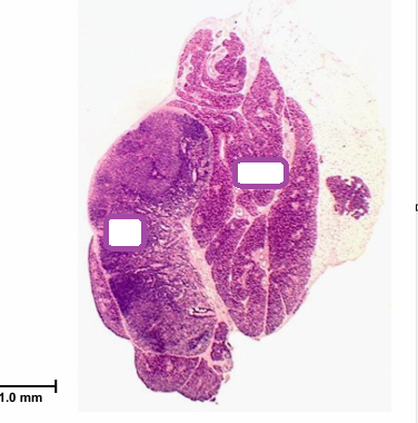
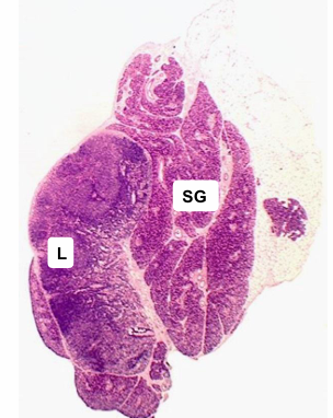
what epithelium lines the interlbular ducts
cuboidl to columnar epithelium
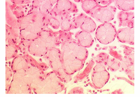
what gland is this and why
foamy appearance of the mucous cells is evident and the darker staining serous cells can be seen around the outside of the acini
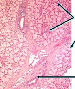
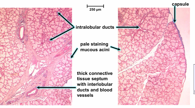
mucous
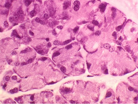
serous
cells are typically cuboidal, pyramidal or crescent shaped
nuclei are psherical and cytoplasm stains well
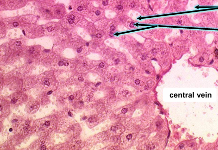
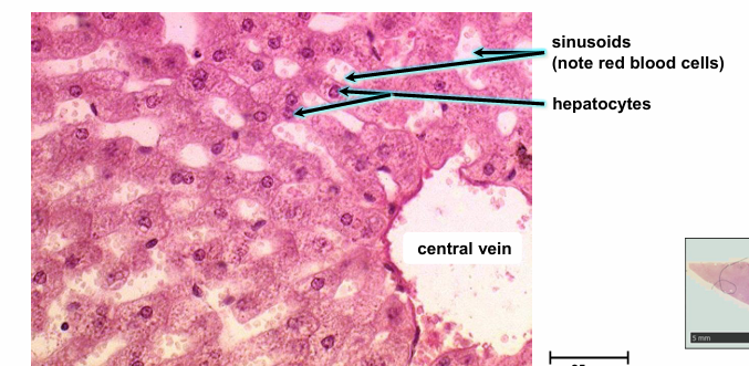
what are reticular fibres composed of
type iii collagen plus proteoglycans and glyctoproteins
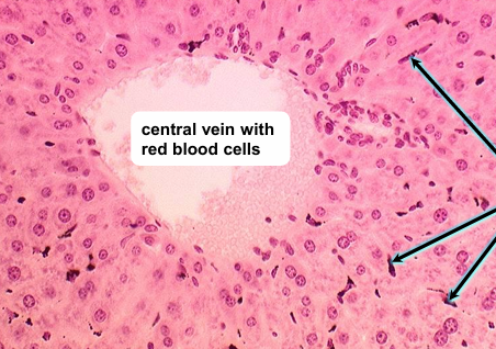
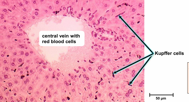
is there a lamina muscularis in the gallbladder
no
development of the gut tube
endoderm folds and elevates. splachnic mesoderm comes from the LPM
endoderm fold around ventrally. endoderm fuses ventrally
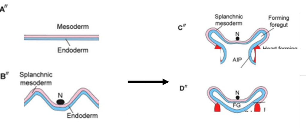
what is the primitive gut tube divided into
foregut
mid gut
hind gut
which membrane is in the oral and anal region
oral- buccopharyngeal
anal- cloacal
layers of the gut- what do endoderm and splanphic mesoderm form
endoderm- epithelial cell adjacent to lumen
splanchnic mesoderm- connective tissue, smooth muscle and mesothelial layers (mesentery and peritoneum)
failure to perforate membranes
persistent buccopharyngeal membrane in human- feeding is impossible, can perforate to treat
persistent cloacal membrane in cow, must be perforated
foregut
forms oesophagus, stomach, liver, gallbladder, bile ducts, pancreas, proximal duodenum
must rotate and twist to elaborate into these structures
rotation of the foregut
what does the greater and lesser omentum contain
what can developmental defects of the omentum causse
greater omentum contains gastrophrenic, gastrocolic, gastrosplenic ligaments
lesser omentum contains hepatophrenic and hepatooesophageal ligaments
developmental defects of the omentum can cause internal hernia
development of the spleen
what type of cells does it form from and what is it also known as
what is it in foetal stages
forms from mesodermal cells in the dorsal mesentery- also called mesogastrium
is a major haemopoetic tissue in foetal stages
what does the midgut consist of
distal half of duodenum
jejunum
ileum
cecum
ascending colon
proximal half of transverse colon
the equine caecum
hindgut fermenter
continues to grow in embryo
in adults roughly 1m in length with a 30L capacity
species differences in ascending colon
horse
ruminant and pig
horse- ascending colon continues to grow forming loop connected by remnants of dorsal mesentery
ruminant and pig- ascending colon continues to grow forming spiral
rotation of the stomach
what does it begin as
what attaches it to the body walls
what lies on the ventral and dorsal surfaces
what happens first and what does it do the the other structures
at the end what is the ventral and dorsal mesentery attachment
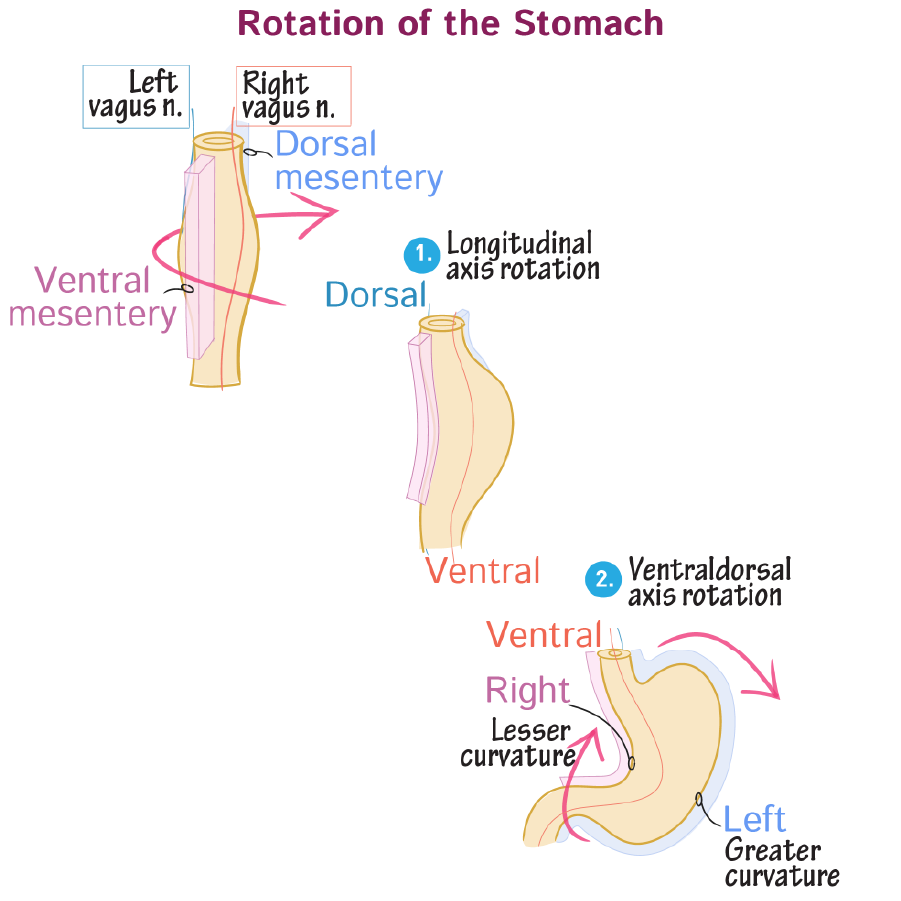
begins as a spindle shaped tube
ventral and dorsal mesenteries attach tube to body walls
branches of the left and right vagus nerves lie on ventral and dorsal surfaces
90 degree clockwise rotation along the longitudinal axis then occurs
this alters the course of the vagus nerve branches: right now innervates the ventral surface and left lies on the dorsal aspect
as the stomach rotates along the ventral dorsal axis, the caudal end is displaced towards the right and cephalic end to the left
ventral and dorsal mesenteries also displaced to right and left respectively
lesser curvature is the ventral mesentery attachment
greater curvature is the dorsal mesentery attachment
what does the ventral and dorsal mesentery give rise to
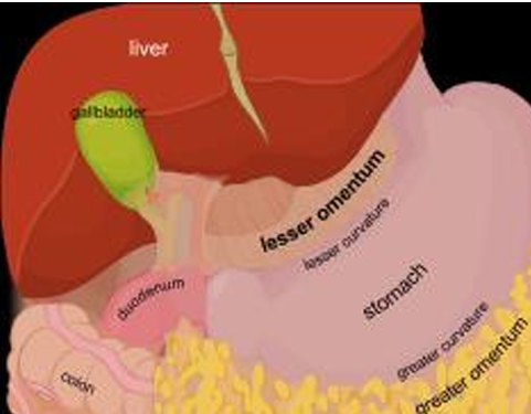
ventral to falciform ligament which secures the liver ventrally
also the lesser omentum which connects the liver and stomach and proximal duodenum
dorsal mesentery gives rise to the greater omentum which is an apron like fold of mesentery that attaches to the greater curvature of the stomach and drapes over the small intestine
final arrangement of the biliary and pancreatic ducts step one
what arise as outgrowths of the foregut
what extend ventrally and dorsally
what do liver buds outgrow into
what happens to the proximal duodenum
abdominal accessory digestive organs arise as outgrowths of foregut prior to stomach and duodenal rotation
dorsal pancreatic duct extends dorsally
ventral pancreatic duct, gallbladder and liver buds extend ventrally
liver buds develoop as an outgrowth of the foregut into the septum transversum
proximal duodenum rotates clockwise
part 2
what happens to the dorsal and ventral pancreatic buds and ducts
then where does it go and exttnd
what does the ventral pancreatic bud form
where does the main and accessory pancreatic duct drain into and how
what does the pancreatic duct join to and why
how does the common bile duct form
what direction does the common bile duct wrap around the duodenum
what does it join with
dorsal and ventral pancreatic buds and their ducts fuse
the now formed complete pancreas nestles in the c curve of the duodenum and extends toward the left side of the body
the uncinate process is the portion derived from the ventral pancreatic bud
main pancreatic duct empties into the duodenum via major papilla
accessory drains via minor
the bile and pancreatic ducts join to drain bile and pancreatic juices at the major papilla
liver drains bile into hepatic duct
hepatic and cystic ducts merge to form the common bile duct
due to the rotation of foregut and displacement of papillae the common bile duct wraps posteriorly around the duodenum
it joins with the main pancreatic duct to drain bile at the major papilla to the duodenum
what are the four steps in midgut development
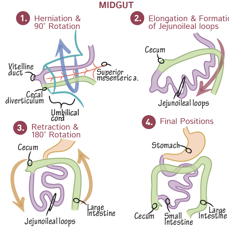
herniation and counterclockwise rotation: counterclockwise rotation about the axis of the superior mesenteric artery. midgut comprises the primary intestinal loop which elongates so rapidly that it temporarily outgrows the abdominal cavity, herniating into the umbilical cord
elongation: the caecum is now on the right hand side, as it has undergone a 270 degree rotation now (in pigs this is 450) . the rotated primary loop elongates rapidly creating the jejunoileal loops
retraction and counterclockwise rotation: loops retract from the umbilical cord into the abdominal cavity, and the intestines rotate 180 degrees counterclockwise as they continue to elongate. caecum now in upper right.
final positions: large intestine frames the small intestine. caecum now in the lower right
3 steps of hindgut development
what closes off the cloaca
what forms between the allantois and the hindgut
what grows towards the cloacal membrane and what does it separate
what happens to the cloacal membrane
what separates the urogenital and anal membranes
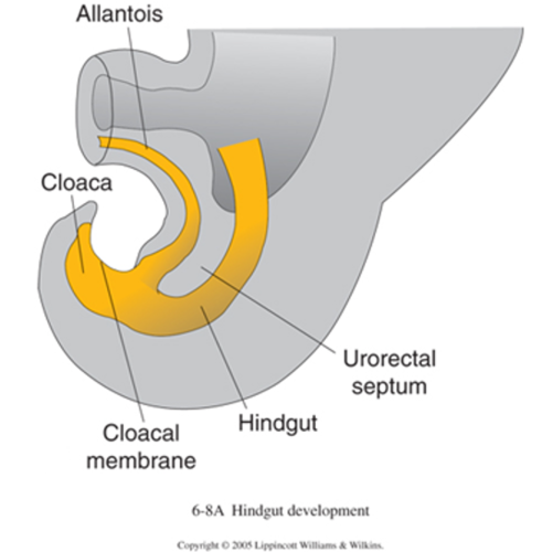
cloaca: initially the cloaca is the common end of the hindgut and urogenital tract. the cloacal membrane (endoderm and ectoderm) close off the cloaca. the urorectal septum forms between the allantois and hindgut
urorectal septum: urorectal septum grows towards the cloacal membrane. it now separates the primitive urogenital sinus which appears as an anterior swelling. cloacal membrane ruptures before the urorectal septum reaches
perineal body: represents the tup of the urorectal septum, separates the urogenital and anal membranes. now completely divides the urinary and digestive tracts so that the urinary bladder lies anteriorly and anorectal canal remains posteirorly
tooth development from embryonic layers
where does tooth development begin
where are signals exchanged between and what does this initiate
what happens and what structure is formed. what does this newly formed structure separate into
what thickens
what happens to the mesenchyme
what forms
…..
tooth development begins in the oral epithelium (ectodermal in origin.) signals exchanged between the oral epithelium and NCC derived mesenchyme underneath initiate odontogenesis
localised thickening of oral epithelium occurs forming the labiogingival band. this separates into the labiogingival lamina and the dental lamina.
thickening of dental laminae on medial aspect of the labiogingival band
mesenchyme underneath each laminae condenses
dental lamina invaginates forming the dental bud
dental bud expands and branches to form an enamel organ to surround a NCC derived dental papilla
this dental papilla enamel organ complex will form the deciduous tooth
a small mass of cells bud off from the dental lamina of the deciduous enamel organ, this cell cluster is the primordium of the permanent tooth.
what do the inner enamel epithelium differentiate into, how are odontoblasts derived, what does dentine and cementoblasts do?
budding off of permanent tooth primordium continues
inner enamel epithelium (derived from oral epithelium) differentiates into ameloblasts.
cells in the dental papilla which are neural crest cell derived mesenchyme differentiate into odontoblasts.
as odondoblasts lay down dentine, central part of the dental papilla remains as the pulp and the dentine surrounds the pulp and extends downwards to form tooth rot.
later epithelial cells near distal part of tooth known as cementoblasts secrete cementum aorund tooth rot
ameloblasts
what do they secrete
how do they maintain contact with new unmineralised enamel
what characteristic do they form
what do they produce
what happens after they erupt
secrete enamel which forms the outer covering of the tooth crown
maintain contact with new unmineralised enamel via cellular projections called thomes fibres
form crown characteristc of each tooth type
produce matrix which is not remodelled
are lost upon tooth eruption
odontoblasts- what do they do and what type of cell are they
secrete dentine which forms before enamel and induces enamel formation
columnar
role of mesenchyme, how are tooth tissues laid down
mesenchymal cells can differentiate but have stable morphogenetic properties
experimentally molar tooth mesenchyme and non oral ectoderm was combined resulting in a tooth shaped structure, showing that mesenchyme carries instructures for tooth morphology
neural crest derived mesenchyme determines tooth type
when molar mesenchyme is paired with incisor epithelium in culture the tooth that forms is a molar
tooth tissues are laid down from the crown to the root
tooth does not reach its final length while still embedded in jaw, achieved after eruption begins
tooth eruption- temporary
eruption after crown is fully formed but before complete formation of the root
eruption provides space for completion of root
epithelial covering is continuous with gums upon eruption
wear removed epithelium
permanent tooth eruption
migrates into socket of temporary tooth
increasing pressure from tooth
resorption of temporary tooth root
loosening
shedding
permanent tooth replaces
what aids tooth eruption?
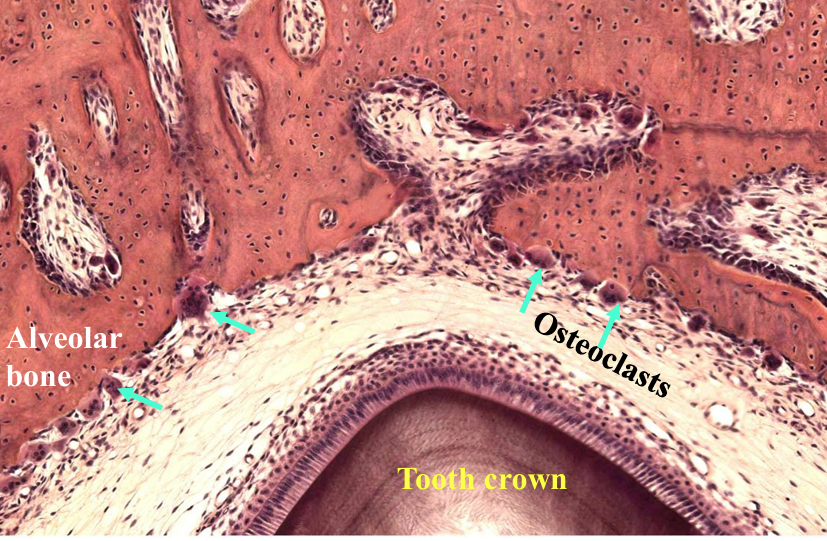
osteoclasts and odontoclasts resorb alveolar bone and root of the deciduous tooth to allow space for permanent tooth to move upward
bone remodeling occurs to allow the tooth to move upward
at the bottom of the developing root there is tissue fluid and blood supply which create hydrostatic pressure pushing the tooth upward as the root grows
the periodontal ligament remodel collagen fibres and generate contractile forces to help pull the tooth upwards
Poll
dentine
nature of production and rate
contents
produced continually
rate increases during repair
innervated
mineralised matrix- collagen type i, dentine, specific protein
odontoblasts recede from newly formed surfaces
like bone but acellular
peridontal ligament
where does it suspend tooth from
what do the PDL fibres bridge
shape
what is it lined by
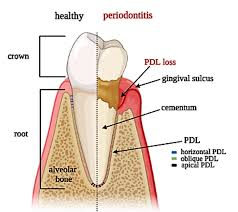
suspends tooth in its alveolar socket
fibres bridge alveolar bone and cementum
shaped to accomodate tooth root-may be branched
lined by compact bone
cementum
what does it surround
properties
what collagen
is it remodelled
surrounds dentine of root
mostly acellular
not readily resorbed
type i collagen
not remodelled
can alveolar bone be remodelled
yes
what is the furcation angle
point where roots diverge in teeth with two or more roots
where does the crown meet the root
cemento enamel junction
what does the enamel consist of
hexagonal prisms or rods of hydroxyapatite crystals held together by cementing organic matrix
pulp
connective tissue, nerves, lymph, blood vessels, collagen and undifferentiated reserve mesenchymal cells
what causes dentine to be sensitie to pain
Odontoblasts line the pulp cavity and branch into the dentine tubules. These branches, together with the fine nerve endings, cause the dentine to be sensitive to temperature and pain.
The odontoblasts lay down dentine and reduce the pulp cavity in size as the animal ages.