Other Shoulder Conditions
 Call Kai
 Learn
Learn Practice Test
 Spaced Repetition
 Match
Match Flashcards
 Knowt Play
1/51

There's no tags or description
Looks like no tags are added yet.
Name | Mastery | Learn | Test | Matching | Spaced | Call with Kai | Chat |
|---|
No analytics yet
Send a link to your students to track their progress
52 Terms
Osteoarthritis of shoulder occurs less commonly than in WBing jts. Primary OA begins on
glenoid surface
proceeds to humeral head over many years
Secondary OA caused by:
instability
previous surgery
trauma
Patients with shoulder Osteoarthritis will have ↓ ROM due to shortening of jt capsule, as well as
atrophy of muscles 2ndary to disuse
Pain/discomfort (sore at 1st, better w/ motion. Worse w/ too much OR little motion)
Crepitus (joint crunch)
labrum degradation
What is the conservative tx for Rheumatoid Arthritis? (progressive & destructive joint disease)
Exercises to improve ROM & strength (back off heavy loading)
Activity modification- prevent shoulder impingement
NSAID’s
What is the surgical tx for Rheumatoid Arthritis (Progressive & destructive joint disease)?
TSA
Arthrodesis: Bony fusion of glenoid fossa, humerus & acromion
rheumatoid arthritis population
40-50s, worse at 60s
primarily females > males
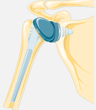
Indications for Total Shoulder Arthroplasty
RA: 24-40%
Complex Fractures: 30%
OA: 20%
Tumors, avascular necrosis: 10%
Cemented TSA is more stable immediately s/p surgery than non-cemented → faster rehab progress, cemented TSA is typically indicated for pts w/
considerable bone loss
Does not allow for biological growth (long term), not good for younger candidates
Cement-less TSA fixation uses metal backed w/ a surface tx to facilitate biological ingrowth. This surgical technique is used for pts w/ adequate bone. Loosening may occur before ingrowth takes place.
More conservative rehab needed, slower progress
Subscap is taken down & repaired for visualization
Subscap precautions for 6 weeks:
avoidance of (passive) ER stretching ~30°
avoid resisted (active) IR
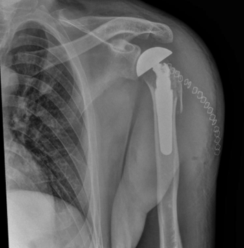
Indications for Hemiarthroplasty
Proximal humerus fracture: Intact glenoid
Mostly intact/functional RC is needed
Poor glenoid bone stock
Osteonecrosis
Hemiarthroplasty has better outcomes than TSA with
OA & RA
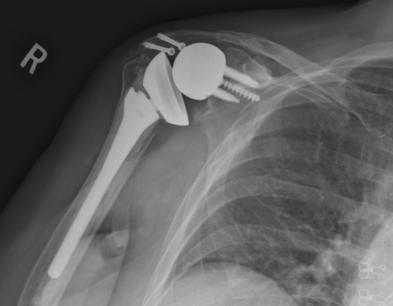
Indications for Reverse Total Shoulder, (advantages are you don’t need RC)
Large irreparable rotator cuff
Poorer Overhead motions compared to TSA
Overall good outcomes
s/p TSA involves immobilization for 1–2 wks in a sling. Early Rehab Goals s/p TSA include
↑ mobility/ROM 1-6 weeks
Immediately do AAROM in pain free range
Protect Subscapularis if repaired
Avoid excessive ER stretching >30°
Goals for >6 weeks s/p TSA
↑ ROM
↑ Strength
↑ Functional activities
Goals for >12 weeks s/p TSA
No limitations
Functional strength
Prognosis after TSA rehab is Fair/Good; restricted motion & some pain but still
significant ROM deficits at end of rehab, but functional
Flexion: ~140º
ER: 25-40º
IR: hand to gluteal region to lumbar spine
Worse Outcomes w/ RA
Clavicular Fractures prevalence
most prevalent in children (1 in every 20 fractures in children)
80% of clavicle fractures occur in the
medial ⅓ portion
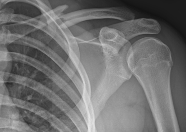
clavicle fracture MOI
Fall on point of shoulder: Children falling off playground equipment
Blow to lateral aspect of shoulder
Direct blow to clavicle
Force to superior shoulder from above
FOOSH
Clavicular Fractures common clinical findings/observation
Edema, Eccyhmosis
Tender to touch, Crepitus
Lateral clavicle is usually depressed (due to weight of arm) & medial clavicle is usually elevated (due to tension of SCM)
Clavicular fractures possible complications
Pneumothorax (puncture pleural cavity)
Damage to ligaments (shoulder separation):
Coracoclavicular—conoid or trapezoid fibers
Acromioclavicular
Clavicle fracture rehab s/p Clavicular Healing includes Stretching, Strengthening, & Functional Activities. What is the prognosis for healing of a clavicle fracture?
heals in 6–8 weeks
Humeral Fractures make up 5-8% of all fractures in body, its most prevalent in
women &/or osteoporosis
Incidence ↑ w/ age (balance, proprioception, falls)
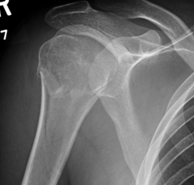
types of humeral fractures
Non-displaced or minimally displaced
Displaced Surgical Neck Fracture (rare) -> (surgery)
Fracture – Dislocation (rare)
Humeral fracture protocols typically involve limiting all ROM for
~6 weeks (some surgeons do less ~2 weeks)
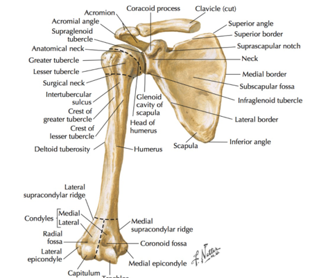
Proximal Humeral Fractures account for 10% of all humeral fractures; Affected proximal locations include
Greater Tuberosity (RC attachment, can’t lift arm)
Lesser Tuberosity
Head
Shaft
Surgical neck is at distal end of greater & lesser tuberosities
Humeral Fractures MOI for proximal fractures
FOOSH
direct blow
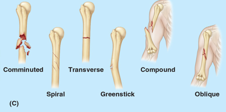
Humeral Midshaft fractures account for 1-3% of all fractures in body, MOI for diaphysis fractures include
Direct blow to humeral shaft: transverse fracture
FOOSH: spiral/comminuted fracture
Common Findings with a Humeral Fracture
Ecchymosis (w/in 24-48 hrs of injury; can spread down to elbow due to gravity)
Shoulder pain
Loss of shoulder motion due to pain
pts present w/ involved UE adducted & supported by opposite hand to protect it
For treatment of Proximal Humerus Fractures, most are
minimally displaced & do not require surgery: A sling is worn; not casted
Closed reduction humeral fracture treatment by
manipulation or traction (reduce fracture) then sling immobilization for 3-4 wks
If Closed Reduction for Humeral Fracture fails, surgeon uses
Percutaneous Pinning
Then 4 wks of immobilization using a sling → development of a stiff shoulder
ORIF for Humeral Fracture if is displaced
>1cm
What Humeral Fracture treatment involves shaft & humeral head replacement?
Hemiarthroplasty (humeral head too damaged to be repaired)
Humerus Fracture Complications: Malunion includes
incomplete union or union that occurred in poor alignment
Humerus Fracture Complications include RC damage based on affected bony structures
Greater tubercle of humeral head: infra/supraspinatus, teres minor
Surgical neck: subscapularis attachment to lesser tubercle
Humerus Fracture Complications include nerve injury, what are the affected structures for a proximal or diaphysis humeral fracture?
Proximal Humerus: Brachial plexus; Axillary nerve most common
Diaphysis (midshaft): Radial Nerve (travels in groove) (Sensory & motor deficits distally)
Humerus Fracture Complications: Arterial Injury can occur w/ a proximal humerus or midshaft fracture affecting the
Proximal: Anterior & Posterior Humeral Circumflex artery (supplies humeral head, can cause AVN)
Diaphysis: Brachial artery (medical emergency, diminished pulses)
Prognosis for proximal & midshaft humeral fracture healing?
Proximal Humerus Fracture: ~6 wks to heal
Diaphysis Fracture: 6–10 wks to heal
Most do not require surgery
Rehabilitation s/p Healing of Proximal humeral fx w/ closed reduction is to focus on prevent loss of ROM, can do
pain-free AROM after 2-4 wks of immobilization
Minimize stiffness, Maximize function
Restrictions are based upon method of immobilization (sling only, pinning, ORIF)
Once healing has occurred (~4-8 wks) can begin: A/PROM
Joint mobilization, Resisted exercise
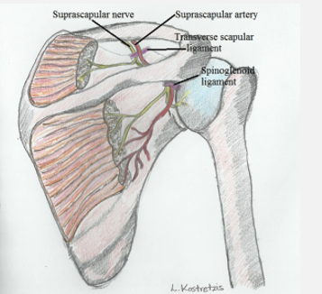
Suprascapular Nerve Palsy- Sites of entrapment
Suprascapular notch: under hypertrophied transverse scapular ligament
Spinoglenoid notch: by hypertrophied spinoglenoid ligament
Spine of scapula & medial tendinous margin of infraspinatus & supraspinatus
Possible causes suprascapular Nerve Palsy include
Compression, Traction, Friction
Repetitive microtrauma
Direct injury
Brachial plexus disorder
Ganglion cysts
Lipomas
Transverse scapular ligament calcification
suprascapular nerve palsy signs/sxs
can’t lift arm: (+) drop arm
limited active ER: (+) ER lag, (+) resisted ER
atrophy of infraspinatus
WEAK & painLESS
(may look like RC tear)
Suprascapular Nerve Palsy clinical presentation
Deep, diffuse, dull aching pain over posterior lateral aspects of shoulder
Supraspinatus &/or infraspinatus muscle atrophy
Weakness of ER & abduction
(+) External Rotation Lag Sign, Drop Arm Sign
Empty Can: (+) for weakness
Suprascapular Nerve Palsy tx
NMES
wait & see (may need relocation of tendons of RC), need nerve to reinnervate
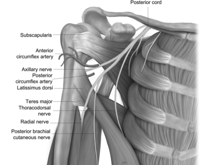
Axillary Nerve passes thru quadrangular space to innervate
deltoid & teres minor
if an Axillary Nerve Palsy occurs more distally then the
posterior deltoid (maybe lateral) & teres minor would be affected but not the anterior deltoid
Axillary Nerve Palsy MOI & sxs
Usually associated w/ trauma
Loss of sensation around deltoid tuberosity
Atrophy of deltoid
Weakness in all shoulder motions: May substitute w/ RC
Long Thoracic Nerve Palsy MOI
traction injury, viral (all these are suspected, but unknown)
Long Thoracic Nerve Palsy signs
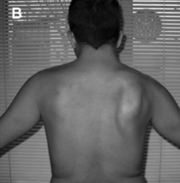
Scapular flip sign
Scapular Winging: Anterior elevation; on a wall push‐ups, Medial Towards spine
Usually a result of poor scapulohumeral mechanics mechanics, generally no pain
Spinal Accessory Nerve Palsy MOI
Medical/Surgical Intervention (head & neck cancer tx)
Viral
Spinal Accessory Nerve Palsy signs/sxs
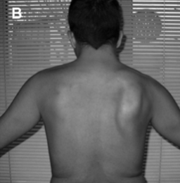
Trapezius Atrophy
Winging Scapula: Lateral elevation; shoulder ER against resistance, Lateral away from spine
No shoulder pain
Usually a result of poor scapulohumeral mechanics mechanics