The Respiratory System (Part 2)
1/45
There's no tags or description
Looks like no tags are added yet.
Name | Mastery | Learn | Test | Matching | Spaced |
|---|
No study sessions yet.
46 Terms
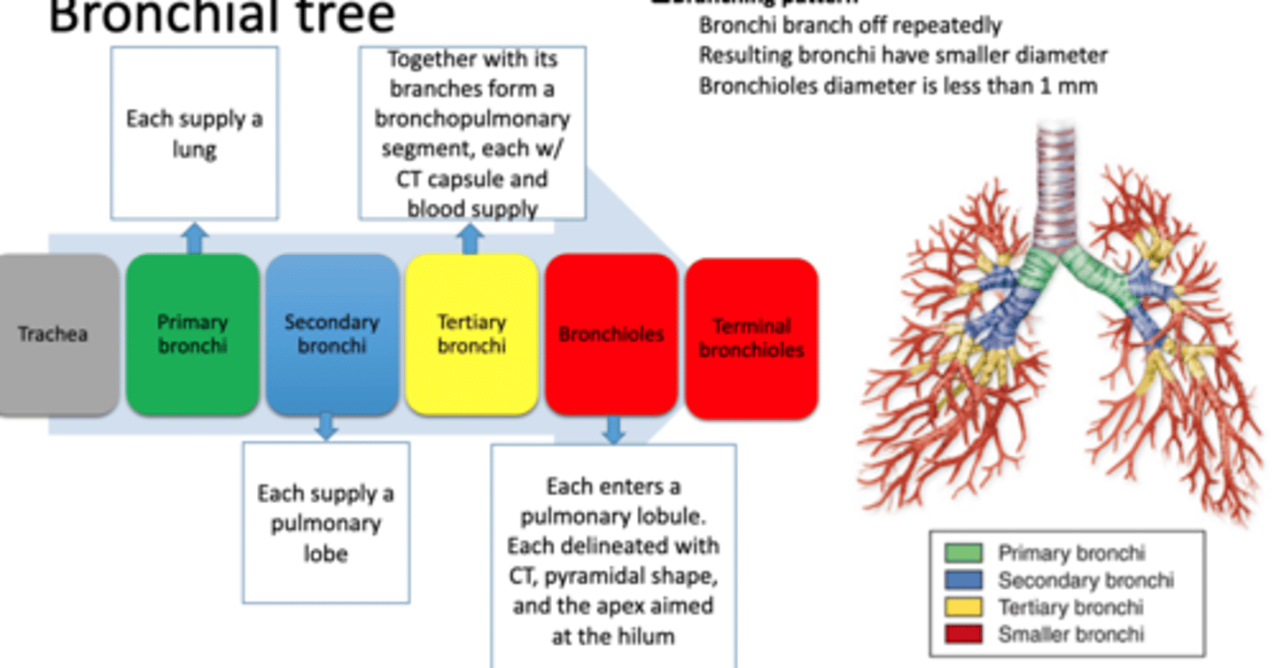
The primary bronchi each supply what?
a lung
What does the secondary bronchi supply?
a pulmonary lobe
Tertiary bronchi and its branches form what?
a bronchopulmonary segment each with connective tissue capsule and blood supply
Bronchioles each enters a pulmonary what?
pulmonary lobule with each delineated with connective tissue, pyramidal shape, and the apex aimed at the hilum
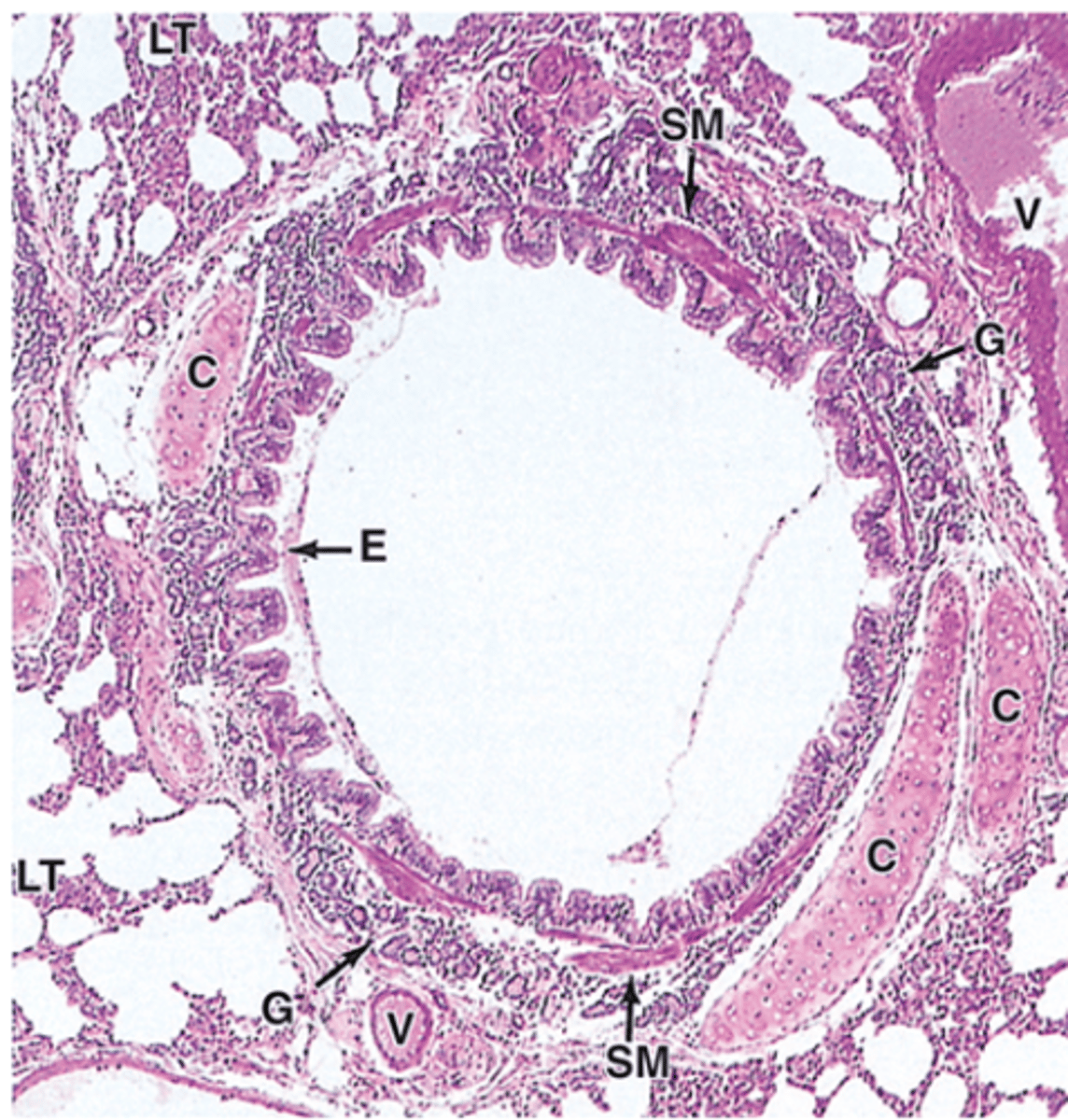
What are the five bronchi layers?
Mucosa, Muscularis, Submucosa, Cartilage layer, and Adventitia
What do the main, secondary, and tertiary bronchi have?
respiratory epithelium
The mucosa of the bronchi becomes thinner as what?
as bronchial diameter decreased
The bronchioles epithelium greadually changed from what?
larger bronchioles to terminal bronchioles. Has pseudostratified, ciliated simple columnar, and ciliated simple cuboidal
Muscularis bronchi is made of what?
smooth muscle. Goes through bronchi, bronchioles, and terminal bronchioles
Submucosa bronchi is made of what?
loose connective tissue. where glands and adipose tissue are in larger bronchi but bronchioles do not have glands
What are cartilage layers of the bronchi?
main bronchi has circular rings of cartilage and the intrapulmonary bronchi has plates instead and become smaller in small bronchi. Theres no cartilage in bronchioles
The adventitia has what kind of tissue?
Dense connective tissue
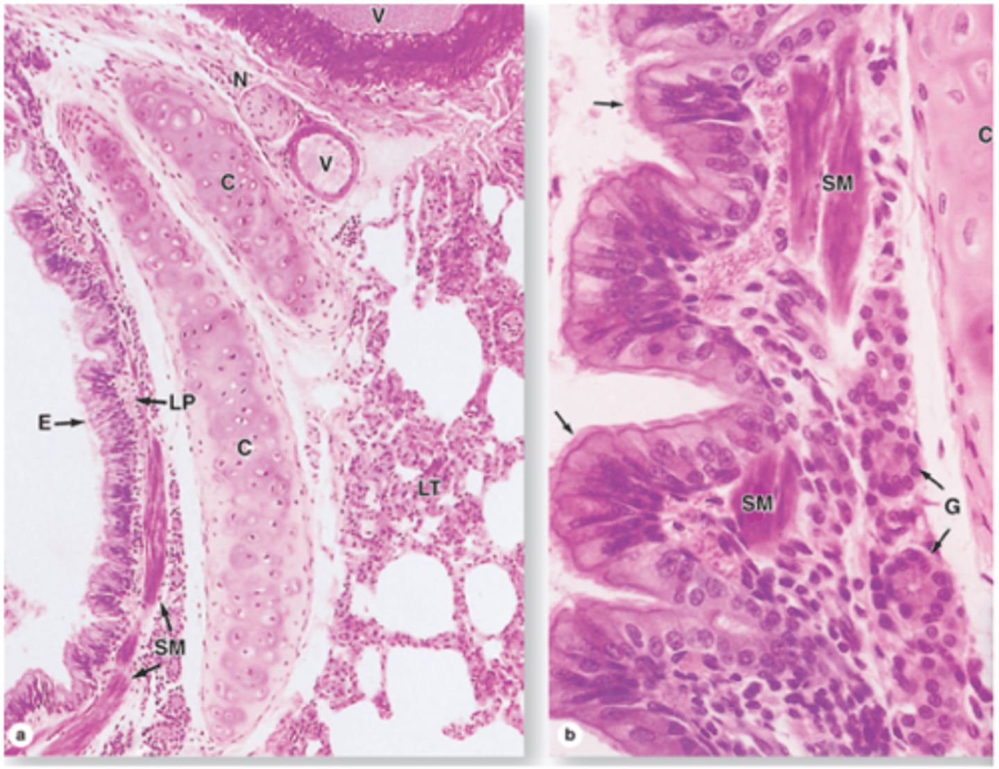
In the main extrapulmonary bronchi it has the same structure as the trachea except what?
except the cartilage forms a circular ring
What are the changes that happen in intrapulmonary bronchi?
Cartilage rings are replaced by cartilage plates. The plates become smaller and less numerous. Once you reach the bronchioles theres no cartilage plates.
Segmental bronchus
In the bronchi the smooth muscle forms a complete circular layer but as the bronchi branch becomes smaller the smooth muscle becomes what?
an increasingly conspicuous layer and the amount of cartilage decreases and the smaller bronchi have discontinuous muscle layer.
Cartilage plates and smooth muscle in intrapulmonary bronchi
What are bronchioles?
A branch of segmental bronchi and give rise to respiratory bronchioles. It doesn't have any mucosal glands nor cartilage. It has thicker smooth muscle layer. Where the epithelium changes gradually
The mucociliary escalator begins at what?
ciliated epithelium of bronchioles
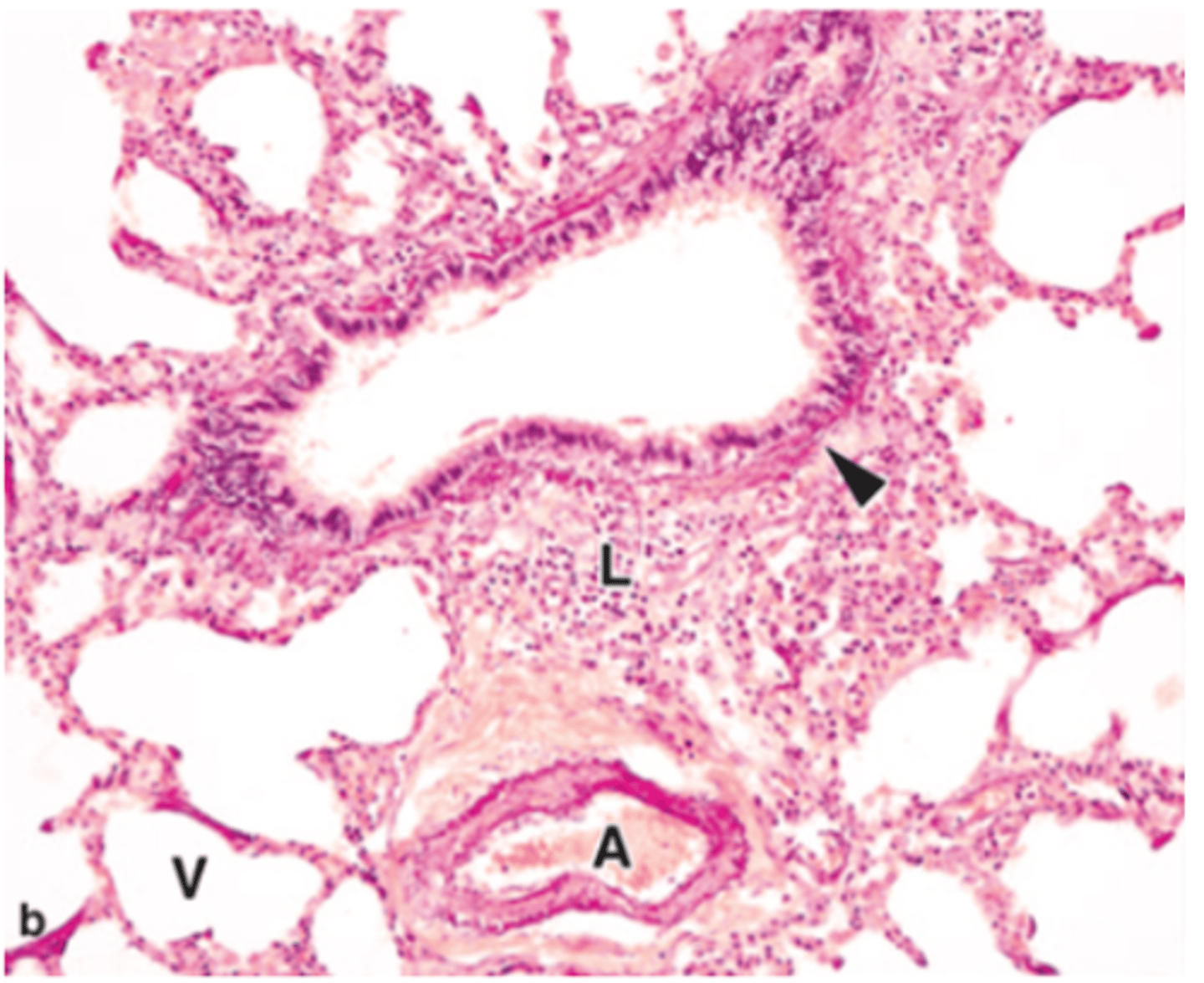
Bronchioles
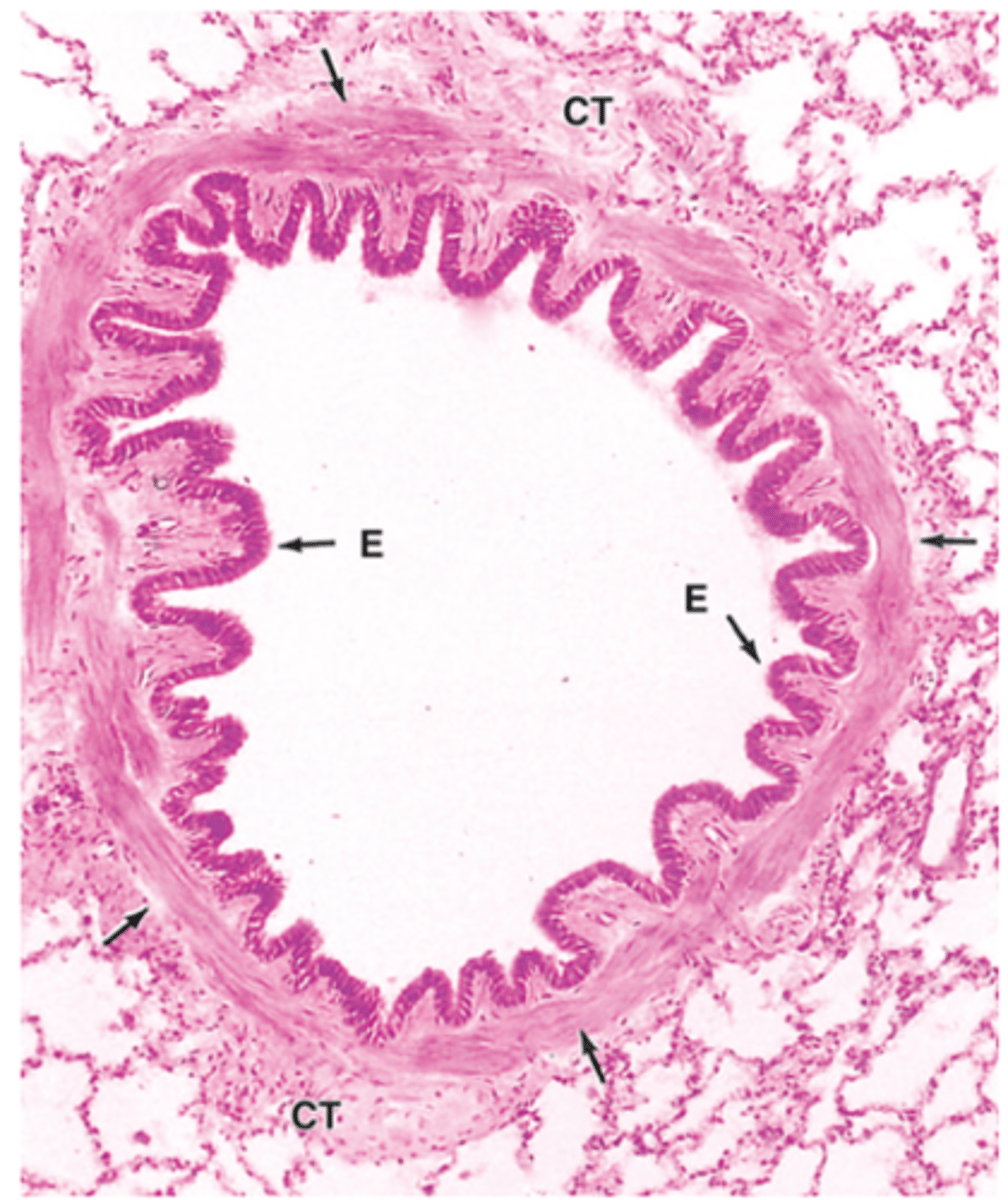
What are mid-sized bronchioles?
It is ciliated epithelium with smooth muscle associated with abundant elastic fibers. Connective tissue, abundant lymphocytes of MALT and has numerous lymphoid nodules
Mis-sized bronchioles
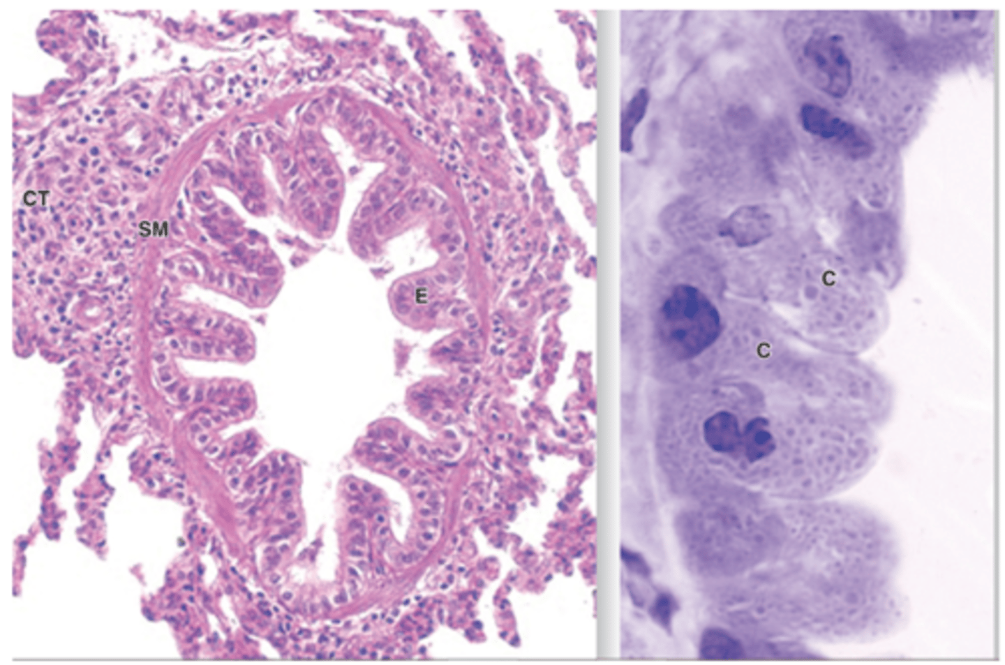
Smaller bronchioles
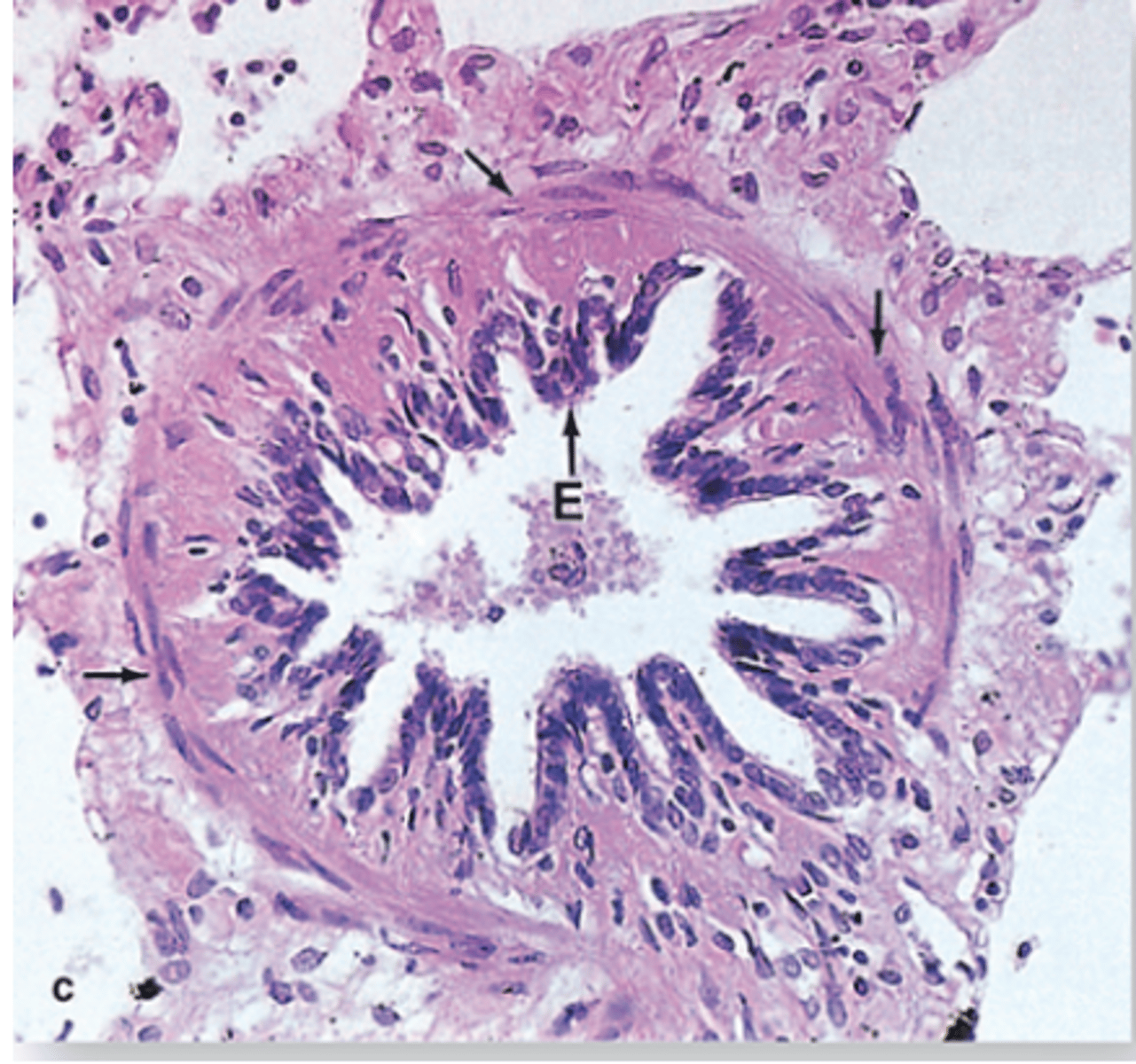
What are the smaller bronchioles?
is ciliated simple cuboidal epithelium, has prominent layers of smooth muscle, and abundant MALT
What is the terminal bronchioles?
is a single layer of simple cuboidal epithelium that has club cells with exocrine functions, apical domain is dome shaped, and the abundant secretory granules.
Terminal bronchioles contains what?
brush cells, DNES, Smaller granular cells, Stem cells, and lamina propria contains elastic fibers as well as smooth muscle
What are the functions of the bronchial exocrine cells of the terminal bronchioles?
Secretion of surfactant lipoproteins and mucins, Detoxification of inhaled xenobiotic compounds by enzymes of the SER, and Secretion of antimicrobial peptides and cytokines
Terminal bronchioles
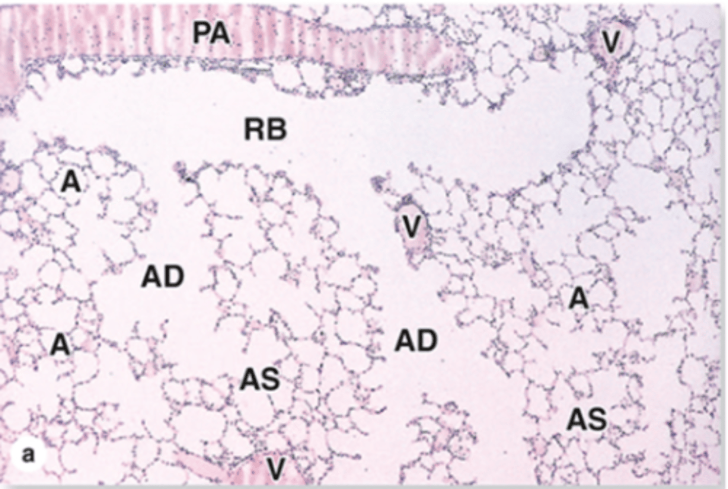
What are the respiratory bronchioles?
It is the first part of respiratory portion, it has alveoli present in their walls, the mucosa is similar to the terminal, has few openings to alveoli, has elastic CT, smooth muscle, lamina propria of loose ct, epithelium has club cells/squamous at alveolar openings, and the club cells are numerous distally when ciliated cells decrease
Respiratory bronchioles
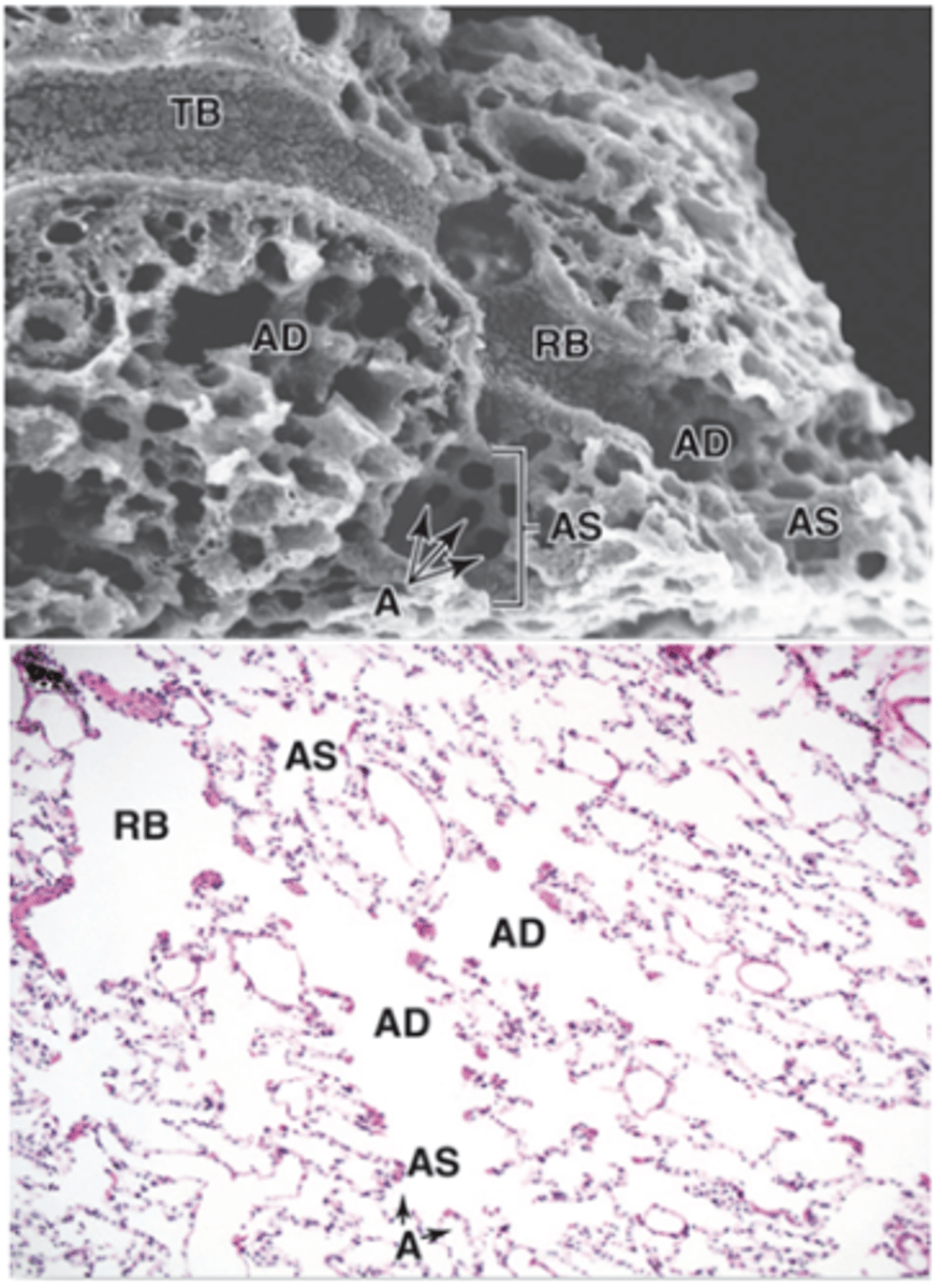
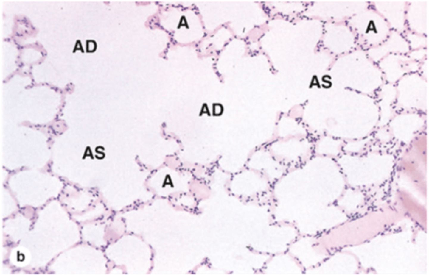
What are Alveolar ducts?
ducts lead into terminal clusters called alveolar sacs. They are located after respiratory bronchioles and are lined by openings of alveoli with squamous epithelium in both the alveolar ducts and alveoli. Contains the thin lamina propria with a strand of smooth muscle around the opening of alveoli. Has elastic and collagen fibers network supports every duct and its alveoli
Alveolar ducts
Alveolar sacs
alveoli
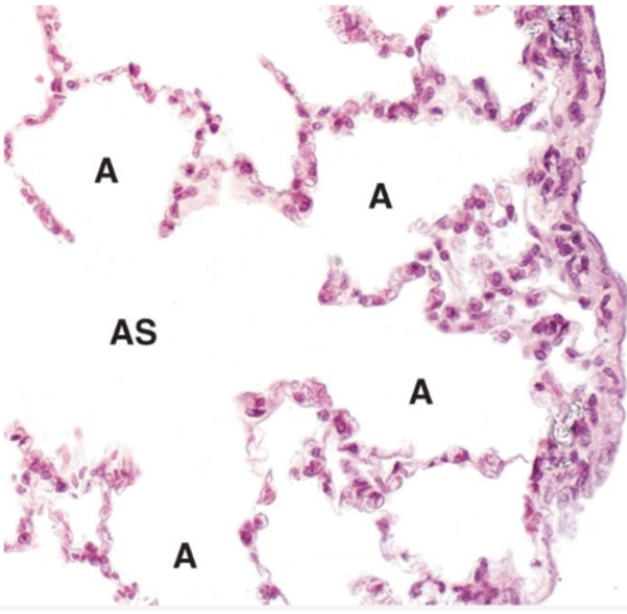
What are alveolar sacs?
They are at the end of alveolar ducts, have spaces surrounded by alveoli opening into this space, is lined by a thin simple squamous epithelium, is supported by very thin lamina propria with elastic and reticular fibers, and abundant capillaries surround each alveolus
What are the alveoli?
saclike evaginations, separated by inter-alveolar septa has scattered fibroblasts. Contaisn sparse ECM and CT and the richest capillary network
Type 1 alveolar cells (Type 1 pneumocytes)
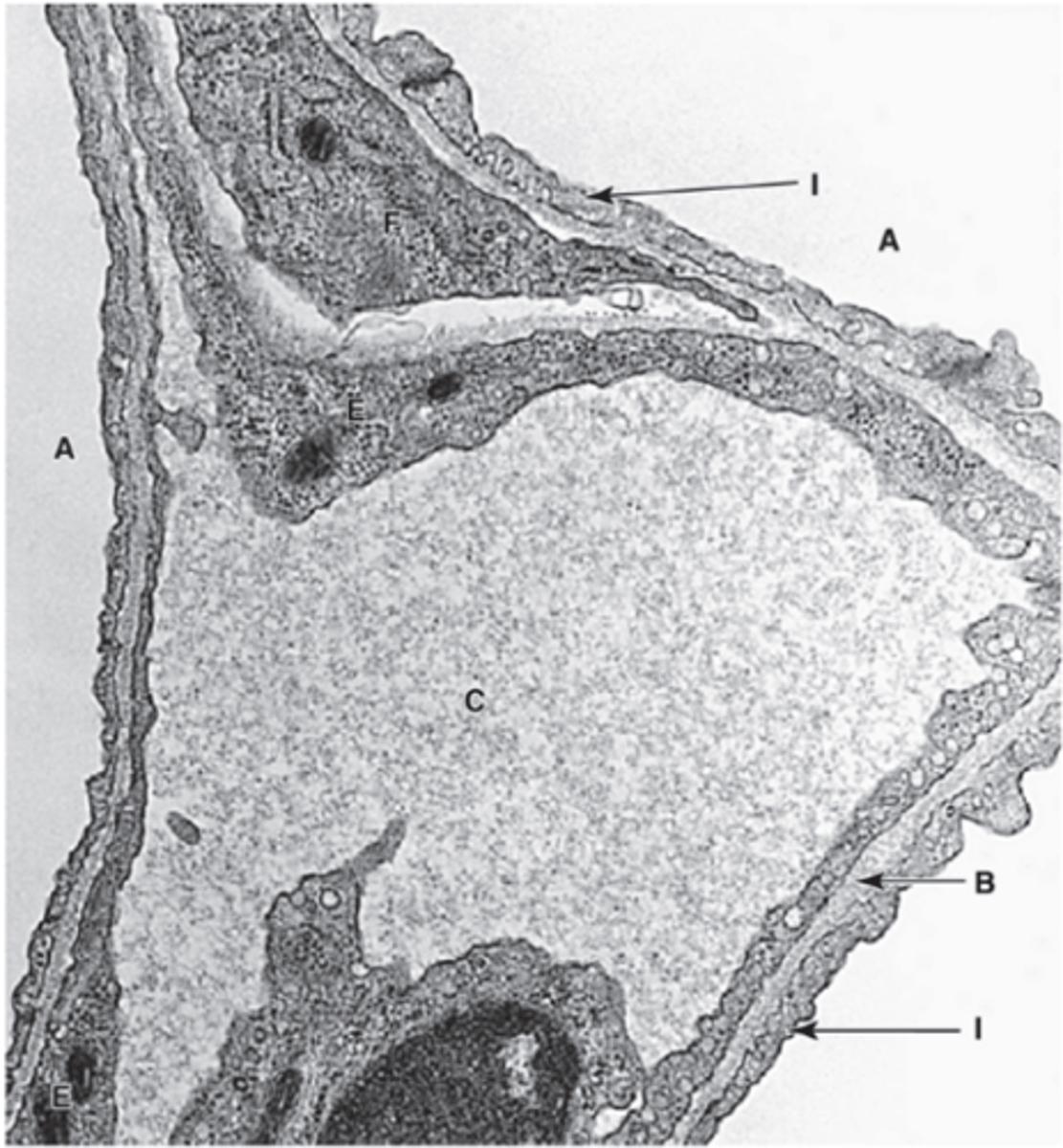
What are the type 1 pneumocytes (alveolar cells)?
They are very thin squamous epithelial cells, contains perinuclear organelles, joined by desmosomes, and contains tight junctions to prevent leakage of interstitial fluid into alveolar lumer
Type 2 alveolar cells (type 2 pneumocytes or septal cells)
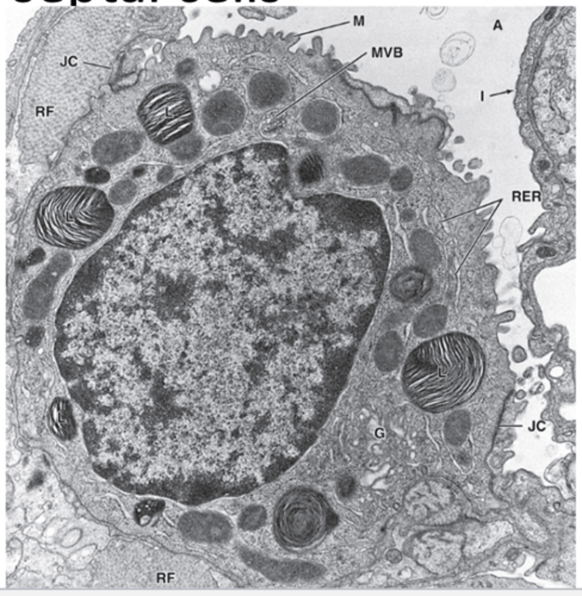
What are Type 2 pneumocytes (alveolar cells or septal cells)?
Contains cuboidal cells, interspersed among type 1 pneumocytes, has tight junctions and desmosomes, has large euchromatic round nucleus with nucleoli. It has lightly stained cytoplasm with many vesicles and contains lamellar bodies and released pulmonary surfactant
Dust cells
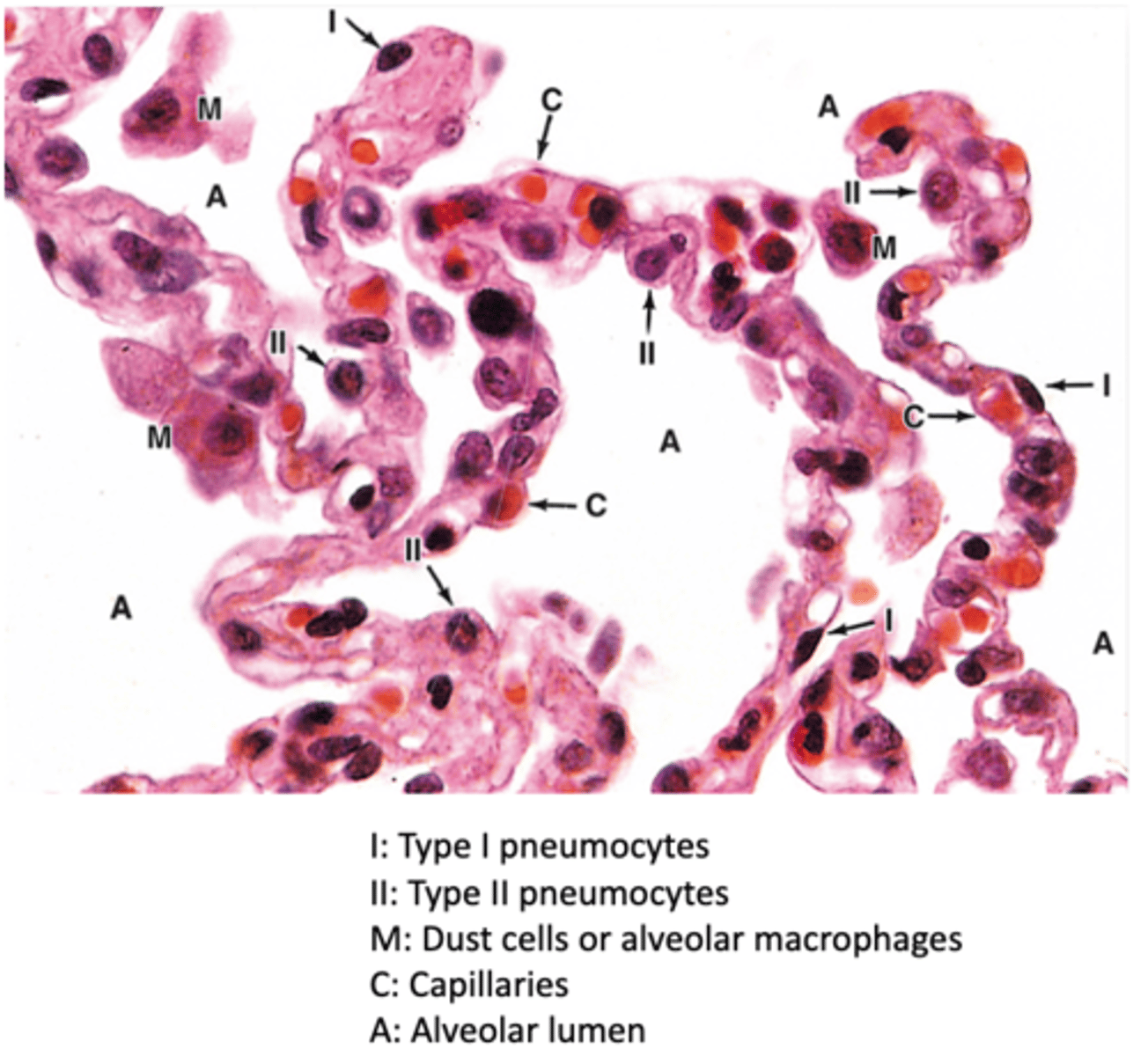
What are dust cells?
they are alveolar macrophages, in alveoli and interalveolar septum, and is slightly darker than type 2 pneumocytes
Pleural membranes
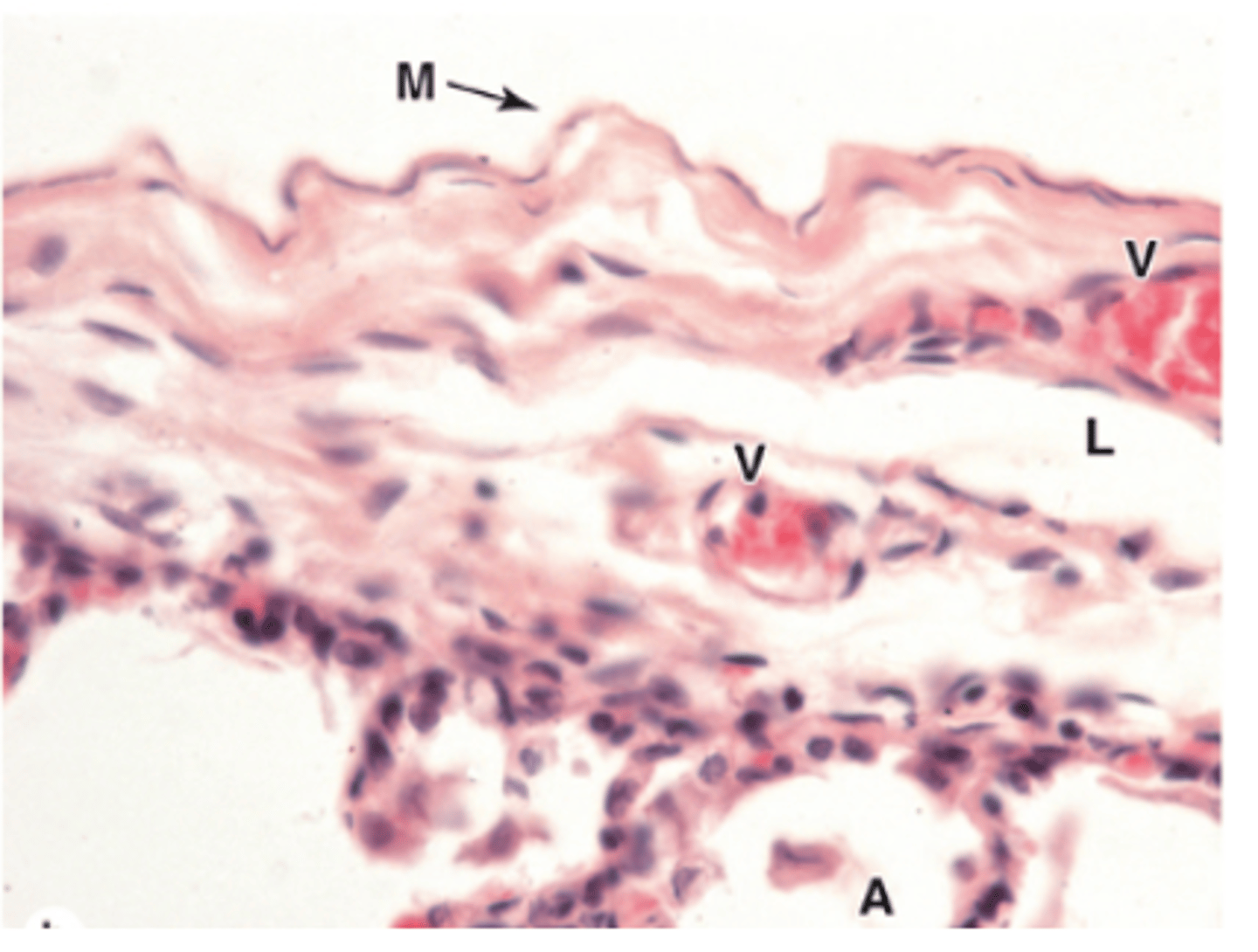
What is the pleural membrane?
Fine serous membrane with the visceral pleura attached to lungs and the parietal pleura lines the thoracic wall which both are continuous at the hilium. Contaisn emothelium, loose connective, collagen and elastic.
What is the pleural cavity?
space between visceral and parietal layers