Lymph Nodes, Spleen, & Thymus
 Call Kai
 Learn
Learn Practice Test
 Spaced Repetition
 Match
Match Flashcards
 Knowt Play
1/66

There's no tags or description
Looks like no tags are added yet.
Name | Mastery | Learn | Test | Matching | Spaced | Call with Kai | Chat |
|---|
No analytics yet
Send a link to your students to track their progress
67 Terms
Lymph
- Interstitial fluid that drains from tissues into lymphatic system → circulates through lymph nodes → drains into subclavian veins
Lymphatic System
- Composed of a network of vessels, ducts, nodes, and organs. Provides defense against infection.

Where does all the lymph in the body eventually drain?
- Into subclavian veins → returned to the venous system of body

What are the types of lymphoid organs?
- Primary
- Secondary
Primary Lymphoid Organs
- Bone marrow and thymus
Thymus is derived from what?
- 3rd pharyngeal pouch
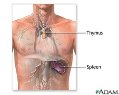
Where is the thymus located?
- Behind the sternum and between the lungs

Thymus: Cortex and Medulla
- Cortex: Peripheral, dark, densely packed with immature T lymphocytes
- Medulla: Central region, light, mature T lymphocytes and hassall corpuscles

What structure is unique to the thymus?
- Hassall Corpuscle

When is the thymus 'large and robust'
- During infancy, as the immune system is still developing and the infant is exposed to a lot of unfamiliar pathogens
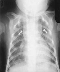
What is seen on a chest X ray of an infant?
- Thymic shadow, appears as a sail boat (normal appearance)

Thymic Aplasia
- The thymus fails to develop so T cells don't mature and aren't functional
What happens to the thymus with age?
- Atrophies and is replaced by fat as the T cells previously made there now circulate and protect the body for life
Primary Lymphoid Organs are important for what?
- Sites of lymphocyte formation → creates B/T cells
Secondary Lymphoid Organs
- Spleen
- Lymph nodes
- Peyer's Patches (Small intestines)
- Tonsiles

Why are secondary lymphoid organs important?
- They are areas where B/T cells can proliferate in response to infection
Lymph nodes
- Bean-shaped filters that cluster along the lymphatic vessels of the body. They function as a cleanser of lymph as wells as a site of T and B cell activation

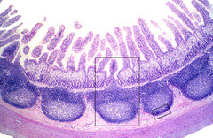
Peyer's Patches
- Large collections of lymphoid tissue found in the submucosa of the small intestine
Tonsils
- Masses of lymphatic tissue in the back of the oropharynx

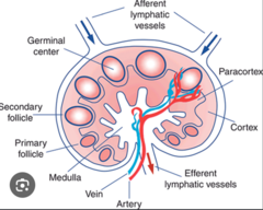
What are the layers of a lymph node?
1. Cortex
2. Paracortex
3. Medulla
Afferent Lymph Vessel
- Carries lymph toward the lymph node → lymph percolates through the 3 layers of the node

General lymph flow through lymph node
- Cortex → Paracortex → Medulla → Efferent Lymph Vessel
Efferent Lymph Vessel
- Small lymphatic vessels that carry lymphatic fluid away from a lymph node

What surface proteins do dendritic cells express? What is their general role?
- MHC I/II
- B7
APCs who take pieces of bacteria → present them on surface to activate other immune cells
Example: There is a site of infection in the tissues/lungs; what occurs after?
1. Lymph fluid drains from site of infection (containing dendritic cells that have antigens on their surface from site of infection as well as free antigens)
2. Lymph with dendritic APCs enter nodes → there are many B/T cells waiting there for a matching antigen
3. Dendritic cells present antigen to T cells // Dendritic cells already inside lymph nodes can process the free antigen and present it to B cells
4. Adaptive immune response
What is the first layer than lymph encounters in the node?
- Cortex
Where does all the immune action occur within the cortex?
- The follicles

Lymphoid Follicles
- Contain follicular dendritic cells (permanent cells, take up free antigen from lymph) → present it to B cells → activates them
Are able to bind a lot of different antigens (velcro)

What is an important reservoir for HIV? What is seen in early infection?
- Follicular dendritic cells in cortex of node
- Early after infection → large amts of HIV particles in FDCs
What are the types of follicles within the cortex?
- Primary and secondary follicles
Which type of follicle is more prominent during inflammation/infection?
- Secondary follicle
Primary Follicles
- Inactive, containing FDCs and B cells
Secondary Follicles
- An activated cluster of B cells that develops a central germinal center after encountering an antigen
Area of B cell growth/differentiation + class switching

Paracortex
- Contains T cells that are activated by dendritic cells + antigen on their surface
- Contains high endothelial venules

What do the high endothelial venules in the paracortex allow?
- Special vascular structures that allow B/T cells to enter the nodes
- Different from normal vessels → has spaces that B/T cells can squeeze through

When is the paracortex engorged?
- During immune response (swollen lymph nodes)

What is underdeveloped in the setting of DiGeorge Syndrome?
- The paracortex of lymph nodes (T cell deficiency disorders)
What is the innermost section of the lymph node?
- Medulla
What are the components of the medulla?
- Medullary cords and sinuses
Medullary Sinuses
- Cavities lined with macrophages → filters lymph → phagocytosis (of bacteria, viruses, or other antigens that get into sinus)
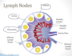
Medullary Cords
- Tissue between the sinuses/cavities that contain plasma cells that secrete antibodies

Spleen
- An organ that is part of the lymphatic system; it produces lymphocytes, filters the blood, stores blood cells, and destroys old blood cells
LUQ
Filters BLOOD, NOT lymph
Order of Blood Flow in the Spleen
- Enters via splenic artery → white pulp → red pulp → splenic vein to drain from the organ
What are the important structures within the white pulp of the spleen?
- Follicles
- Pariarteriolar Lymphocyte sheaths (PALS)
- Marginal Zone
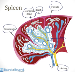
What are the important structures within the red pulp of the spleen?
- Sinusouds

White Pulp vs Red Pulp of Spleen
- White: Acts as part of the immune system to fight infections; blood is exposed to B/T cells & Macrophages
- Red: Filters the blood through sinusoids and removes old or damaged blood cells; area of platelet storage

Marginal Zone
Contains:
- Macrophages to remove debris from blood
- Dendritic cells to process antigens from blood
What do the follicles of the spleen contain?
- B cells
What does the PALS of the spleen contain?
- T cells
Sinusoids of spleen
- Lines the red pulp
- Open endothelium → cells can pass in/out
What is the order of blood flow to the sinusoids?
- Capillaries → cords → sinusoids ( → then to venous system)
Cords contain macrophages for filtering/removal of dying RBCs from circulation
Splenic Dysfunction causes an increased risk of what?
- Spleen uses macrophages → when lost in splenic dysfunction, cannot process/phagocytes encapsulated organisms → ↑ risk of infection
Encapsulated organisms
- Microbes shielded by an outer protective layer, usually a polysaccharide capsule, that helps them hide from the host immune system
Ex: Pneumoniae, Influenza, Meningitides, Cryptococcus etc
Sepsis
- Dangerous infection of the blood
What is the predominant pathogen that causes sepsis
- Strep Pneumonia (death in >50% of pts with splenic dysfunction)
What are the most common reasons for splenic dysfunction?
- Splenectomy: d/t trauma, can also elective when wanting to ↓ phagocytosis of platelets and to ↓ anemia
- Functional asplenia (sickle cell)
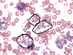
Howell Jolly Bodies
- Some RBCs leave marrow w/ nuclear remnants; this is normally cleared by the spleen but their presence can be seen in peripheral blood and can indicate splenic dysfunction

Target Cells
- Appear on a blood smear when red blood cells have an excess membrane relative to their internal volume

What happens to platelets in a patient with splenic dysfunction?
- Thrombocytosis bc/ spleen fails to remove them
SCID
- Severe combined immunodeficiency disease
- Both T/B cells affected
SCID Symptoms
- Failure to thrive
- Chronic diarrhea
- Recurrent viral/bacterial/fungal infection
Direction of Lymph Flow
- Afferent lymphatic vessel → subcapsular sinus → Trabecular sinus → Medulla → efferent lymphatic vessel
Functions of the Lymphatic System
- Immune surveillance
- Transport large molecules (hormones, lipids)
- Fluid Balance (returns fluid → heart)
Flow of lymph to drainage
- Lymph capillaries → vessels → trunks → ducts → subclavian vein
Right lymphatic duct vs thoracic duct
Right lymphatic duct drains much smaller area (right upper quarter).
Thoracic duct drains entire left side and both LEs.

Lymphedema
- Swelling due to an abnormal accumulation of lymph fluid within the tissues (drainage obstruction)
How can we improve lymphatic drainage?
- Compression garments
- Exercise
- Manual lymphatic drainage