Terminology and Biomechanical considerations
 Call Kai
 Learn
Learn Practice Test
 Spaced Repetition
 Match
Match Flashcards
 Knowt Play
1/87

Earn XP
Description and Tags
Operative Dentistry Exam 1
Name | Mastery | Learn | Test | Matching | Spaced | Call with Kai | Chat |
|---|
No analytics yet
Send a link to your students to track their progress
88 Terms
Operative dentistry
Operative dentistry is the art and science of the prevention, diagnosis and treatment of defects in the enamel, dentin and cementum of teeth
Restorative dentistry
Restorative dentistry is the study, diagnosis and integrated management of
diseases of the teeth and their supporting structures and the rehabilitation of
the dentition to functional and aesthetic requirements of the individual.
Preventive dentistry
Aspects of dentistry concerned with promoting good oral health and
function by preventing or reducing the onset and/or development of oral
diseases or deformities and the occurrence of oro-facial injuries.
Cosmetic dentistry
Cosmetic dentistry is generally used to refer to any dental work that
improves the appearance of teeth, gums and/or bite. It primarily focuses on
improvement in dental aesthetics in color, position, shape, size, alignment
and overall smile appearance
Caries
Commonly used term for tooth decay
Carious Lesion
A cavity caused by caries
Cavity
Missing tooth structure. A cavity may be due to decay, erosion or abrasion.
If caused by caries; also referred to as carious lesion
Decay
Lay term for caries
Filling
A lay term used for the restoring of lost tooth structure by using materials
such as metal, alloy, plastic or porcelain
Direct restoration
A restoration of any type (e.g., “filling”; crown) fabricated inside the mouth
A procedure where the service is delivered completely in the patient’s oral
cavity and without the use of a dental laboratory..
Indirect restoration
A procedure that involves activity that occurs away from the patient, such
as creating a restorative prosthesis. An indirect procedure is also known as a
laboratory procedure, and the laboratory’s location can be within or
separate from the dentist’s practice.
Temporary restoration
A restoration or prosthesis placed for a shorter time interval for use while a
definitive restoration or prosthesis is being fabricated –
see interim and provisional.
Preparation
Is the mechanical alteration of a defective, injured, or diseased tooth to
receive a restorative material that re-establishes a healthy state for
the tooth, including esthetic corrections where indicated and normal form
and function.
Treatment Plan
The sequential guide for the patient's care as determined by the dentist's
diagnosis and is used by the dentist for the restoration to and/or
maintenance of optimal oral health.
Diagnostic imaging
A visual display of structural or functional patterns for the purpose of
diagnostic evaluation. May be photographic or radiographic.
Radiograph
An image or picture produced on a radiation sensitive film, phosphorous
plate, emulsion or digital sensor by exposure to ionizing radiation
Anatomy of the Tooth Crown
Enamel
Dentin
Dentino Enamel Junction
Pulp horn
Pulp Chamber
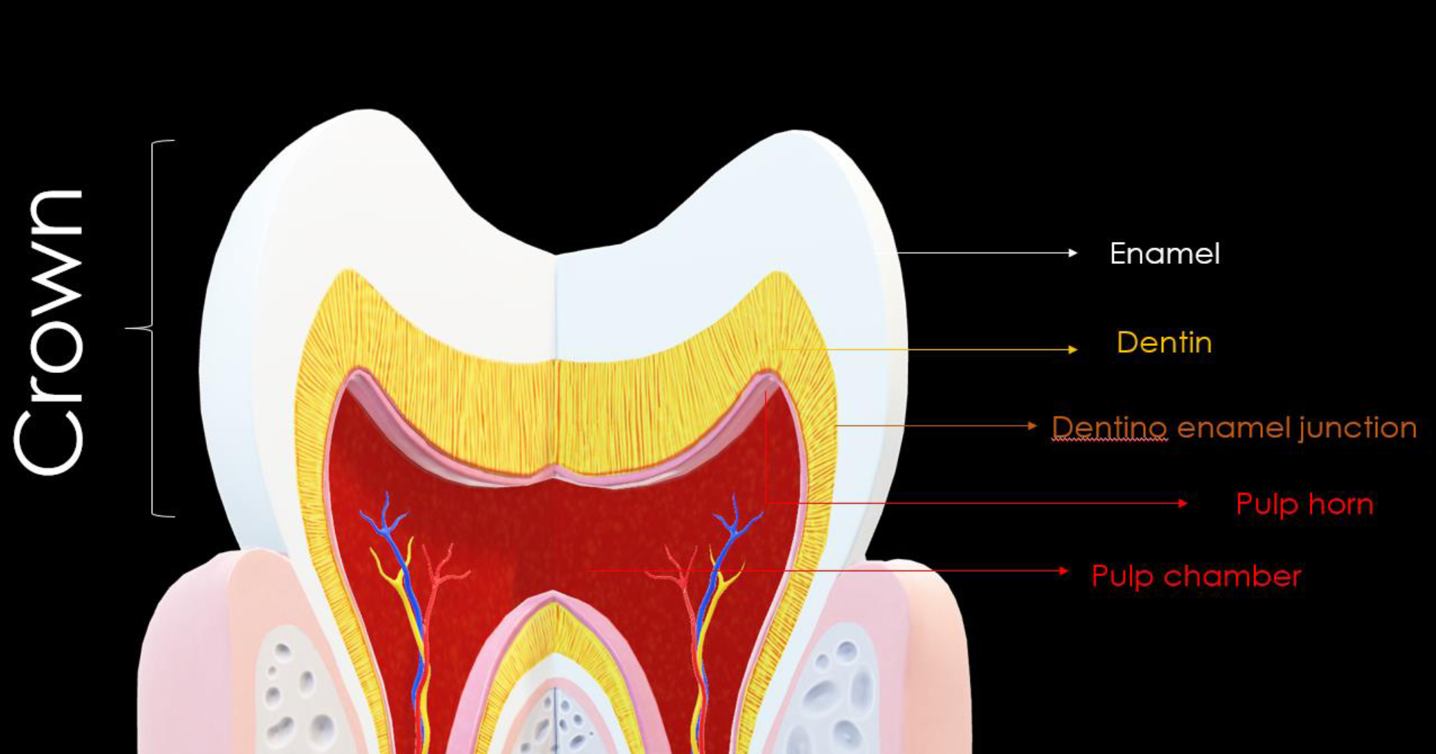
Enamel
Dentin
Dentino Enamel Junction
Pulp horn
Pulp Chamber
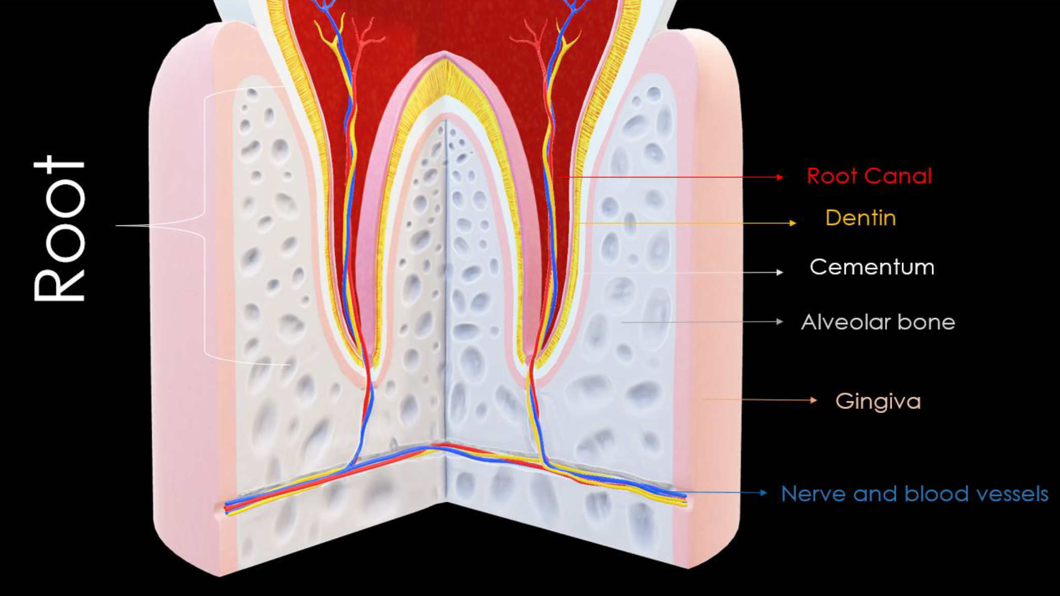
Anatomy of the Tooth Root
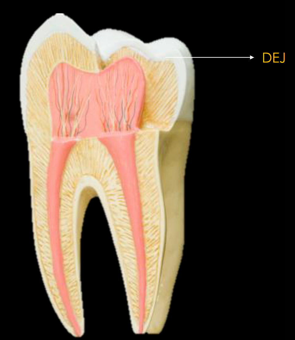
Dentino-Enamel Junction (DEJ)
Where enamel meets dentin
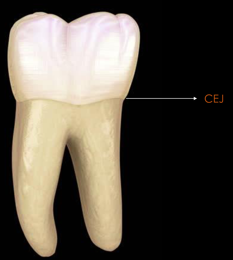
Cemento-Enamel Junction (CEJ)
: Where enamel meets cementum
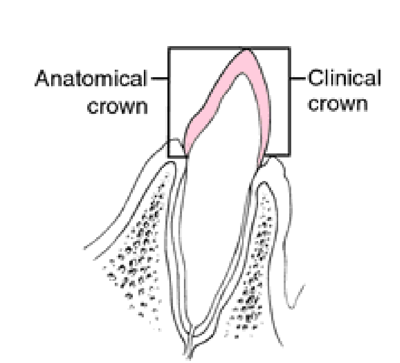
Anatomical Crown
is that part of the tooth that is covered by Enamel
or outer covering of the tooth. Its up to the cementoenamel junction.
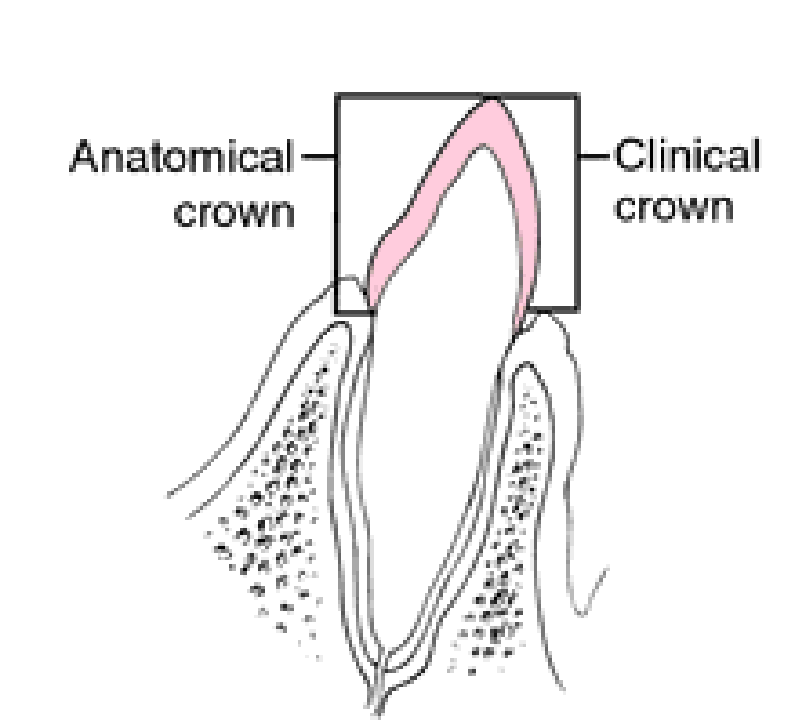
Clinical crown
is that part that is visible in the oral cavity.
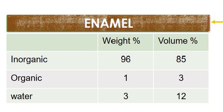
Hard Tissue Composition (Enamal)
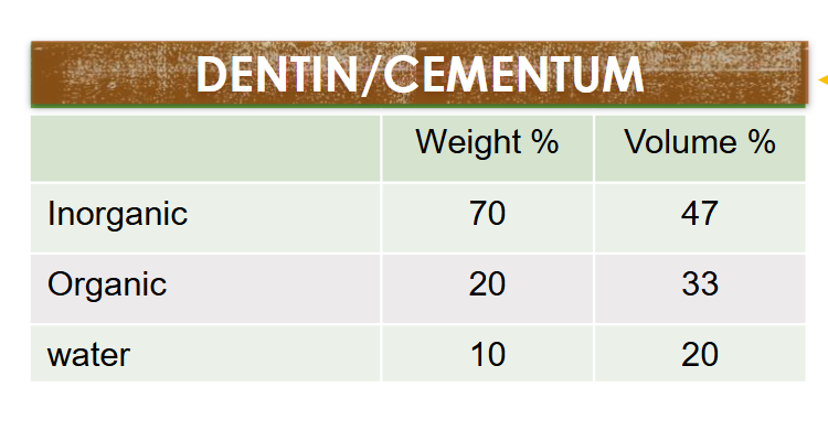
Hard Tissue Composition (Dentin & Cementum)
Inorganic Hard tissue composition (3)
Carbonated apatite
• Hydroxy-apatite
• Fluro-apatite
Organic Hard tissue composition (3)
• Lipids
• Proteins
• Collagen
enamel rod
An enamel rod is the basic unit of tooth enamel. It is a tightly packed, highly organized mass of
hydroxyapatite crystals, which are hexagonal in shape and provide rigidity and strengthen the enamel
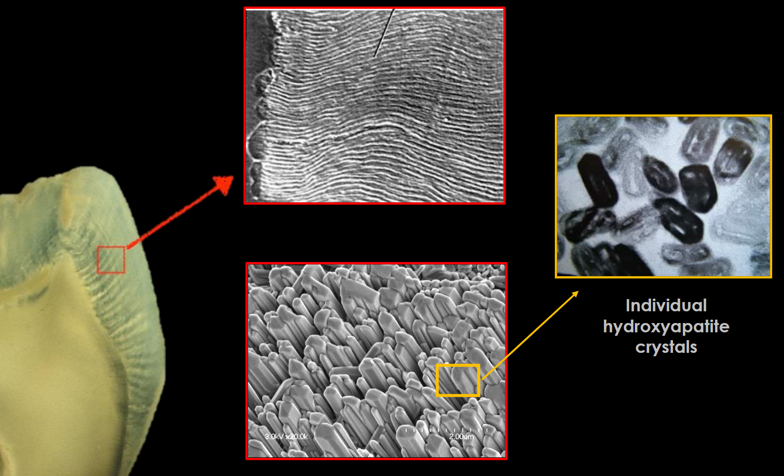
Perikymata
are incremental growth lines that appear on the surface of tooth enamel as
a series of linear grooves.
Enamel Has …
Enamel has diffusion channels that allow for acid or ionic exchange
Enamel
Enamel rods
• Interrod enamel:
- Located around the rod
enamel
- Same composition as enamel
rod
- Different direction
• Rod sheath: Contains proteins,
water, and lipids
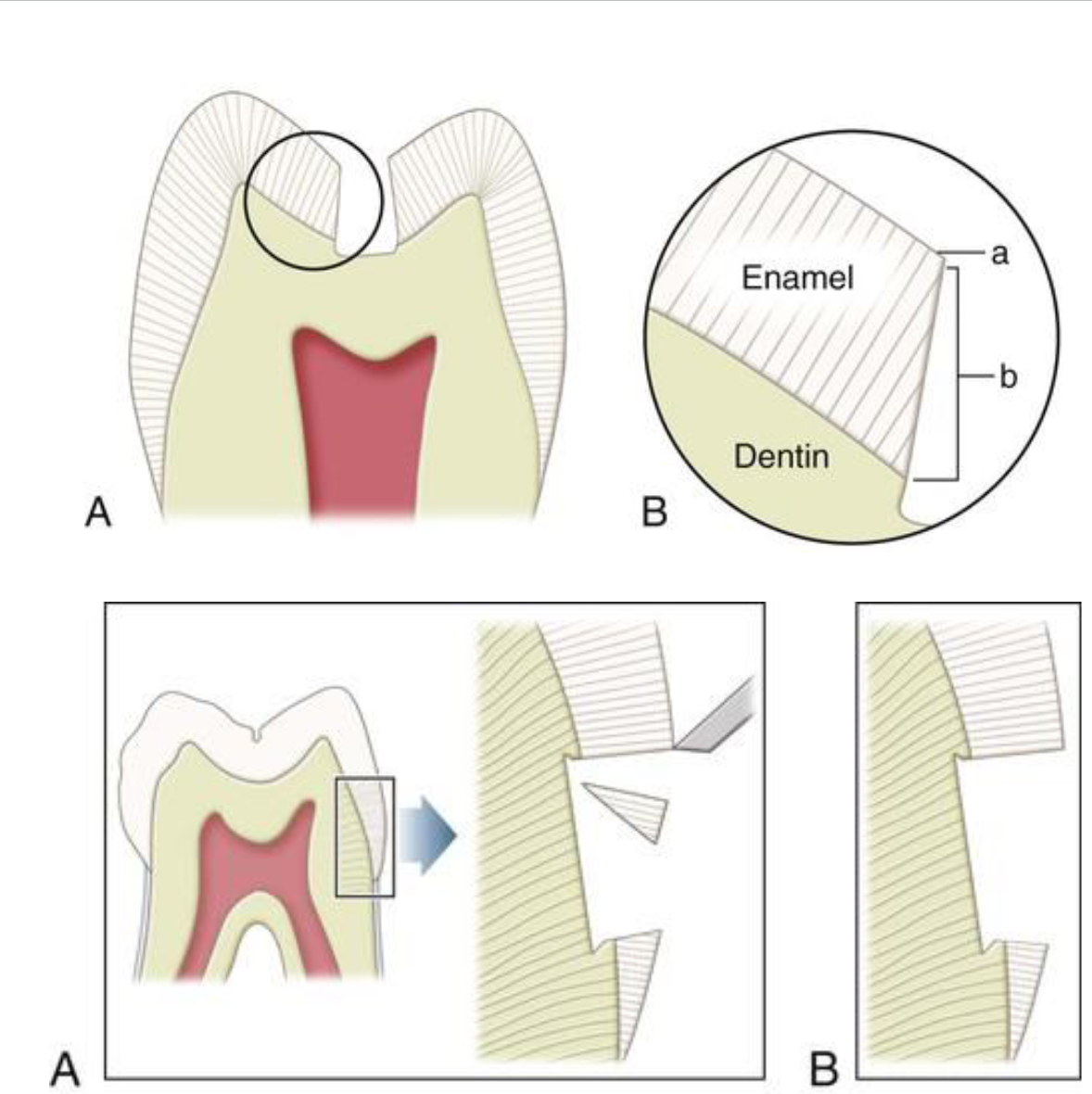
Enamel rods/prisms Location
Enamel rods/prisms exit
perpendicular to tooth surface
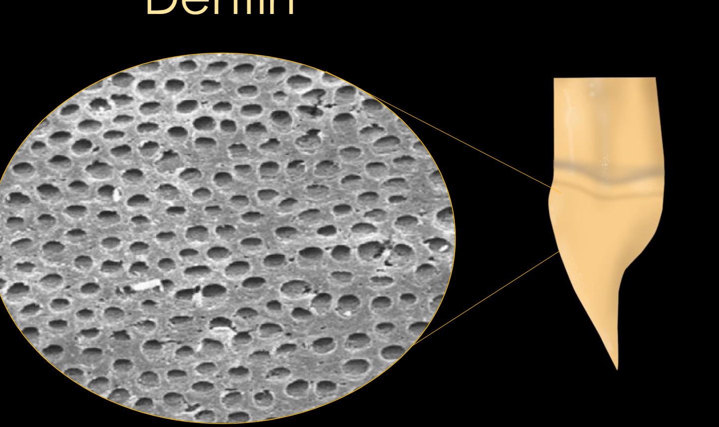
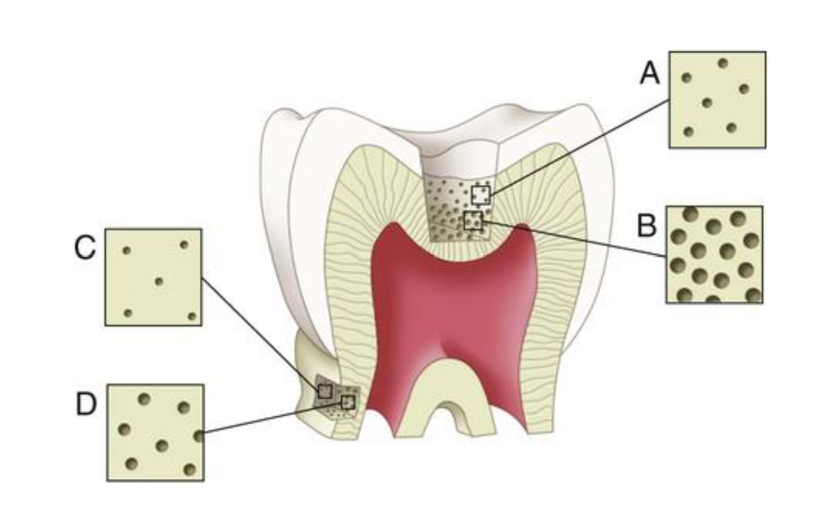
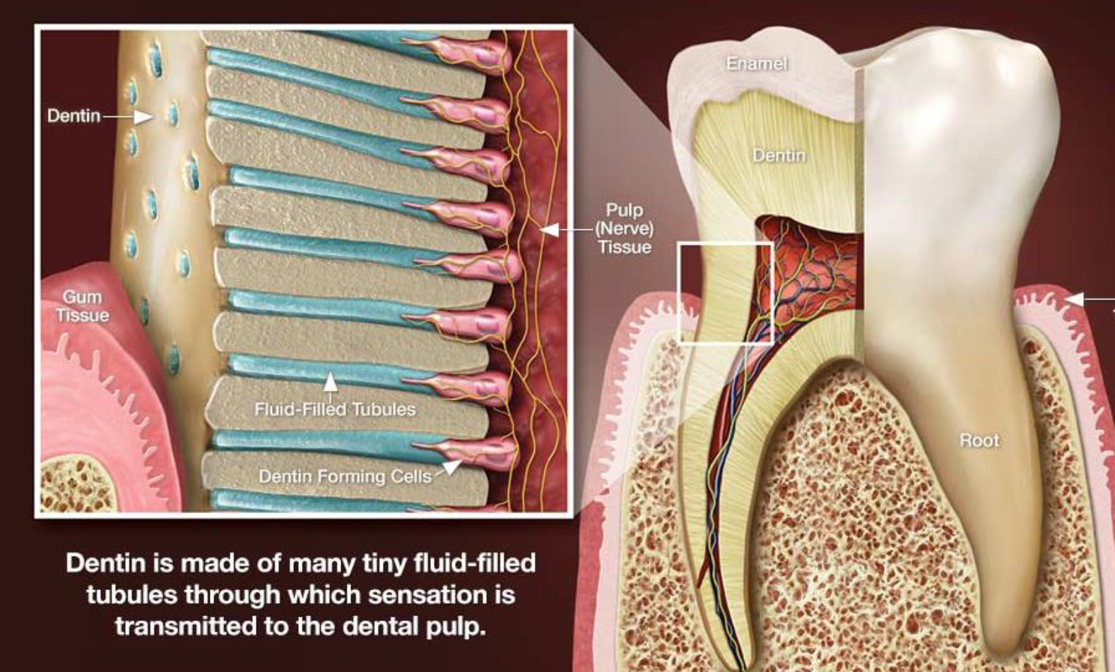
dentinal tubules
Dentin consists of microscopic channels, called dentinal tubules, which radiate outward through the dentin from
the pulp to the exterior cementum or enamel border.
Dentin vs Enamel
Hydroxyapatite crystals are 30 times smaller, making
dentin somewhat softer than enamel.
Dentin is duller and darker in appearance
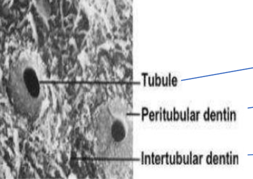
Peritubular dentin
is a relatively dense mineralized tissue that surrounds the dentinal
tubules. It is composed mainly of crystals of carbonated/hydroxy apatite together
with a small amount of collagen
intratubular dentin
The intertubular dentin, which lies between the tubules, is a less-calcified matrix that
consists of some apatite crystals embedded within a collagen matrix.
intratubular dentin vs Peritubular dentin
Very Important about Dentin Tubules
The number and diameter of the tubules increase from DEJ to the pulp
Types of Dentin (4)
Primary dentin
• Secondary dentin
• Tertiary dentin:
• Sclerotic dentin: Hypermineralized dentin, Darker, Non-carious, not necessary to remove
Primary dentin:
First dentin deposited during development
• Secondary dentin
: Dentin deposited as a process of normal aging
Tertiary dentin:
Dentin deposited in response to trauma
Sclerotic dentin:
Hypermineralized dentin, Darker, Non-carious, not necessary to remove
Pulp
Connective tissue that contains blood vessels and nerve tissue which
occupies the pulp cavity of a tooth.
Function of Pulp
Formative: Generates primary, secondary, and tertiary dentin
(dentinogenesis)
• Nutritive: Provides the vascular supply and ground substance transfer
medium for metabolic functions and maintenance of cells and organic
matrix
• Sensory: Transmits afferent pain sensation
• Protective: Coordinates inflammatory responses to injury
Hydrodynamic Theory of Pulpal Pain
Inflammation→
Swelling of odontoblasts→
Occlude dentinal tubules→
Increased pressure and sensitivity to fluid movements in the tubules
Causes of Pulpal Inflammation (4)
Bacterial Influences
Desiccation
Overly drying the dentin especially after etching dentin
Heat Damage
• Bur friction
• Polymerization of temporary & composite materials
Osmotic Changes
• Sweets
Biomechanical Factors affecting Pulpal Health (4)
Depth and extent of preparation
• Bur used (dull bur)
• Bur rotation (too much pressure, leaving the bur static
when drilling)
• Use of coolants (water)
Universal numbering system
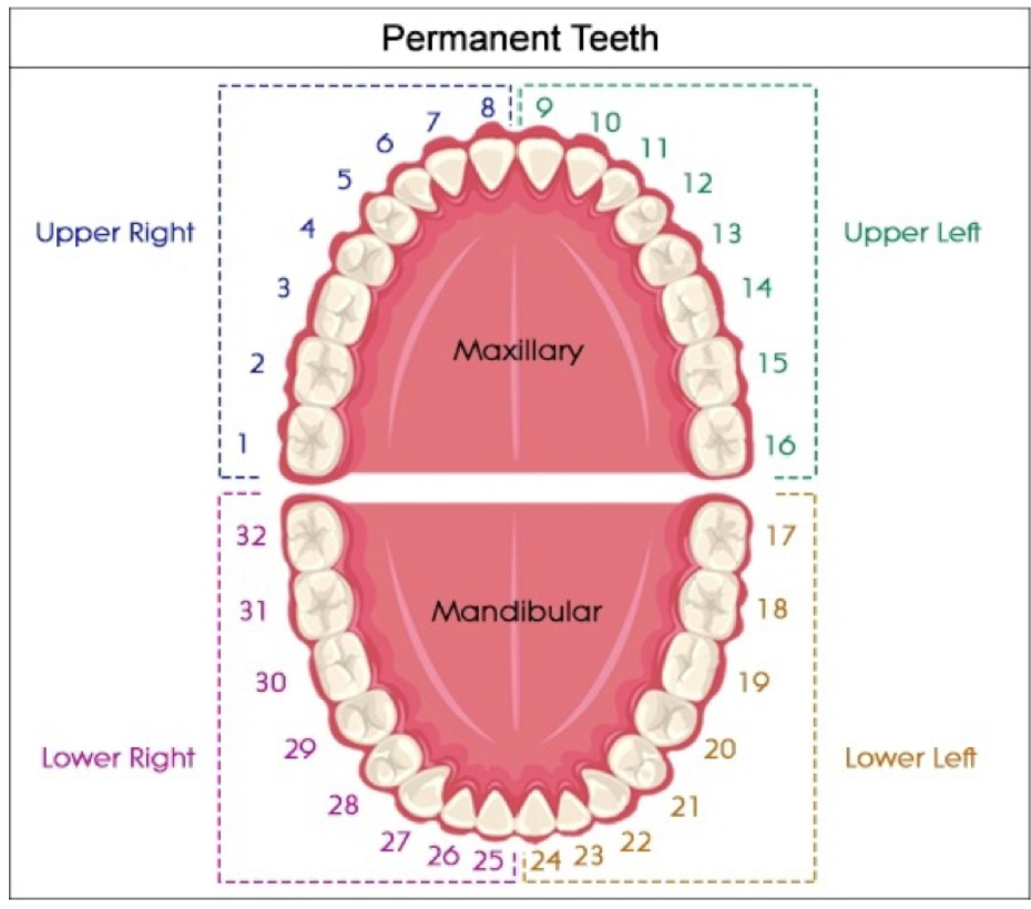
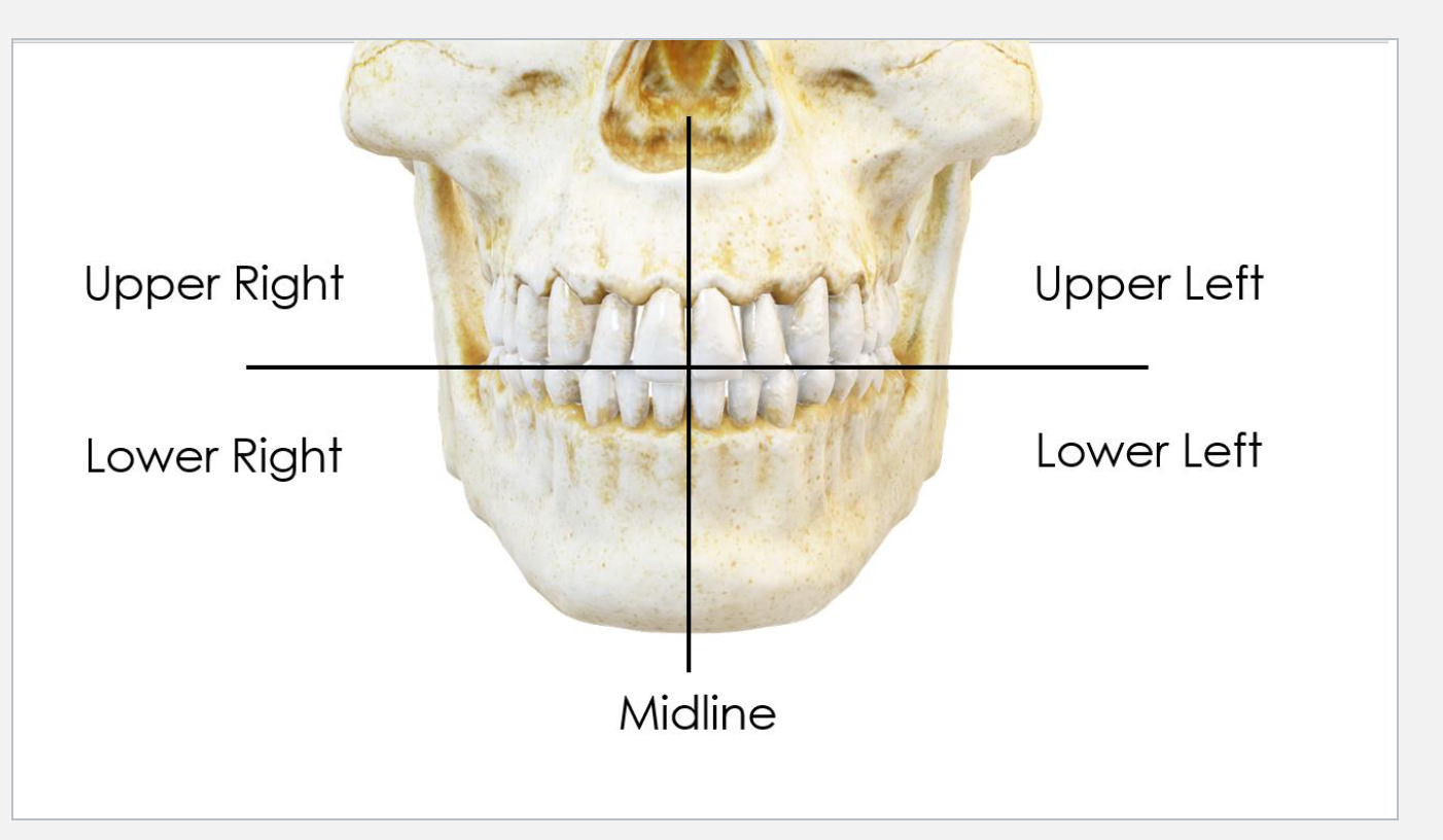
Four Quadrants of the Mouth
Tooth surfaces (8)
B buccal surface
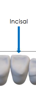
I incisal edge
F facial surface
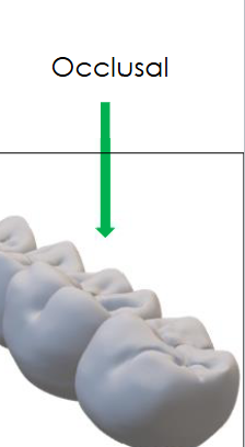
O occlusal surface
L lingual surface
D distal surface
.
M mesial surface
P Palatal surface
O occlusal surface
I incisal edge
F facial surface
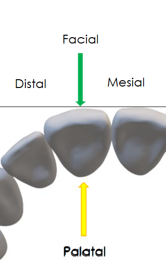
P Palatal surface
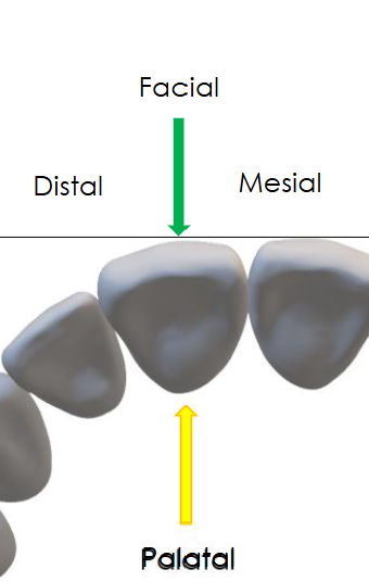
M mesial surface
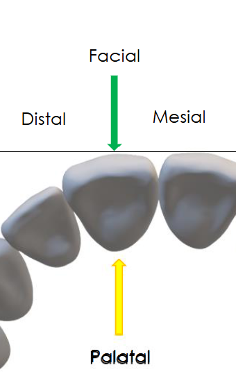
D distal surface
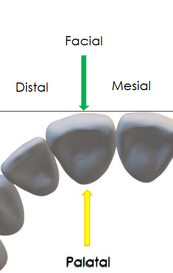
L lingual surface
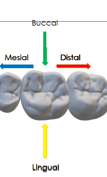
B buccal surface
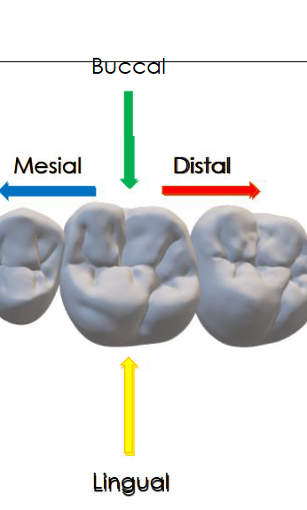
G.V. Black classes
6 of Them
Based on location only
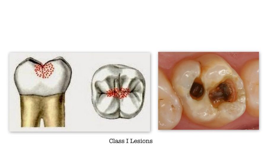
G.V. Black Class I (Three Types)
Pits and fissures of occlusal surfaces of posterior
teeth (premolars and molars)
Pits in the occlusal 2/3 of facial and
lingual surfaces of molars
Smooth proximal surfaces of posterior teeth
(premolars and molars)
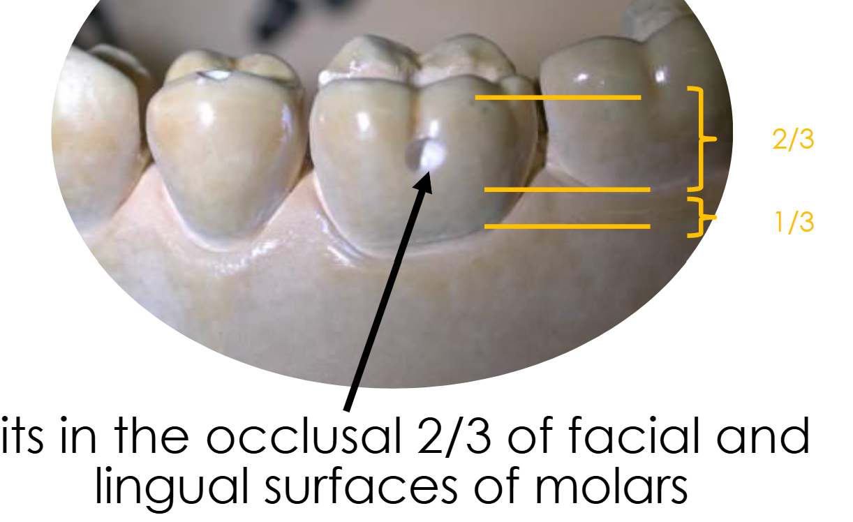
Pits in the occlusal 2/3 of facial and
lingual surfaces of molars
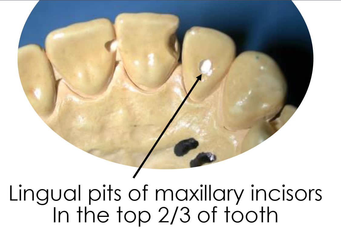
Smooth proximal surfaces of posterior teeth
(premolars and molars)
G.V. Black Class II
Smooth proximal surfaces of posterior teeth
(premolars and molars)
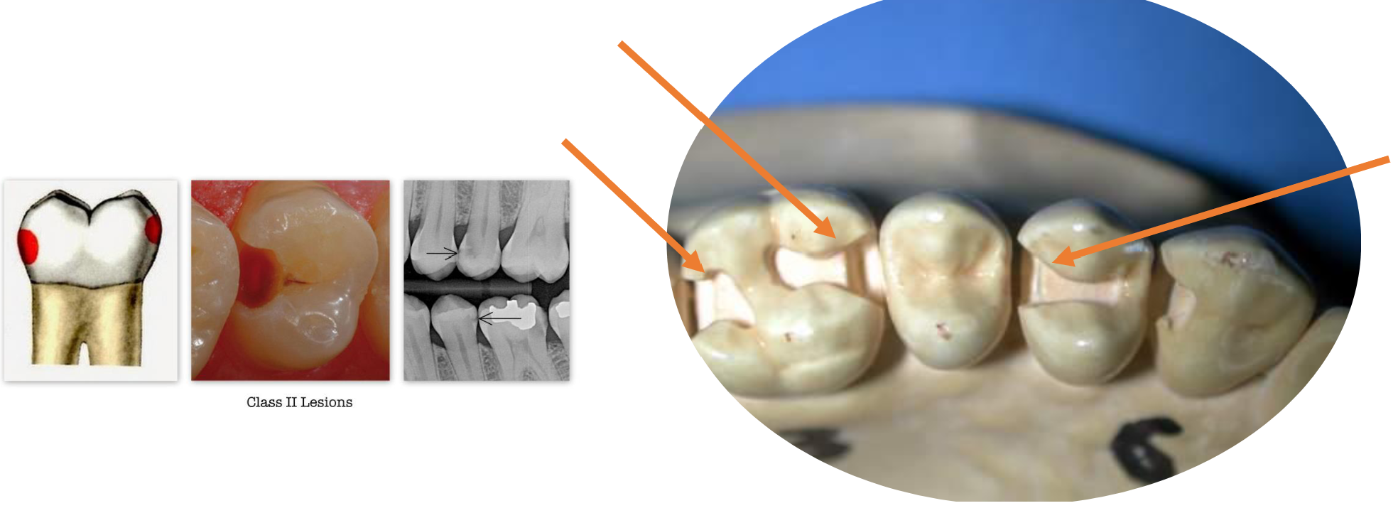
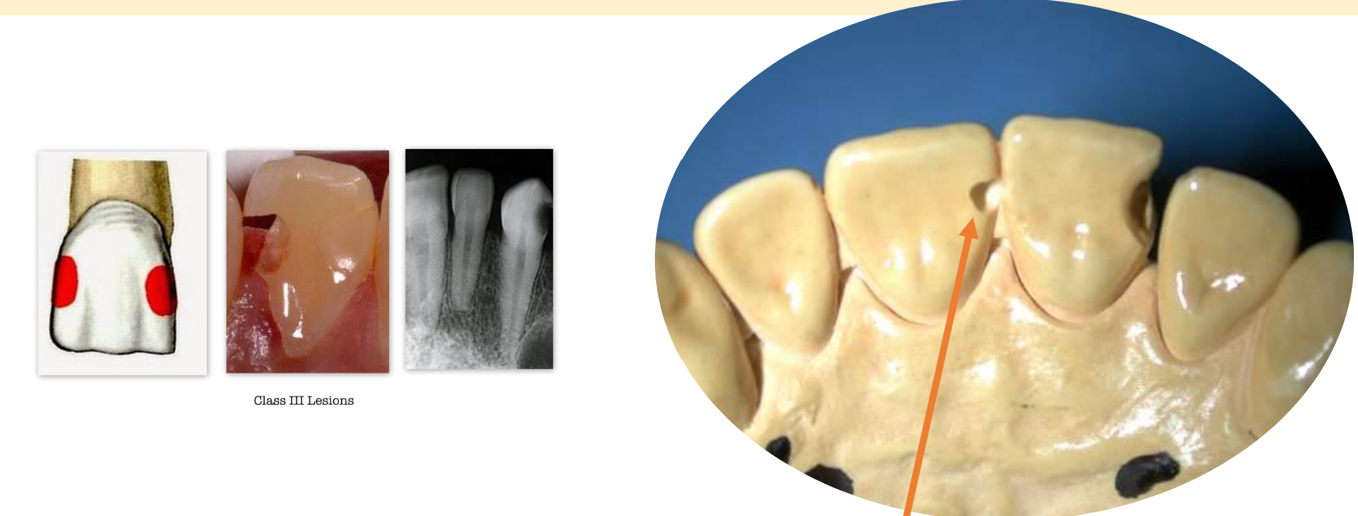
G.V. Black Class III
Smooth proximal surfaces of anterior teeth
(incisors and canines)
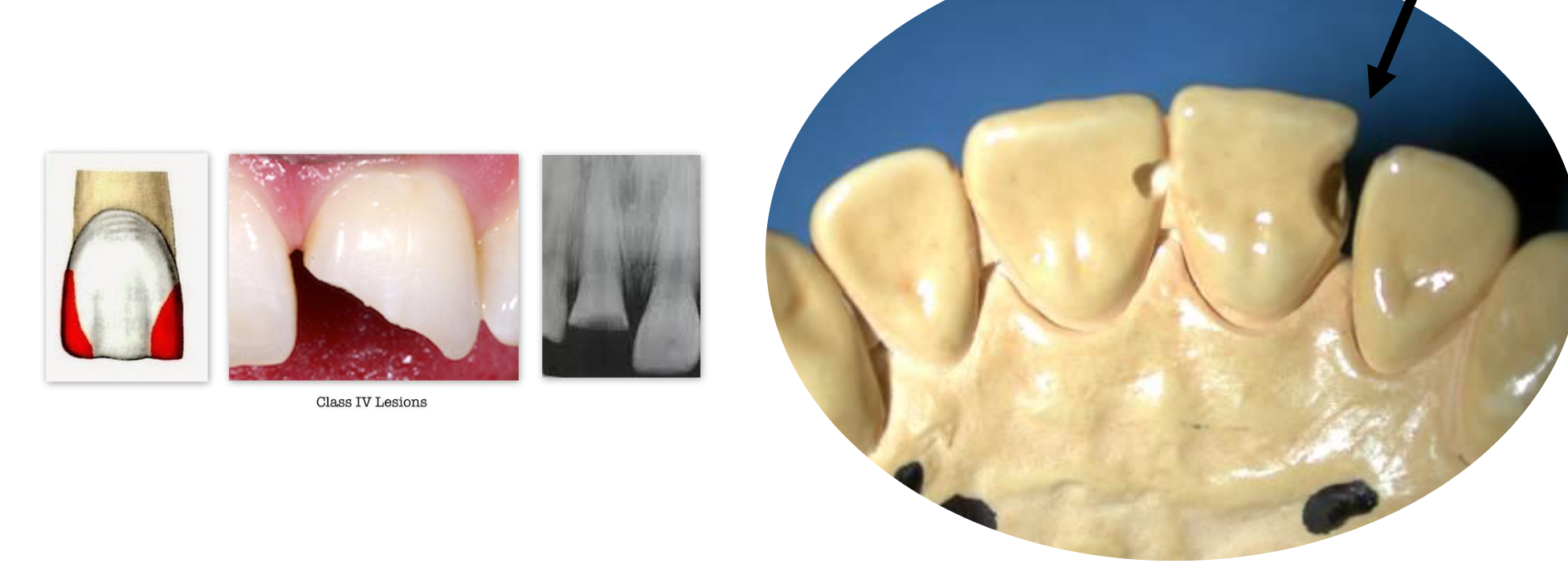
G.V. Black Class IV
Smooth proximal surfaces of anterior teeth (incisors and
canines) which involve the incisal edge as well
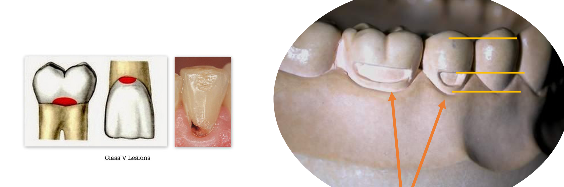
G.V. Black Class V
Smooth surfaces of all teeth in gingival 1/3 of facial and lingual surfaces Class V
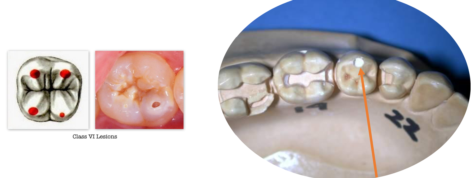
G.V. Black Class VI
Incisal edges of anterior teeth and occlusal cusp tips of posterior teeth
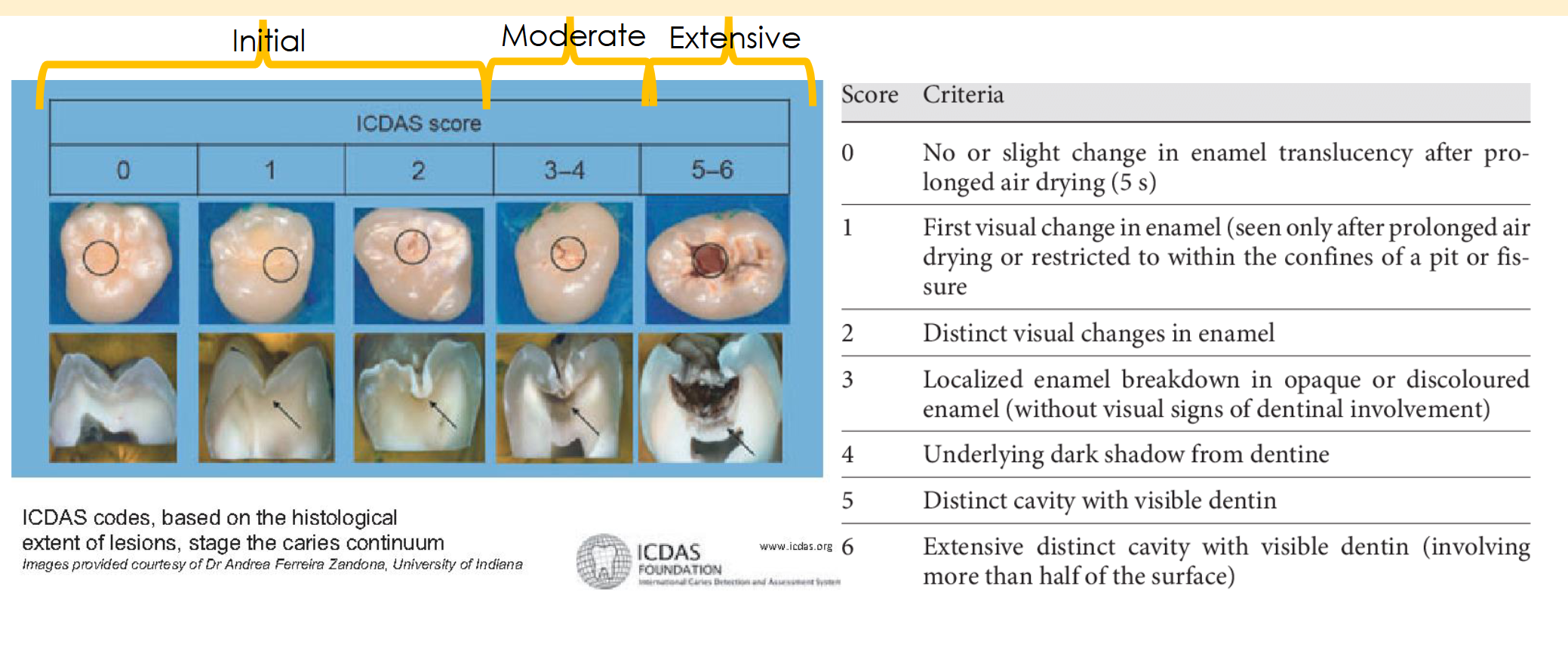
ICDAS
(International Caries detection and assessment system)
From 1 to 6 → How strong is the bacterial caries infection
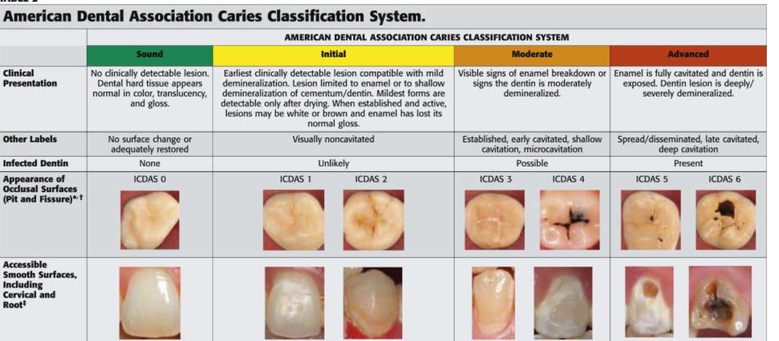
ADA Codes for Caires
1 to 6
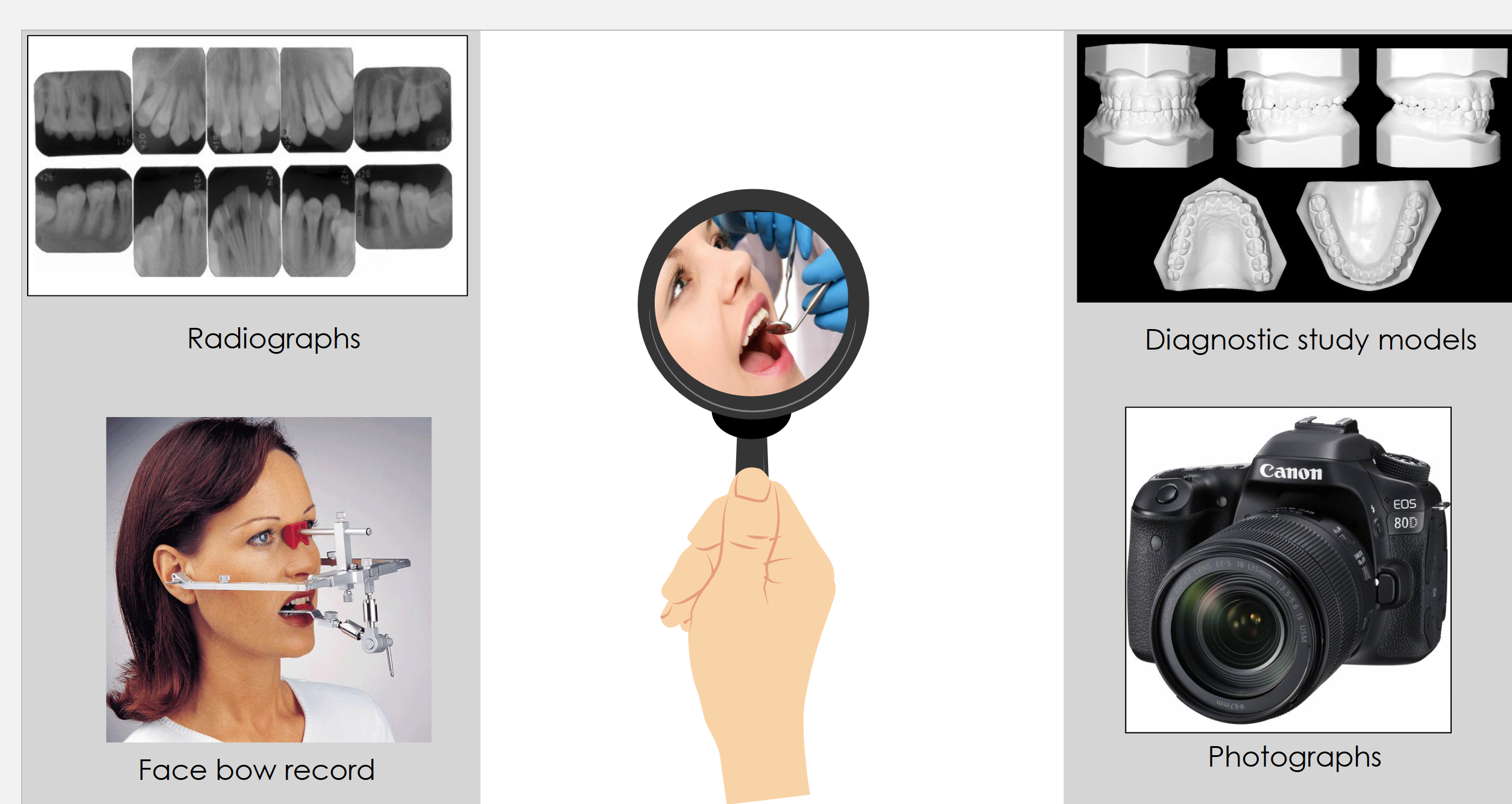
Diagnostic data needed (6)
Caries risk assessment
Medical and dental
history
Patient exam and
charting
Intra and extra oral exam
Diet
Occlusion
Four Tests for Data Gathering Information
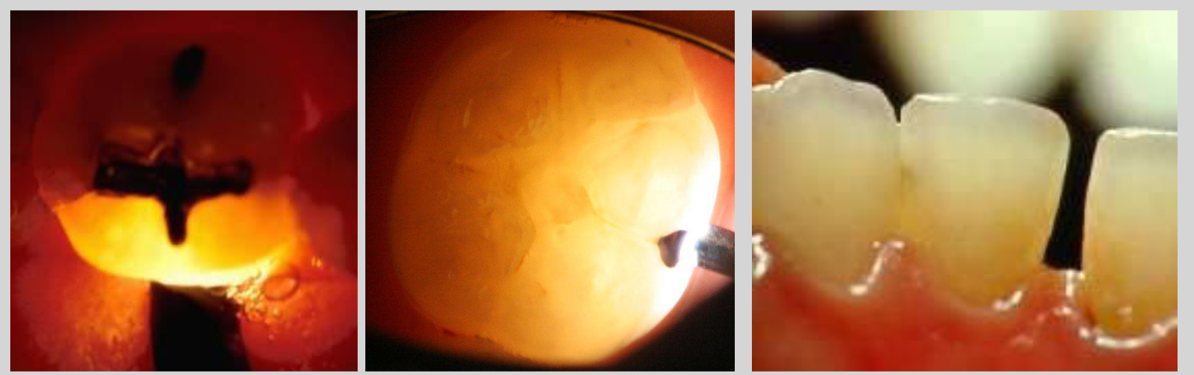
Trans-illumination
Trans-illumination of a cracked tooth and
proximal caries
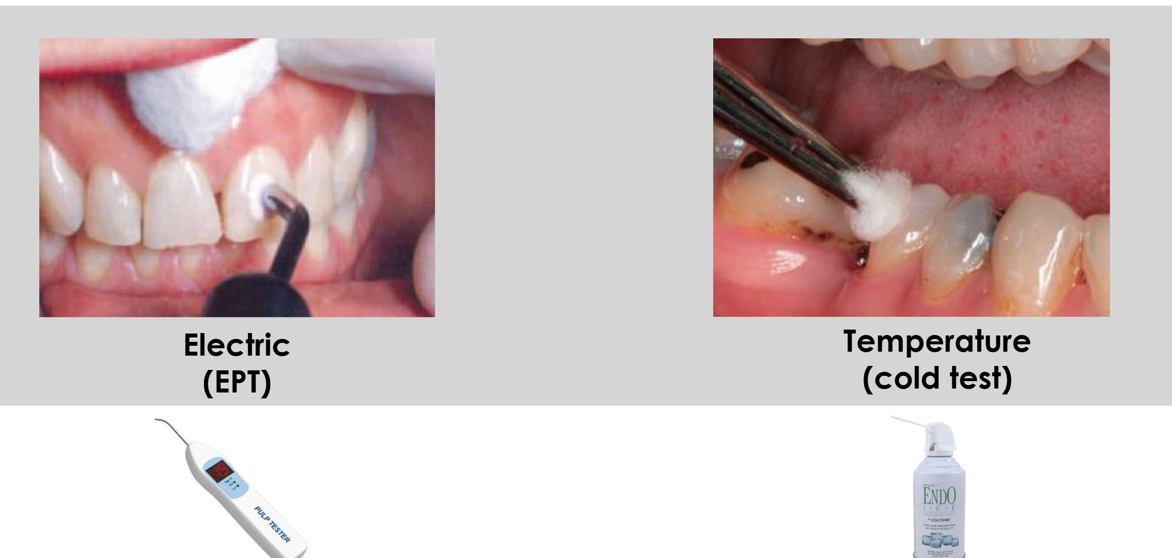
Pulp Vitality Testing (2)
Prioritizing treatment of Caries (3 Phases)
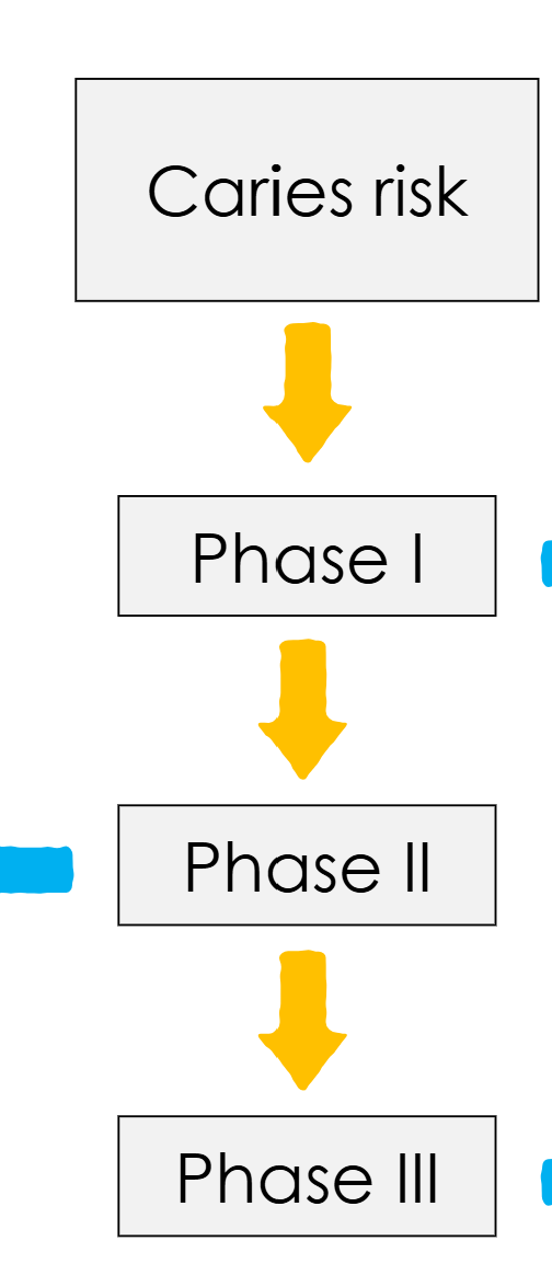
Phase I
Emergencies
• Infections
• Prevention
• Gingival health
• Sealants
Phase II
Simple restorations
Phase III
Complex restorations